|
Indirect Fluorescent Antibody Technique
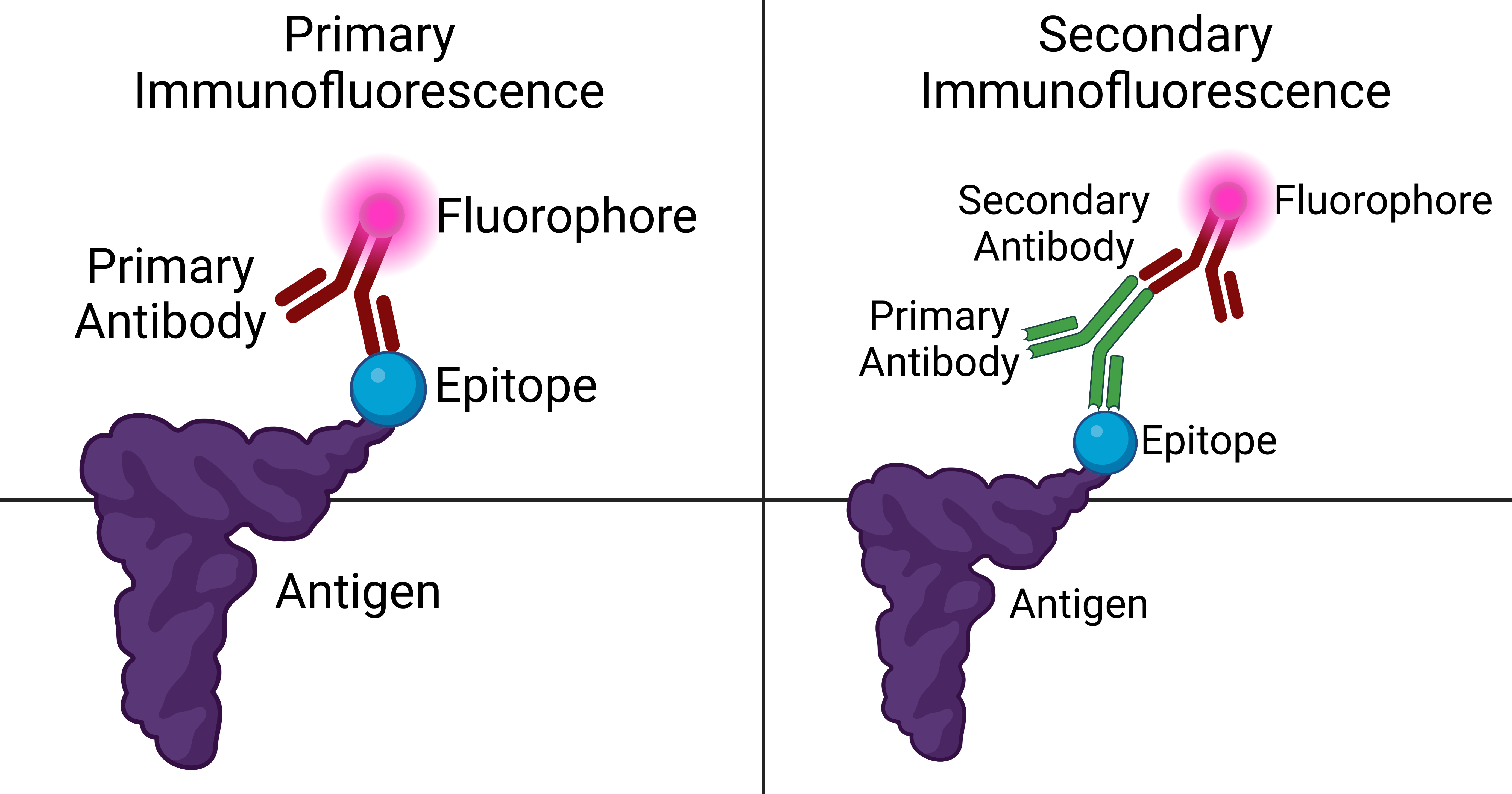
Immunofluorescence is a technique used for light microscopy with a fluorescence microscope and is used primarily on microbiological samples. This technique uses the specificity of antibodies to their antigen to target fluorescent dyes to specific biomolecule targets within a cell, and therefore allows visualization of the distribution of the target molecule through the sample. The specific region an antibody recognizes on an antigen is called an epitope. There have been efforts in epitope mapping since many antibodies can bind the same epitope and levels of binding between antibodies that recognize the same epitope can vary. Additionally, the binding of the fluorophore to the antibody itself cannot interfere with the immunological specificity of the antibody or the binding capacity of its antigen. Immunofluorescence is a widely used example of immunostaining (using antibodies to stain proteins) and is a specific example of immunohistochemistry (the use of the antibody-antige ... [...More Info...] [...Related Items...] OR: [Wikipedia] [Google] [Baidu] |
HSP IF IgA
HSP may refer to: Biology, chemistry, and medicine *Hansen solubility parameters *Heat shock protein *Henoch–Schönlein purpura *Hereditary spastic paraplegia *Highly sensitive person, with high sensory processing sensitivity Mathematics, software, and technology *Hidden subgroup problem, in mathematics *High Speed Photometer, Hubble Space Telescope instrument *Host signal processing, software emulating hardware *Hot Soup Processor, a programming language *High-Scoring Segment Pair, in the BLAST algorithm *List of Bluetooth profiles#Headset Profile (HSP) Education *Harvard Sussex Program, an inter-university collaboration *Holy Spirit Preparatory School, in Atlanta, Georgia, United States Political parties * Croatian Party of Rights (Croatian: ') * People's Voice Party (Turkish: '), Turkey Other uses *Halal snack pack, an Australian dish {{disambiguation ... [...More Info...] [...Related Items...] OR: [Wikipedia] [Google] [Baidu] |
Epifluorescence Microscope
A fluorescence microscope is an optical microscope that uses fluorescence instead of, or in addition to, scattering, reflection, and attenuation or absorption, to study the properties of organic or inorganic substances. "Fluorescence microscope" refers to any microscope that uses fluorescence to generate an image, whether it is a simple set up like an epifluorescence microscope or a more complicated design such as a confocal microscope, which uses optical sectioning to get better resolution of the fluorescence image. Principle The specimen is illuminated with light of a specific wavelength (or wavelengths) which is absorbed by the fluorophores, causing them to emit light of longer wavelengths (i.e., of a different color than the absorbed light). The illumination light is separated from the much weaker emitted fluorescence through the use of a spectral emission filter. Typical components of a fluorescence microscope are a light source (xenon arc lamp or mercury-vapor lamp are co ... [...More Info...] [...Related Items...] OR: [Wikipedia] [Google] [Baidu] |
Patching And Capping
The aggregation of fluorescently tagged antibodies that are associated with proteins on membranes of living cells. The aggregation appears as a cap or a patch in the fluorescence microscope and is due to the bivalent nature of antibodies. Patching and capping were critical in demonstrating the fluid nature of plasma membranes. Variations in density within the specimen are amplified to enhance contrast in unstained cells which is especially useful for examining living unpigmented cells. In other words, phase contrast is a contrast-enhancing optical technique that can be used to produce high contrast images such as living cells and subcellular including nuclei and other organelles. One of the major advantages of using phase contrast microscopy is that living cells can be examined in their natural state without being killed, fixed, or especially stained. As a result, biological processes in the cell can be observed and recorded in high contrast with sharp clarity of minute specimen det ... [...More Info...] [...Related Items...] OR: [Wikipedia] [Google] [Baidu] |
Immunochemistry
Immunochemistry is the study of the chemistry of the immune system. This involves the study of the properties, functions, interactions and production of the chemical components (antibodies/immunoglobulins, toxin, epitopes of proteins like CD4, antitoxins, cytokines/chemokines, antigens) of the immune system. It also include immune responses and determination of immune materials/products by immunochemical assays. In addition, immunochemistry is the study of the identities and functions of the components of the immune system. Immunochemistry is also used to describe the application of immune system components, in particular antibodies, to chemically labelled antigen molecules for visualization. Various methods in immunochemistry have been developed and refined, and used in scientific study, from virology to molecular evolution. Immunochemical techniques include: enzyme-linked immunosorbent assay, immunoblotting (e.g., Western blot assay), precipitation and agglutination reaction ... [...More Info...] [...Related Items...] OR: [Wikipedia] [Google] [Baidu] |
Cutaneous Conditions With Immunofluorescence Findings
Several cutaneous conditions can be diagnosed with the aid of immunofluorescence studies. Cutaneous conditions with positive direct or indirect immunofluorescence when using salt-split skin include: For several subtypes of pemphigus a variety of substrates are used for indirect immunofluorescence: See also * List of cutaneous conditions * List of genes mutated in cutaneous conditions * List of cutaneous conditions caused by mutations in keratins There are many different keratin proteins normally expressed in the human integumentary system. Mutations in keratin proteins in the skin can cause disease. Of note, other structural proteins in the epidermis of the skin that are closely rel ... References * * {{DEFAULTSORT:Immunofluorescence findings for autoimmune bullous conditions Cutaneous conditions Dermatology-related lists ... [...More Info...] [...Related Items...] OR: [Wikipedia] [Google] [Baidu] |
Green Fluorescent Protein
The green fluorescent protein (GFP) is a protein that exhibits bright green fluorescence when exposed to light in the blue to ultraviolet range. The label ''GFP'' traditionally refers to the protein first isolated from the jellyfish ''Aequorea victoria'' and is sometimes called ''avGFP''. However, GFPs have been found in other organisms including corals, sea anemones, zoanithids, copepods and lancelets. The GFP from ''A. victoria'' has a major excitation peak at a wavelength of 395 nm and a minor one at 475 nm. Its emission peak is at 509 nm, which is in the lower green portion of the visible spectrum. The fluorescence quantum yield (QY) of GFP is 0.79. The GFP from the sea pansy (''Renilla reniformis'') has a single major excitation peak at 498 nm. GFP makes for an excellent tool in many forms of biology due to its ability to form an internal chromophore without requiring any accessory cofactors, gene products, or enzymes / substrates other than mo ... [...More Info...] [...Related Items...] OR: [Wikipedia] [Google] [Baidu] |
Recombinant Protein
Recombinant DNA (rDNA) molecules are DNA molecules formed by laboratory methods of genetic recombination (such as molecular cloning) that bring together genetic material from multiple sources, creating sequences that would not otherwise be found in the genome. Recombinant DNA is the general name for a piece of DNA that has been created by combining at least two fragments from two different sources. Recombinant DNA is possible because DNA molecules from all organisms share the same chemical structure, and differ only in the nucleotide sequence within that identical overall structure. Recombinant DNA molecules are sometimes called chimeric DNA, because they can be made of material from two different species, like the mythical chimera. R-DNA technology uses palindromic sequences and leads to the production of sticky and blunt ends. The DNA sequences used in the construction of recombinant DNA molecules can originate from any species. For example, plant DNA may be joined to bacte ... [...More Info...] [...Related Items...] OR: [Wikipedia] [Google] [Baidu] |
DyLight Fluor
The DyLight Fluor family of fluorescent dyes are produced by Dyomics in collaboration with Thermo Fisher Scientific. DyLight dyes are typically used in biotechnology and research applications as biomolecule, cell and tissue labels for fluorescence microscopy, cell biology or molecular biology. Historically, fluorophores such as fluorescein, rhodamine, Cy3 and Cy5 have been used in a wide variety of applications. These dyes have limitations for use in microscopy and other applications that require exposure to an intense light source such as a laser, because they photobleach quickly (however, bleaching can be reduced at least 10 fold using oxygen scavenging). DyLight Fluors have comparable excitation and emission spectra and are claimed to be more photostable, brighter, and less pH-sensitive. The excitation and emission spectra of the DyLight Fluor series cover much of the visible spectrum and extend into the infrared region, allowing detection using most fluorescence microsco ... [...More Info...] [...Related Items...] OR: [Wikipedia] [Google] [Baidu] |
Alexa Fluor
The Alexa Fluor family of fluorescent dyes is a series of dyes invented by Molecular Probes, now a part of Thermo Fisher Scientific, and sold under the Invitrogen brand name. Alexa Fluor dyes are frequently used as cell and tissue labels in fluorescence microscopy and cell biology. Alexa Fluor dyes can be conjugated directly to primary antibodies or to secondary antibodies to amplify signal and sensitivity or other biomolecules. The excitation and emission spectra of the Alexa Fluor series cover the visible spectrum and extend into the infrared. The individual members of the family are numbered according roughly to their excitation maxima in nanometers. History Richard and Rosaria Haugland, the founders of Molecular Probes, are well known in biology and chemistry for their research into fluorescent dyes useful in biological research applications. At the time that Molecular Probes was founded, such products were largely unavailable commercially. A number of fluorescent dyes ... [...More Info...] [...Related Items...] OR: [Wikipedia] [Google] [Baidu] |
Photobleaching
In optics, photobleaching (sometimes termed fading) is the photochemical alteration of a dye or a fluorophore molecule such that it is permanently unable to fluoresce. This is caused by cleaving of covalent bonds or non-specific reactions between the fluorophore and surrounding molecules. Such irreversible modifications in covalent bonds are caused by transition from a singlet state to the triplet state of the fluorophores. The number of excitation cycles to achieve full bleaching varies. In microscopy, photobleaching may complicate the observation of fluorescent molecules, since they will eventually be destroyed by the light exposure necessary to stimulate them into fluorescing. This is especially problematic in time-lapse microscopy. However, photobleaching may also be used prior to applying the (primarily antibody-linked) fluorescent molecules, in an attempt to quench autofluorescence. This can help improve the signal-to-noise ratio. Photobleaching may also be exploited to study ... [...More Info...] [...Related Items...] OR: [Wikipedia] [Google] [Baidu] |
Blood Vessels In Porcine Skin - SMA A488 - 20x
Blood is a body fluid in the circulatory system of humans and other vertebrates that delivers necessary substances such as nutrients and oxygen to the cells, and transports metabolic waste products away from those same cells. Blood in the circulatory system is also known as ''peripheral blood'', and the blood cells it carries, ''peripheral blood cells''. Blood is composed of blood cells suspended in blood plasma. Plasma, which constitutes 55% of blood fluid, is mostly water (92% by volume), and contains proteins, glucose, mineral ions, hormones, carbon dioxide (plasma being the main medium for excretory product transportation), and blood cells themselves. Albumin is the main protein in plasma, and it functions to regulate the colloidal osmotic pressure of blood. The blood cells are mainly red blood cells (also called RBCs or erythrocytes), white blood cells (also called WBCs or leukocytes) and platelets (also called thrombocytes). The most abundant cells in vertebrate blood ar ... [...More Info...] [...Related Items...] OR: [Wikipedia] [Google] [Baidu] |
Fluorophore
A fluorophore (or fluorochrome, similarly to a chromophore) is a fluorescent chemical compound that can re-emit light upon light excitation. Fluorophores typically contain several combined aromatic groups, or planar or cyclic molecules with several π bonds. Fluorophores are sometimes used alone, as a tracer in fluids, as a dye for staining of certain structures, as a substrate of enzymes, or as a probe or indicator (when its fluorescence is affected by environmental aspects such as polarity or ions). More generally they are covalently bonded to a macromolecule, serving as a marker (or dye, or tag, or reporter) for affine or bioactive reagents (antibodies, peptides, nucleic acids). Fluorophores are notably used to stain tissues, cells, or materials in a variety of analytical methods, i.e., fluorescent imaging and spectroscopy. Fluorescein, via its amine-reactive isothiocyanate derivative fluorescein isothiocyanate (FITC), has been one of the most popular fluorophores. Fro ... [...More Info...] [...Related Items...] OR: [Wikipedia] [Google] [Baidu] |


