|
Ileal Conduit
An ileal conduit urinary diversion is one of various surgical techniques for urinary diversion. It has sometimes been referred to as the Bricker ileal conduit after its inventor, Eugene M. Bricker. It is a form of incontinent urostomy, and was developed during the 1940s and is still one of the most used techniques for the diversion of urine after a patient has had their bladder removed, due to its low complication rate and high patient satisfaction level. It is usually used in conjunction with radical cystectomy in order to control invasive bladder cancer. To create an ileal conduit, the ureters are surgically resected from the bladder and a ureteroenteric anastomosis is made in order to drain the urine into a detached section of ileum at the distal small intestine, though the distal most 25 cm of terminal ileum are avoided as this is where bile salts are reabsorbed. The end of the ileum is then brought out through an opening (a stoma) in the abdominal wall. The residual small bo ... [...More Info...] [...Related Items...] OR: [Wikipedia] [Google] [Baidu] |
Urinary Diversion
Urinary diversion is any one of several surgical procedures to reroute urine flow from its normal pathway. It may be necessary for diseased or defective ureters, bladder or urethra, either temporarily or permanently. Some diversions result in a stoma. Types * Nephrostomy from the renal pelvis * Urostomy from more distal origins along the urinary tract, with subtypes including: ** Ileal conduit urinary diversion (Bricker conduit) ** Indiana pouch * Neobladder to urethra diversion Ureteroenteric anastomosis A common feature of the three first, and most common, types of urinary diversion is the ureteroenteric anastomosis. This is the joining site of the ureters and the section of intestine used for the diversion. The ureteroenteric anastomosis can be created in a number of different ways. There is the option of a refluxing or a non-refluxing type, and the two ureters can be joined into the intestinal segment either together or separately. The non-refluxing type has been associ ... [...More Info...] [...Related Items...] OR: [Wikipedia] [Google] [Baidu] |
Terminal Ileum
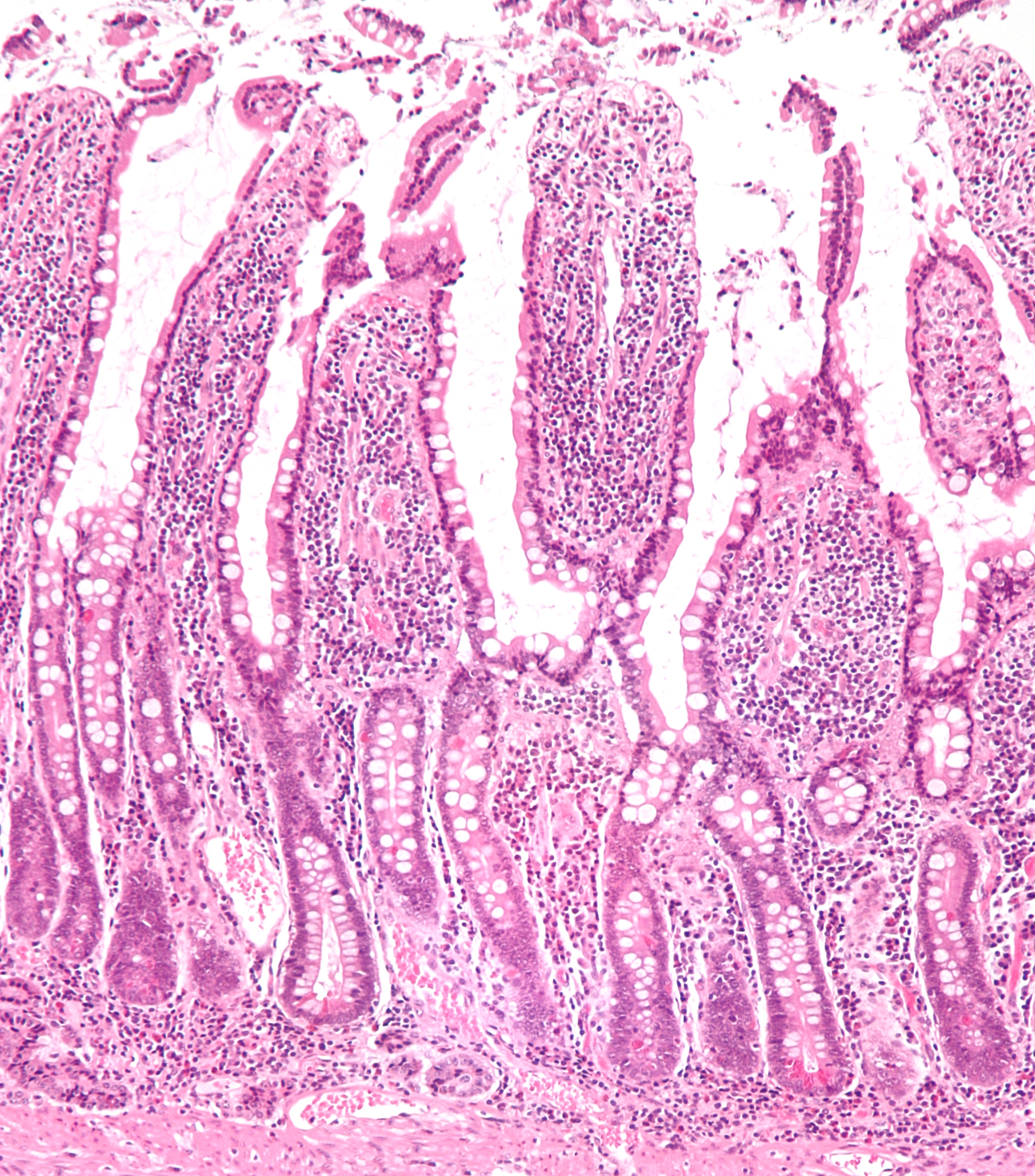
The ileum () is the final section of the small intestine in most higher vertebrates, including mammals, reptiles, and birds. In fish, the divisions of the small intestine are not as clear and the terms posterior intestine or distal intestine may be used instead of ileum. Its main function is to absorb vitamin B12, bile salts, and whatever products of digestion that were not absorbed by the jejunum. The ileum follows the duodenum and jejunum and is separated from the cecum by the ileocecal valve (ICV). In humans, the ileum is about 2–4 m long, and the pH is usually between 7 and 8 (neutral or slightly basic). ''Ileum ''is derived from the Greek word ''eilein'', meaning "to twist up tightly". Structure The ileum is the third and final part of the small intestine. It follows the jejunum and ends at the ileocecal junction, where the terminal ileum communicates with the cecum of the large intestine through the ileocecal valve. The ileum, along with the jejunum, is suspended ... [...More Info...] [...Related Items...] OR: [Wikipedia] [Google] [Baidu] |
Mitrofanoff Procedure
The Mitrofanoff procedure, also known as the Mitrofanoff appendicovesicostomy, is a surgical procedure in which the appendix is used to create a conduit, or channel, between the skin surface and the urinary bladder. The small opening on the skin surface, or the stoma, is typically located either in the navel or nearby the navel on the right lower side of the abdomen. Originally developed by Professor Paul Mitrofanoff in 1980, the procedure represents an alternative to urethral catheterization and is sometimes used by people with urethral damage or by those with severe autonomic dysreflexia. An intermittent catheter, or a catheter that is inserted and then removed after use, is typically passed through the channel every 3–4 hours and the urine is drained into a toilet or a bottle. As the bladder fills, rising pressure compresses the channel against the bladder wall, creating a one-way valve that prevents leakage of urine between catheterizations. Description of procedure A surgeo ... [...More Info...] [...Related Items...] OR: [Wikipedia] [Google] [Baidu] |
Urostomy
A urostomy is a surgical procedure that creates a stoma (artificial opening) for the urinary system. A urostomy is made to avail for urinary diversion in cases where drainage of urine through the bladder and urethra is not possible, e.g. after extensive surgery or in case of obstruction. fro Cancer.org (American Cancer Society).Last Medical Review: 03/17/2011. Last Revised: 03/17/2011 Techniques  Techniques include:
:::::::* Techniques include:
:::::::*
|
Excoriation
A skin condition, also known as cutaneous condition, is any medical condition that affects the integumentary system—the organ system that encloses the body and includes skin, nails, and related muscle and glands. The major function of this system is as a barrier against the external environment. Conditions of the human integumentary system constitute a broad spectrum of diseases, also known as dermatoses, as well as many nonpathologic states (like, in certain circumstances, melanonychia and racquet nails). While only a small number of skin diseases account for most visits to the physician, thousands of skin conditions have been described. Classification of these conditions often presents many nosological challenges, since underlying causes and pathogenetics are often not known. Therefore, most current textbooks present a classification based on location (for example, conditions of the mucous membrane), morphology ( chronic blistering conditions), cause (skin conditions resu ... [...More Info...] [...Related Items...] OR: [Wikipedia] [Google] [Baidu] |
Abdominal Wall
In anatomy, the abdominal wall represents the boundaries of the abdominal cavity. The abdominal wall is split into the anterolateral and posterior walls. There is a common set of layers covering and forming all the walls: the deepest being the visceral peritoneum, which covers many of the abdominal organs (most of the large and small intestines, for example), and the parietal peritoneum- which covers the visceral peritoneum below it, the extraperitoneal fat, the transversalis fascia, the internal and external oblique and transversus abdominis aponeurosis, and a layer of fascia, which has different names according to what it covers (e.g., transversalis, psoas fascia). In medical vernacular, the term 'abdominal wall' most commonly refers to the layers composing the anterior abdominal wall which, in addition to the layers mentioned above, includes the three layers of muscle: the transversus abdominis (transverse abdominal muscle), the internal (obliquus internus) and the external o ... [...More Info...] [...Related Items...] OR: [Wikipedia] [Google] [Baidu] |
Stoma (medicine)
In anatomy, a stoma (plural stomata or stomas) is any opening in the body. For example, a mouth, a nose, and an anus are natural stomata. Any hollow organ can be manipulated into an artificial stoma as necessary. This includes the esophagus, stomach, duodenum, ileum, colon, pleural cavity, ureters, urinary bladder, and renal pelvis. Such a stoma may be permanent or temporary. Surgical procedures that involve the creation of an artificial stoma have names that typically end with the suffix "-ostomy", and the same names are also often used to refer to the stoma thus created. For example, the word "colostomy" often refers either to an artificial anus or the procedure that creates one. Accordingly, it is not unusual for a stoma to be called an ostomy (plural ostomies), as is the norm in wound, ostomy, and continence nursing. Gastrointestinal stomata Stomata are created in particular in surgical procedures involving the gastrointestinal tract (GIT) or gastrointestinal system ( ... [...More Info...] [...Related Items...] OR: [Wikipedia] [Google] [Baidu] |
Bile Salts
Bile acids are steroid acids found predominantly in the bile of mammals and other vertebrates. Diverse bile acids are synthesized in the liver. Bile acids are conjugated with taurine or glycine residues to give anions called bile salts. Primary bile acids are those synthesized by the liver. Secondary bile acids result from bacterial actions in the colon. In humans, taurocholic acid and glycocholic acid (derivatives of cholic acid) and taurochenodeoxycholic acid and glycochenodeoxycholic acid (derivatives of chenodeoxycholic acid) are the major bile salts. They are roughly equal in concentration. The salts of their 7-alpha-dehydroxylated derivatives, deoxycholic acid and lithocholic acid, are also found, with derivatives of cholic, chenodeoxycholic and deoxycholic acids accounting for over 90% of human biliary bile acids. Bile acids comprise about 80% of the organic compounds in bile (others are phospholipids and cholesterol). An increased secretion of bile acids produces an inc ... [...More Info...] [...Related Items...] OR: [Wikipedia] [Google] [Baidu] |
Small Intestine
The small intestine or small bowel is an organ in the gastrointestinal tract where most of the absorption of nutrients from food takes place. It lies between the stomach and large intestine, and receives bile and pancreatic juice through the pancreatic duct to aid in digestion. The small intestine is about long and folds many times to fit in the abdomen. Although it is longer than the large intestine, it is called the small intestine because it is narrower in diameter. The small intestine has three distinct regions – the duodenum, jejunum, and ileum. The duodenum, the shortest, is where preparation for absorption through small finger-like protrusions called villi begins. The jejunum is specialized for the absorption through its lining by enterocytes: small nutrient particles which have been previously digested by enzymes in the duodenum. The main function of the ileum is to absorb vitamin B12, bile salts, and whatever products of digestion that were not absorbed by the ... [...More Info...] [...Related Items...] OR: [Wikipedia] [Google] [Baidu] |
Urostomy
A urostomy is a surgical procedure that creates a stoma (artificial opening) for the urinary system. A urostomy is made to avail for urinary diversion in cases where drainage of urine through the bladder and urethra is not possible, e.g. after extensive surgery or in case of obstruction. fro Cancer.org (American Cancer Society).Last Medical Review: 03/17/2011. Last Revised: 03/17/2011 Techniques  Techniques include:
:::::::* Techniques include:
:::::::*
|
Ileum
The ileum () is the final section of the small intestine in most higher vertebrates, including mammals, reptiles, and birds. In fish, the divisions of the small intestine are not as clear and the terms posterior intestine or distal intestine may be used instead of ileum. Its main function is to absorb vitamin B12, bile salts, and whatever products of digestion that were not absorbed by the jejunum. The ileum follows the duodenum and jejunum and is separated from the cecum by the ileocecal valve (ICV). In humans, the ileum is about 2–4 m long, and the pH is usually between 7 and 8 (neutral or slightly basic). ''Ileum ''is derived from the Greek word ''eilein'', meaning "to twist up tightly". Structure The ileum is the third and final part of the small intestine. It follows the jejunum and ends at the ileocecal junction, where the terminal ileum communicates with the cecum of the large intestine through the ileocecal valve. The ileum, along with the jejunum, is suspended ... [...More Info...] [...Related Items...] OR: [Wikipedia] [Google] [Baidu] |
Ureteroenteric Anastomosis
Urinary diversion is any one of several surgical procedures to reroute urine flow from its normal pathway. It may be necessary for diseased or defective ureters, bladder or urethra, either temporarily or permanently. Some diversions result in a stoma. Types * Nephrostomy from the renal pelvis * Urostomy from more distal origins along the urinary tract, with subtypes including: ** Ileal conduit urinary diversion (Bricker conduit) ** Indiana pouch * Neobladder to urethra diversion Ureteroenteric anastomosis A common feature of the three first, and most common, types of urinary diversion is the ureteroenteric anastomosis. This is the joining site of the ureters and the section of intestine used for the diversion. The ureteroenteric anastomosis can be created in a number of different ways. There is the option of a refluxing or a non-refluxing type, and the two ureters can be joined into the intestinal segment either together or separately. The non-refluxing type has been associate ... [...More Info...] [...Related Items...] OR: [Wikipedia] [Google] [Baidu] |


