|
Ischemic Optic Neuropathy
Ischemic optic neuropathy (ION) is the loss of structure and function of a portion of the optic nerve due to obstruction of blood flow to the nerve (i.e. ischemia). Ischemic forms of optic neuropathy are typically classified as either anterior ischemic optic neuropathy or posterior ischemic optic neuropathy according to the part of the optic nerve that is affected. People affected will often complain of a loss of visual acuity and a visual field, the latter of which is usually in the superior or inferior field. When ION occurs in patients below the age of 50 years old, other causes should be considered, such as juvenile diabetes mellitus, antiphospholipid antibody-associated clotting disorders, collagen-vascular disease, and migraines. Rarely, complications of intraocular surgery or acute blood loss may cause an ischemic event in the optic nerve. Presentation Anterior ION presents with sudden, painless visual loss, developing over hours to days. Diagnosis Examination finding ... [...More Info...] [...Related Items...] OR: [Wikipedia] [Google] [Baidu] |
Optic Nerve
In neuroanatomy, the optic nerve, also known as the second cranial nerve, cranial nerve II, or simply CN II, is a paired cranial nerve that transmits visual system, visual information from the retina to the brain. In humans, the optic nerve is derived from optic stalks during the seventh week of development and is composed of retinal ganglion cell axons and glial cells; it extends from the optic disc to the optic chiasma and continues as the optic tract to the lateral geniculate nucleus, Pretectal area, pretectal nuclei, and superior colliculus. Structure The optic nerve has been classified as the second of twelve paired cranial nerves, but it is technically part of the central nervous system, rather than the peripheral nervous system because it is derived from an out-pouching of the diencephalon (optic stalks) during embryonic development. As a consequence, the fibers of the optic nerve are covered with myelin produced by oligodendrocytes, rather than Schwann cells of the per ... [...More Info...] [...Related Items...] OR: [Wikipedia] [Google] [Baidu] |
Migraine
Migraine (, ) is a common neurological disorder characterized by recurrent headaches. Typically, the associated headache affects one side of the head, is pulsating in nature, may be moderate to severe in intensity, and could last from a few hours to three days. Non-headache symptoms may include nausea, vomiting, and photophobia, sensitivity to light, hyperacusis, sound, or Osmophobia, smell. The pain is generally made worse by physical activity during an attack,as PDF although regular physical exercise may prevent future attacks. Up to one-third of people affected have Aura (symptom), aura: typically, it is a short period of visual disturbance that signals that the headache will soon occur. Occasionally, aura can occur with little or no headache follow ... [...More Info...] [...Related Items...] OR: [Wikipedia] [Google] [Baidu] |
Edema
Edema, also spelled oedema, and also known as fluid retention, dropsy, hydropsy and swelling, is the build-up of fluid in the body's Tissue (biology), tissue. Most commonly, the legs or arms are affected. Symptoms may include skin which feels tight, the area may feel heavy, and joint stiffness. Other symptoms depend on the underlying cause. Causes may include Chronic venous insufficiency, venous insufficiency, heart failure, kidney problems, hypoalbuminemia, low protein levels, liver problems, deep vein thrombosis, infections, angioedema, certain medications, and lymphedema. It may also occur after prolonged sitting or standing and during menstruation or pregnancy. The condition is more concerning if it starts suddenly, or pain or shortness of breath is present. Treatment depends on the underlying cause. If the underlying mechanism involves Hypernatremia, sodium retention, decreased salt intake and a diuretic may be used. Elevating the legs and support stockings may be useful ... [...More Info...] [...Related Items...] OR: [Wikipedia] [Google] [Baidu] |
Optic Disc
The optic disc or optic nerve head is the point of exit for ganglion cell axons leaving the eye. Because there are no rods or cones overlying the optic disc, it corresponds to a small blind spot in each eye. The ganglion cell axons form the optic nerve after they leave the eye. The optic disc represents the beginning of the optic nerve and is the point where the axons of retinal ganglion cells come together. The optic disc is also the entry point for the major blood vessels that supply the retina. The optic disc in a normal human eye carries 1–1.2 million afferent nerve fibers from the eye towards the brain. Structure The optic disc is placed 3 to 4 mm to the nasal side of the fovea. It is a vertical oval, with average dimensions of 1.76mm horizontally by 1.92mm vertically. There is a central depression, of variable size, called the optic cup. This depression can be a variety of shapes from a shallow indentation to a bean pot—this shape can be significant for diagn ... [...More Info...] [...Related Items...] OR: [Wikipedia] [Google] [Baidu] |
Relative Afferent Pupillary Defect
A relative afferent pupillary defect (RAPD), also known as a Marcus Gunn pupil, is a medical sign observed during the swinging-flashlight test whereupon the patient's pupils dilate when a bright light is swung from the unaffected eye to the affected eye. The affected eye still senses the light and produces pupillary sphincter constriction to some degree, albeit reduced. Depending on severity, different symptoms may appear during the swinging flash light test: Mild RAPD will presents as a weak pupil constriction initially, after which dilation continues to happen. When RAPD is moderate, pupil size will remain, after which it dilates When RAPD is severe, the pupil will dilate quickly Cause The most common cause of Marcus Gunn pupil is a lesion of the optic nerve (between the retina and the optic chiasm) due to glaucoma, or severe retinal disease, or due to multiple sclerosis. It is named after Scottish ophthalmologist Robert Marcus Gunn. A second common cause of Marcus Gunn pup ... [...More Info...] [...Related Items...] OR: [Wikipedia] [Google] [Baidu] |
Color Vision
Color vision, a feature of visual perception, is an ability to perceive differences between light composed of different wavelengths (i.e., different spectral power distributions) independently of light intensity. Color perception is a part of the larger visual system and is mediated by a complex process between neurons that begins with differential stimulation of different types of photoreceptors by light entering the eye. Those photoreceptors then emit outputs that are propagated through many layers of neurons and then ultimately to the brain. Color vision is found in many animals and is mediated by similar underlying mechanisms with common types of biological molecules and a complex history of evolution in different animal taxa. In primates, color vision may have evolved under selective pressure for a variety of visual tasks including the foraging for nutritious young leaves, ripe fruit, and flowers, as well as detecting predator camouflage and emotional states in other pr ... [...More Info...] [...Related Items...] OR: [Wikipedia] [Google] [Baidu] |
Visual Impairment
Visual impairment, also known as vision impairment, is a medical definition primarily measured based on an individual's better eye visual acuity; in the absence of treatment such as correctable eyewear, assistive devices, and medical treatment– visual impairment may cause the individual difficulties with normal daily tasks including reading and walking. Low vision is a functional definition of visual impairment that is chronic, uncorrectable with treatment or correctable lenses, and impacts daily living. As such low vision can be used as a disability metric and varies based on an individual's experience, environmental demands, accommodations, and access to services. The American Academy of Ophthalmology defines visual impairment as the best-corrected visual acuity of less than 20/40 in the better eye, and the World Health Organization defines it as a presenting acuity of less than 6/12 in the better eye. The term blindness is used for complete or nearly complete vision loss. In ... [...More Info...] [...Related Items...] OR: [Wikipedia] [Google] [Baidu] |
Bleeding
Bleeding, hemorrhage, haemorrhage or blood loss, is blood escaping from the circulatory system from damaged blood vessels. Bleeding can occur internally, or externally either through a natural opening such as the mouth, nose, ear, urethra, vagina or anus, or through a puncture in the skin. Hypovolemia is a massive decrease in blood volume, and death by excessive loss of blood is referred to as exsanguination. Typically, a healthy person can endure a loss of 10–15% of the total blood volume without serious medical difficulties (by comparison, blood donation typically takes 8–10% of the donor's blood volume). The stopping or controlling of bleeding is called hemostasis and is an important part of both first aid and surgery. Types * Upper head ** Intracranial hemorrhage – bleeding in the skull. ** Cerebral hemorrhage – a type of intracranial hemorrhage, bleeding within the brain tissue itself. ** Intracerebral hemorrhage – bleeding in the brain caused by the ruptur ... [...More Info...] [...Related Items...] OR: [Wikipedia] [Google] [Baidu] |
Eye Surgery
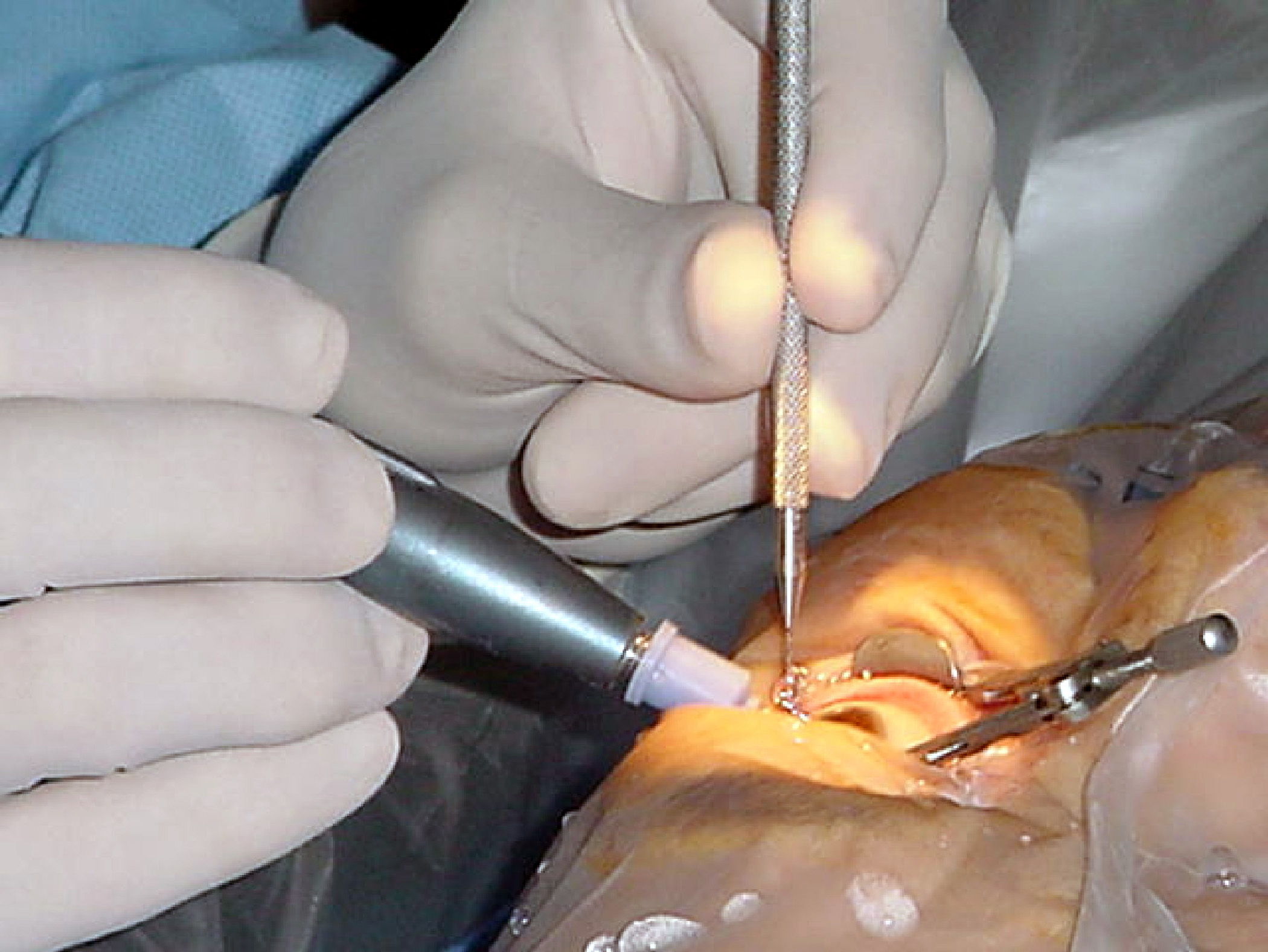
Eye surgery, also known as ophthalmic or ocular surgery, is surgery performed on the eye or its adnexa, by an ophthalmologist or sometimes, an optometrist. Eye surgery is synonymous with ophthalmology. The eye is a very fragile organ, and requires extreme care before, during, and after a surgical procedure to minimize or prevent further damage. An expert eye surgeon is responsible for selecting the appropriate surgical procedure for the patient, and for taking the necessary safety precautions. Mentions of eye surgery can be found in several ancient texts dating back as early as 1800 BC, with cataract treatment starting in the fifth century BC. Today it continues to be a widely practiced type of surgery, with various techniques having been developed for treating eye problems. Preparation and precautions Since the eye is heavily supplied by nerves, anesthesia is essential. Local anesthesia is most commonly used. Topical anesthesia using lidocaine topical gel is often used fo ... [...More Info...] [...Related Items...] OR: [Wikipedia] [Google] [Baidu] |
Coagulopathy
Coagulopathy (also called a bleeding disorder) is a condition in which the blood's ability to coagulate (form clots) is impaired. This condition can cause a tendency toward prolonged or excessive bleeding (bleeding diathesis), which may occur spontaneously or following an injury or medical and dental procedures. Coagulopathies are sometimes erroneously referred to as "clotting disorders", but a clotting disorder is the opposite, defined as a predisposition to excessive clot formation (thrombus), also known as a hypercoagulable state or thrombophilia. Signs and symptoms Coagulopathy may cause uncontrolled internal or external bleeding. Left untreated, uncontrolled bleeding may cause damage to joints, muscles, or internal organs and may be life-threatening. People should seek immediate medical care for serious symptoms, including heavy external bleeding, blood in the urine or stool, double vision, severe head or neck pain, repeated vomiting, difficulty walking, convulsions, or se ... [...More Info...] [...Related Items...] OR: [Wikipedia] [Google] [Baidu] |
Ischemia
Ischemia or ischaemia is a restriction in blood supply to any tissue, muscle group, or organ of the body, causing a shortage of oxygen that is needed for cellular metabolism (to keep tissue alive). Ischemia is generally caused by problems with blood vessels, with resultant damage to or dysfunction of tissue i.e. hypoxia and microvascular dysfunction. It also implies local hypoxia in a part of a body resulting from constriction (such as vasoconstriction, thrombosis, or embolism). Ischemia causes not only insufficiency of oxygen, but also reduced availability of nutrients and inadequate removal of metabolic wastes. Ischemia can be partial (poor perfusion) or total blockage. The inadequate delivery of oxygenated blood to the organs must be resolved either by treating the cause of the inadequate delivery or reducing the oxygen demand of the system that needs it. For example, patients with myocardial ischemia have a decreased blood flow to the heart and are prescribed with medi ... [...More Info...] [...Related Items...] OR: [Wikipedia] [Google] [Baidu] |
Antiphospholipid Syndrome
Antiphospholipid syndrome, or antiphospholipid antibody syndrome (APS or APLS), is an autoimmune, hypercoagulable state caused by antiphospholipid antibodies. APS provokes blood clots (thrombosis) in both arteries and veins as well as pregnancy-related complications such as miscarriage, stillbirth, preterm delivery, and severe preeclampsia. Although the exact etiology of APS is still not clear, genetics is believed to play a key role in the development of the disease. The diagnostic criteria require one clinical event (i.e. thrombosis or pregnancy complication) and two positive blood test results spaced at least three months apart that detect lupus anticoagulant, anti-apolipoprotein antibodies, or anti-cardiolipin antibodies. Antiphospholipid syndrome can be primary or secondary. Primary antiphospholipid syndrome occurs in the absence of any other related disease. Secondary antiphospholipid syndrome occurs with other autoimmune diseases, such as systemic lupus erythematosu ... [...More Info...] [...Related Items...] OR: [Wikipedia] [Google] [Baidu] |

_1.jpg)


