|
Intertrochanteric Crest
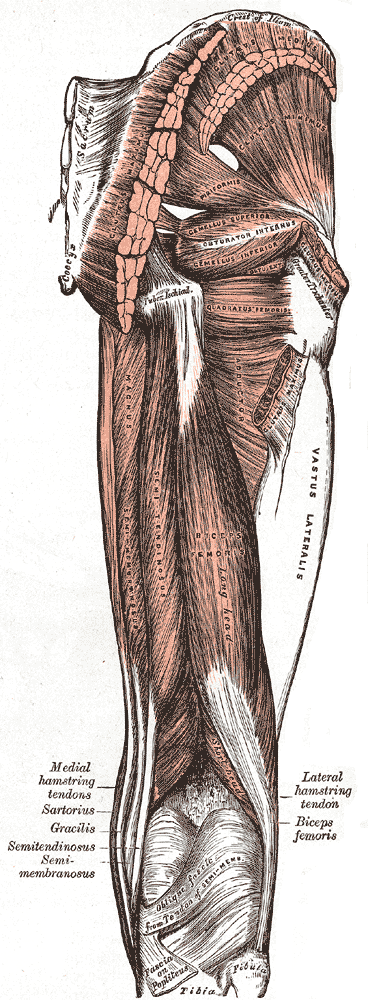
The intertrochanteric crest is a prominent bony ridge upon the posterior surface of the femur at the junction of the neck and the shaft of the femur. It extends between the greater trochanter superiorly, and the lesser trochanter inferiorly. Anatomy The intertrochanteric crest is a prominent smooth bony ridge upon the posterior surface of the femur at the junction of the neck and the shaft of the femur; together with the intertrochanteric line on the anterior side of the head, the intertrochanteric crest marks the transition between the femoral neck and shaft. The intertrochanteric crest extends between the greater trochanter superiorly, and the lesser trochanter inferiorly; it passes obliquely inferomedially from the greater trochanter to the lesser trochanter. An elevation between the middle and proximal third of the crest is known as the quadrate tubercle. Relations The distal capsular attachment on the femur follows the shape of the irregular rim between the head and th ... [...More Info...] [...Related Items...] OR: [Wikipedia] [Google] [Baidu] |
Femur
The femur (; ), or thigh bone, is the proximal bone of the hindlimb in tetrapod vertebrates. The head of the femur articulates with the acetabulum in the pelvic bone forming the hip joint, while the distal part of the femur articulates with the tibia (shinbone) and patella (kneecap), forming the knee joint. By most measures the two (left and right) femurs are the strongest bones of the body, and in humans, the largest and thickest. Structure The femur is the only bone in the upper leg. The two femurs converge medially toward the knees, where they articulate with the proximal ends of the tibiae. The angle of convergence of the femora is a major factor in determining the femoral-tibial angle. Human females have thicker pelvic bones, causing their femora to converge more than in males. In the condition ''genu valgum'' (knock knee) the femurs converge so much that the knees touch one another. The opposite extreme is ''genu varum'' (bow-leggedness). In the general popu ... [...More Info...] [...Related Items...] OR: [Wikipedia] [Google] [Baidu] |
Neck Of Femur
The femoral neck (femur neck or neck of the femur) is a flattened pyramidal process of bone, connecting the femoral head with the femoral shaft, and forming with the latter a wide angle opening medialward. Structure The neck is flattened from before backward, contracted in the middle, and broader laterally than medially. The vertical diameter of the lateral half is increased by the obliquity of the lower edge, which slopes downward to join the body at the level of the lesser trochanter, so that it measures one-third more than the antero-posterior diameter. The medial half is smaller and of a more circular shape. The anterior surface of the neck is perforated by numerous vascular foramina. Along the upper part of the line of junction of the anterior surface with the head is a shallow groove, best marked in elderly subjects; this groove lodges the orbicular fibers of the capsule of the hip joint. The posterior surface is smooth, and is broader and more concave than the anteri ... [...More Info...] [...Related Items...] OR: [Wikipedia] [Google] [Baidu] |
Shaft Of Femur
The body of femur (or shaft of femur) is the almost cylindrical, long part of the femur. It is a little broader above than in the center, broadest and somewhat flattened from before backward below. It is slightly arched, so as to be convex in front, and concave behind, where it is strengthened by a prominent longitudinal ridge, the linea aspera. It presents for examination three borders, separating three surfaces. Of the borders, one, the linea aspera, is posterior, one is medial, and the other, lateral. Borders The borders of the femur are the linea aspera, a medial border, and a lateral border. Linea aspera border The linea aspera is a prominent longitudinal ridge or crest, on the middle third of the bone, presenting a medial and a lateral lip, and a narrow rough, intermediate line. Above, the linea aspera is prolonged by three ridges. The lateral ridge termed the gluteal tuberosity is very rough, and runs almost vertically upward to the base of the greater trochanter. It ... [...More Info...] [...Related Items...] OR: [Wikipedia] [Google] [Baidu] |
Greater Trochanter
The greater trochanter of the femur is a large, irregular, quadrilateral eminence and a part of the skeletal system. It is directed lateral and medially and slightly posterior. In the adult it is about 2–4 cm lower than the femoral head.Standring, Susan, editor. ''Gray’s Anatomy: The Anatomical Basis of Clinical Practice''. Forty-First edition, Elsevier Limited, 2016, p. 1327. Because the pelvic outlet in the female is larger than in the male, there is a greater distance between the greater trochanters in the female. It has two surfaces and four borders. It is a traction epiphysis. Surfaces The ''lateral surface'', quadrilateral in form, is broad, rough, convex, and marked by a diagonal impression, which extends from the postero-superior to the antero-inferior angle, and serves for the insertion of the tendon of the gluteus medius. Above the impression is a triangular surface, sometimes rough for part of the tendon of the same muscle, sometimes smooth for the interpo ... [...More Info...] [...Related Items...] OR: [Wikipedia] [Google] [Baidu] |
Lesser Trochanter
The lesser trochanter is a conical posteromedial bony projection of the femoral shaft. it serves as the principal insertion site of the iliopsoas muscle. Structure The lesser trochanter is a conical posteromedial projection of the shaft of the femur, projecting from the posteroinferior aspect of its junction with the femoral neck. The summit and anterior surface of the lesser trochanter are rough, whereas its posterior surface is smooth. From its apex three well-marked borders extend: * two of these are above ** a medial continuous with the lower border of the femur neck ** a lateral with the intertrochanteric crest * the inferior border is continuous with the middle division of the linea aspera Attachments The summit of the lesser trochanter gives insertion to the tendon of the psoas major muscle and the iliacus muscle; the lesser trochanter represents the principal attachment of the iliopsoas. Anatomical relations The intertrochanteric crest (which demarcates the j ... [...More Info...] [...Related Items...] OR: [Wikipedia] [Google] [Baidu] |
Intertrochanteric Line
The intertrochanteric line (or ''spiral line of the femur''White (2005), p 256 ) is a line located on the anterior side of the proximal end of the femur. Structure The rough, variable ridge stretches between the lesser trochanter and the greater trochanter forming the base of the neck of the femur, roughly following the direction of the shaft of the femur. The iliofemoral ligament — the largest ligament of the human body — attaches above the line which also strengthens the capsule of the hip joint. The lower half, less prominent than the upper half, gives origin to the upper part of the Vastus medialis. Just like the intertrochanteric crest on the posterior side of the femoral head, the intertrochanteric line marks the transition between the femoral neck and shaft.Platzer (2004), p 192 The distal capsular attachment on the femur follows the shape of the irregular rim between the head and the neck. As a consequence, the capsule of the hip joint attaches in the ... [...More Info...] [...Related Items...] OR: [Wikipedia] [Google] [Baidu] |
Quadrate Tubercle
The quadrate tubercle is a small tubercle found upon the upper part of the femur. It serves as a point of insertion of the quadratus femoris muscle, along with the intertrochanteric crest and the linea quadrata. Structure The quadrate tubercle is located about the junction of the upper one-third and lower two-thirds, on the intertrochanteric crest The intertrochanteric crest is a prominent bony ridge upon the posterior surface of the femur at the junction of the neck and the shaft of the femur. It extends between the greater trochanter superiorly, and the lesser trochanter inferiorly. Ana .... In a small anatomical study, it was shown that the epiphysial line passes directly through the quadrate tubercle. The quadrate tubercle is the site of insertion of the quadratus femoris muscle. Variation The size of the quadrate tubercle varies. It is not always located on the intertrochanteric crest. Adjacent areas can also be part of the quadrate tubercle, such as the posterior su ... [...More Info...] [...Related Items...] OR: [Wikipedia] [Google] [Baidu] |
Thieme Medical Publishers
Thieme Medical Publishers is a German medical and science publisher in the Thieme Publishing Group. It produces professional journals, textbooks, atlases, monographs and reference books in both German and English covering a variety of medical specialties, including neurosurgery, orthopaedics, endocrinology, urology, radiology, anatomy, chemistry, otolaryngology, ophthalmology, audiology and speech-language pathology, complementary and alternative medicine. Thieme has more than 1,000 employees and maintains offices in seven cities worldwide, including New York City, Beijing, Delhi, Stuttgart, and three other cities in Germany. History Georg Thieme Verlag was founded in 1886 in Leipzig, Germany, by Georg Thieme when he was 26 years old. Thieme remains privately held and family-owned. The company received some early success in 1896 by publishing Wilhelm Röntgen's famous picture of his wife's hand in what is still one of Thieme's and Germany's oldest journals, the '' Deut ... [...More Info...] [...Related Items...] OR: [Wikipedia] [Google] [Baidu] |
Capsule Of Hip Joint
The articular capsule (capsular ligament) is strong and dense. Anterosuperiorly, it is attached to the margin of the acetabulum 5 to 6 mm. beyond the Acetabular labrum, labrum behind; but in front, it is attached to the outer margin of the labrum, and, opposite to the notch where the margin of the cavity is deficient, it is connected to the transverse acetabular ligament, transverse ligament, and by a few fibers to the edge of the obturator foramen. It surrounds the neck of the femur, and is attached, in front, to the intertrochanteric line; above, to the base of the neck; behind, to the neck, about 1.25 cm. above the intertrochanteric crest; below, to the lower part of the neck, close to the lesser trochanter. From its femoral attachment some of the fibers are reflected upward along the neck as longitudinal bands, termed ''retinacula''. The capsule is much thicker at the upper and forepart of the joint, where the most resistance is required; behind and below, it is thi ... [...More Info...] [...Related Items...] OR: [Wikipedia] [Google] [Baidu] |
Finger (unit)
A finger (sometimes fingerbreadth or finger's breadth) is any of several units of measurement that are approximately the width of an adult human finger, including: The digit, also known as digitus or digitus transversus (Latin), dactyl (Greek) or dactylus, or finger's breadth — of an inch or of a foot. In medicine and related disciplines (anatomy, radiology, etc.) the fingerbreadth (literally the width of a finger) is an informal but widely used unit of measure. In the measurement of distilled spirits, a finger of whiskey refers to the amount of whiskey that would fill a glass to the level of one finger wrapped around the glass at the bottom. Another definition (from Noah Webster): "nearly an inch." Finger is also the name of a longer unit of length used in cloth measurement, specifically, one eighth of a yard or 4 inches. In English these units have mostly fallen out of use, apart from the common use in distilled drinks and drinking games. See also * Digit (unit) ... [...More Info...] [...Related Items...] OR: [Wikipedia] [Google] [Baidu] |
Bones Of The Lower Limb
A bone is a rigid organ that constitutes part of the skeleton in most vertebrate animals. Bones protect the various other organs of the body, produce red and white blood cells, store minerals, provide structure and support for the body, and enable mobility. Bones come in a variety of shapes and sizes and have complex internal and external structures. They are lightweight yet strong and hard and serve multiple functions. Bone tissue (osseous tissue), which is also called bone in the uncountable sense of that word, is hard tissue, a type of specialized connective tissue. It has a honeycomb-like matrix internally, which helps to give the bone rigidity. Bone tissue is made up of different types of bone cells. Osteoblasts and osteocytes are involved in the formation and mineralization of bone; osteoclasts are involved in the resorption of bone tissue. Modified (flattened) osteoblasts become the lining cells that form a protective layer on the bone surface. The mineralized m ... [...More Info...] [...Related Items...] OR: [Wikipedia] [Google] [Baidu] |


