|
Internal Cerebral Veins
The internal cerebral veins (deep cerebral veins) drain the deep parts of the hemisphere and are two in number; each internal cerebral vein is formed near the interventricular foramina by the union of the superior thalamostriate vein and the superior choroid vein. They run backward parallel with one another, between the layers of the tela chorioidea of the third ventricle, and beneath the splenium of the corpus callosum, where they unite to form a short trunk, the great cerebral vein of Galen; just before their union each receives the corresponding basal vein The basal vein is a vein in the brain. It is formed at the anterior perforated substance by the union of * (a) a ''small anterior cerebral vein'' which accompanies the anterior cerebral artery and supplies the medial surface of the frontal lobe b .... References External links Diagram at radnet.ucla.edu* http://neuroangio.org/venous-brain-anatomy/deep-venous-system/ Veins of the head and neck {{circulat ... [...More Info...] [...Related Items...] OR: [Wikipedia] [Google] [Baidu] |
Great Cerebral Vein
The great cerebral vein is one of the large blood vessels in the skull draining the cerebrum of the brain. It is also known as the "vein of Galen", named for its discoverer, the Greek physician Galen. However, it is not the only vein with this eponym. Structure The great cerebral vein is considered one of the deep cerebral veins. Other deep cerebral veins are the internal cerebral veins, formed by the union of the superior thalamostriate vein and the superior choroid vein at the interventricular foramina. The internal cerebral veins can be seen on the superior surfaces of the caudate nuclei and thalami just under the corpus callosum. The veins at the anterior poles of the thalami merge posterior to the pineal gland to form the great cerebral vein. Most of the blood in the deep cerebral veins collects into the great cerebral vein. This comes from the inferior side of the posterior end of the corpus callosum and empties ie similarities, there are also differences between these ... [...More Info...] [...Related Items...] OR: [Wikipedia] [Google] [Baidu] |
Cerebral Arteries
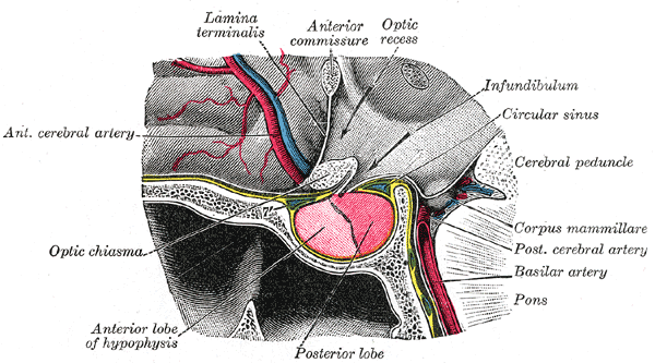
The cerebral arteries describe three main pairs of arteries and their branches, which perfuse the cerebrum of the brain. The three main arteries are the: * ''Anterior cerebral artery'' (ACA) * ''Middle cerebral artery'' (MCA) * ''Posterior cerebral artery'' (PCA) Both the ACA and MCA originate from the cerebral portion of internal carotid artery, while PCA branches from the intersection of the posterior communicating artery and the anterior portion of the basilar artery. The three pairs of arteries are linked via the anterior communicating artery and the posterior communicating arteries In human anatomy, the left and right posterior communicating arteries are arteries at the base of the brain that form part of the circle of Willis The circle of Willis (also called Willis' circle, loop of Willis, cerebral arterial circle, and W .... All three arteries send out arteries that perforate brain in the medial central portions prior to branching and bifurcating further. The arteries ... [...More Info...] [...Related Items...] OR: [Wikipedia] [Google] [Baidu] |
Cerebral Veins
In human anatomy, the cerebral veins are blood vessels which drain blood from the cerebrum of the human brain. They are divisible into ''external'' (superficial cerebral veins) and ''internal'' (internal cerebral veins) groups according to the outer or inner parts of the hemispheres they drain into. External veins The external cerebral veins known as the superficial cerebral veins are the superior cerebral veins, inferior cerebral veins, and middle cerebral veins. The superior cerebral veins on the upper side surfaces of the hemispheres drain into the superior sagittal sinus. The superior cerebral veins include the superior anastomotic vein. Internal veins The internal cerebral veins The internal cerebral veins (deep cerebral veins) drain the deep parts of the hemisphere and are two in number; each internal cerebral vein is formed near the interventricular foramina by the union of the superior thalamostriate vein and the ... are also known as the ''deep cerebral veins'' ... [...More Info...] [...Related Items...] OR: [Wikipedia] [Google] [Baidu] |
Interventricular Foramina (neuroanatomy)
In the brain, the interventricular foramina (or foramina of Monro) are channels that connect the paired lateral ventricles with the third ventricle at the midline of the brain. As channels, they allow cerebrospinal fluid (CSF) produced in the lateral ventricles to reach the third ventricle and then the rest of the brain's ventricular system. The walls of the interventricular foramina also contain choroid plexus, a specialized CSF-producing structure, that is continuous with that of the lateral and third ventricles above and below it. Structure The interventricular foramina are two holes ( la, foramen, pl. ''foramina'') that connect the left and the right lateral ventricles to the third ventricle. They are located on the underside near the midline of the lateral ventricles, and join the third ventricle where its roof meets its anterior surface. In front of the foramen is the fornix and behind is the thalamus. The foramen is normally crescent-shaped, but rounds and increases in s ... [...More Info...] [...Related Items...] OR: [Wikipedia] [Google] [Baidu] |
Superior Thalamostriate Vein
The superior thalamostriate vein or terminal vein commences in the groove between the corpus striatum and thalamus, receives numerous veins from both of these parts, and unites behind the crus of the fornix with the superior choroid vein to form each of the internal cerebral veins The internal cerebral veins (deep cerebral veins) drain the deep parts of the hemisphere and are two in number; each internal cerebral vein is formed near the interventricular foramina by the union of the superior thalamostriate vein and the .... References Veins of the head and neck Thalamus {{circulatory-stub ... [...More Info...] [...Related Items...] OR: [Wikipedia] [Google] [Baidu] |
Choroid Veins
The choroid veins are the superior choroid vein, and the inferior choroid vein of the lateral ventricle. Both veins drain different parts of the choroid plexus. Superior choroid vein The superior choroid vein runs along the length of the choroid plexus in the lateral ventricle. It drains the choroid plexus, and also the hippocampus, fornix, and corpus callosum. It unites with the superior thalamostriate vein to form the internal cerebral vein. Inferior choroid vein The inferior choroid vein drains the inferior choroid plexus into the basal vein The basal vein is a vein in the brain. It is formed at the anterior perforated substance by the union of * (a) a ''small anterior cerebral vein'' which accompanies the anterior cerebral artery and supplies the medial surface of the frontal lobe by .... References Veins of the head and neck {{circulatory-stub ... [...More Info...] [...Related Items...] OR: [Wikipedia] [Google] [Baidu] |
Tela Chorioidea
The tela choroidea (or tela chorioidea) is a region of meningeal pia mater that adheres to the underlying ependyma, and gives rise to the choroid plexus in each of the brain’s four ventricles. ''Tela'' is Latin for ''woven'' and is used to describe a web-like membrane or layer. The tela choroidea is a very thin part of the loose connective tissue of pia mater overlying and closely adhering to the ependyma. It has a rich blood supply. The ependyma and vascular pia mater – the tela choroidea, form regions of minute projections known as a choroid plexus that projects into each ventricle. The choroid plexus produces most of the cerebrospinal fluid of the central nervous system that circulates through the ventricles of the brain, the central canal of the spinal cord, and the subarachnoid space. The tela choroidea in the ventricles forms from different parts of the roof plate in the development of the embryo. Structure In the lateral ventricles the tela choroidea–a double-layere ... [...More Info...] [...Related Items...] OR: [Wikipedia] [Google] [Baidu] |
Third Ventricle
The third ventricle is one of the four connected ventricles of the ventricular system within the mammalian brain. It is a slit-like cavity formed in the diencephalon between the two thalami, in the midline between the right and left lateral ventricles, and is filled with cerebrospinal fluid (CSF). Running through the third ventricle is the interthalamic adhesion, which contains thalamic neurons and fibers that may connect the two thalami. Structure The third ventricle is a narrow, laterally flattened, vaguely rectangular region, filled with cerebrospinal fluid, and lined by ependyma. It is connected at the superior anterior corner to the lateral ventricles, by the interventricular foramina, and becomes the cerebral aqueduct (''aqueduct of Sylvius'') at the posterior caudal corner. Since the interventricular foramina are on the lateral edge, the corner of the third ventricle itself forms a bulb, known as the ''anterior recess'' (it is also known as the ''bulb of the ventricl ... [...More Info...] [...Related Items...] OR: [Wikipedia] [Google] [Baidu] |
Splenium
The corpus callosum (Latin for "tough body"), also callosal commissure, is a wide, thick nerve tract, consisting of a flat bundle of commissural fibers, beneath the cerebral cortex in the brain. The corpus callosum is only found in placental mammals. It spans part of the longitudinal fissure, connecting the left and right cerebral hemispheres, enabling communication between them. It is the largest white matter structure in the human brain, about in length and consisting of 200–300 million axonal projections. A number of separate nerve tracts, classed as subregions of the corpus callosum, connect different parts of the hemispheres. The main ones are known as the genu, the rostrum, the trunk or body, and the splenium. Structure The corpus callosum forms the floor of the longitudinal fissure that separates the two cerebral hemispheres. Part of the corpus callosum forms the roof of the lateral ventricles. The corpus callosum has four main parts – individual nerve tracts ... [...More Info...] [...Related Items...] OR: [Wikipedia] [Google] [Baidu] |
Corpus Callosum
The corpus callosum (Latin for "tough body"), also callosal commissure, is a wide, thick nerve tract, consisting of a flat bundle of commissural fibers, beneath the cerebral cortex in the brain. The corpus callosum is only found in placental mammals. It spans part of the longitudinal fissure, connecting the left and right cerebral hemispheres, enabling communication between them. It is the largest white matter structure in the human brain, about in length and consisting of 200–300 million axonal projections. A number of separate nerve tracts, classed as subregions of the corpus callosum, connect different parts of the hemispheres. The main ones are known as the genu, the rostrum, the trunk or body, and the splenium. Structure The corpus callosum forms the floor of the longitudinal fissure that separates the two cerebral hemispheres. Part of the corpus callosum forms the roof of the lateral ventricles. The corpus callosum has four main parts – individual nerve tracts ... [...More Info...] [...Related Items...] OR: [Wikipedia] [Google] [Baidu] |
Basal Vein
The basal vein is a vein in the brain. It is formed at the anterior perforated substance by the union of * (a) a ''small anterior cerebral vein'' which accompanies the anterior cerebral artery and supplies the medial surface of the frontal lobe by the fronto-basal vein. * (b) the ''deep middle cerebral vein'' (''deep Sylvian vein''), which receives tributaries from the insula and neighboring gyri, and runs in the lower part of the lateral cerebral fissure, and * (c) the ''inferior striate veins'', which leave the corpus striatum through the anterior perforated substance. The basal vein passes backward around the cerebral peduncle, and ends in the great cerebral vein; it receives tributaries from the interpeduncular fossa, the inferior horn of the lateral ventricle, the hippocampal gyrus, and the mid-brain The midbrain or mesencephalon is the forward-most portion of the brainstem and is associated with vision, hearing, motor control, sleep and wakefulness, arousal (alertness) ... [...More Info...] [...Related Items...] OR: [Wikipedia] [Google] [Baidu] |


