|
Inoderma Nipponicum
''Inoderma'' is a genus of lichen-forming fungi in the family Arthoniaceae. It was resurrected for use in 2015 for a small group of species with the following features: elevated, white pruinose pycnidia, immersed to adnate white pruinose apothecia, and a weakly gelatinized hymenium. '' Inoderma byssaceum'' was assigned as the type species for the genus. Description ''Inoderma'' is a genus of lichens that typically forms broad, pale, whitish to light olive-grey patches on its substrate. These patches (the thallus) can be continuous but often develop a network of fine cracks or appear as tiny, powdery . The surface is usually not covered by a protective "skin" () and can feel slightly soft or powdery. The lichen's green partner cells (the ) belong to '' Trentepohlia'', a genus of green algae. Some ''Inoderma'' species produce apothecia—small, often rounded, and slightly raised structures where spores are formed. These may lie flush with the lichen's surface or sit just above it, ... [...More Info...] [...Related Items...] OR: [Wikipedia] [Google] [Baidu] |
Samuel Frederick Gray
Samuel Frederick Gray (10 December 1766 – 12 April 1828) was a British botanist, mycologist, and pharmacologist. He was the father of the zoologists John Edward Gray and George Robert Gray. Background He was the son of Samuel Gray, a London Seed company, seedsman. He received no inheritance and, after failing to qualify for medicine, turned to medical and botanical writing. He married Elizabeth Forfeit in 1794 and moved to Walsall, Staffordshire, where he established an assay office before he moved back to London in 1800. He set up an apothecary business in Wapping, which failed within a few years. Then, he seems to have maintained himself by writing and lecturing. Medical writings Gray wrote a ''Supplement to the Pharmacopoeia'', published in 1818 with several subsequent editions. In 1819, he became co-editor of the ''London Medical Repository'', to which he contributed many articles on medical, botanical, and other topics. He published, in 1823, ''The Elements of Pharmacy'' ... [...More Info...] [...Related Items...] OR: [Wikipedia] [Google] [Baidu] |
Pycnidia
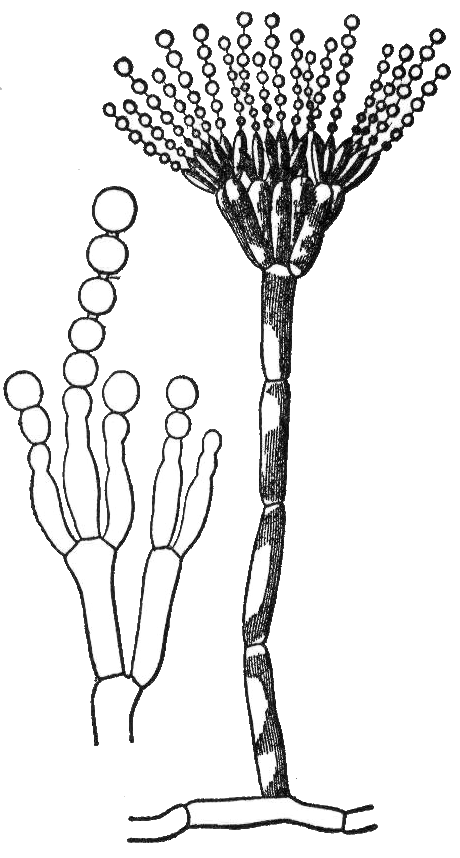
A pycnidium (plural pycnidia) is an asexual fruiting body produced by mitosporic fungi, for instance in the order Sphaeropsidales ( Deuteromycota, Coelomycetes) or order Pleosporales (Ascomycota, Dothideomycetes). It is often spherical or inversely pearshaped ( obpyriform) and its internal cavity is lined with conidiophore A conidium ( ; : conidia), sometimes termed an asexual chlamydospore or chlamydoconidium (: chlamydoconidia), is an Asexual reproduction, asexual, non-motility, motile spore of a fungus. The word ''conidium'' comes from the Ancient Greek word f ...s. When ripe, an opening generally appears at the top, through which the pycnidiospores escape. References {{reflist Further reading *Kulik, Martin M. "Symptomless infection, persistence, and production of pycnidia in host and non-host plants by Phomopsis batatae, Phomopsis phaseoli, and Phomopsis sojae, and the taxonomic implications." Mycologia(1984): 274–291. *Calpouzos, L., and D. B. Lapis. "Effects of l ... [...More Info...] [...Related Items...] OR: [Wikipedia] [Google] [Baidu] |
Conidia
A conidium ( ; : conidia), sometimes termed an asexual chlamydospore or chlamydoconidium (: chlamydoconidia), is an asexual, non- motile spore of a fungus. The word ''conidium'' comes from the Ancient Greek word for dust, ('). They are also called mitospores due to the way they are generated through the cellular process of mitosis. They are produced exogenously. The two new haploid cells are genetically identical to the haploid parent, and can develop into new organisms if conditions are favorable, and serve in biological dispersal. Asexual reproduction in ascomycetes (the phylum Ascomycota) is by the formation of conidia, which are borne on specialized stalks called conidiophores. The morphology of these specialized conidiophores is often distinctive between species and, before the development of molecular techniques at the end of the 20th century, was widely used for identification of (''e.g.'' '' Metarhizium'') species. The terms microconidia and macroconidia are some ... [...More Info...] [...Related Items...] OR: [Wikipedia] [Google] [Baidu] |
Conidiomata
Conidiomata (singular: Conidioma) are blister-like fruiting structures produced by a specific type of fungus called a coelomycete. They are formed as a means of dispersing asexual spores call conidia, which they accomplish by creating the blister-like formations which then rupture to release the contained spores. Structure Conidiomata mainly consist of a mass of densely packed hypha which develops below the surface of the host cuticle, and the fungus may or may not use some of the host’s own tissue to construct the structure. Development of these structures can either occur just below the cuticle or below the epidermal layer of the tissue. Formation on the differing levels is dependent upon the type of conidiomata being formed. Five types of conidioma have been found and are classified as acervuli, pycnidia, sporodochia, synnemata and corenima. Types Acervuli Acervuli is one of the two major groups of conidiomata (the other being pycnidia). Conidiomata of this type for ... [...More Info...] [...Related Items...] OR: [Wikipedia] [Google] [Baidu] |
Septum
In biology, a septum (Latin language, Latin for ''something that encloses''; septa) is a wall, dividing a Body cavity, cavity or structure into smaller ones. A cavity or structure divided in this way may be referred to as septate. Examples Human anatomy * Interatrial septum, the wall of tissue that is a sectional part of the left and right atria of the heart * Interventricular septum, the wall separating the left and right ventricles of the heart * Lingual septum, a vertical layer of fibrous tissue that separates the halves of the tongue *Nasal septum: the cartilage wall separating the nostrils of the nose * Alveolar septum: the thin wall which separates the Pulmonary alveolus, alveoli from each other in the lungs * Orbital septum, a palpebral ligament in the upper and lower eyelids * Septum pellucidum or septum lucidum, a thin structure separating two fluid pockets in the brain * Uterine septum, a malformation of the uterus * Septum of the penis, Penile septum, a fibrous w ... [...More Info...] [...Related Items...] OR: [Wikipedia] [Google] [Baidu] |
Iodine
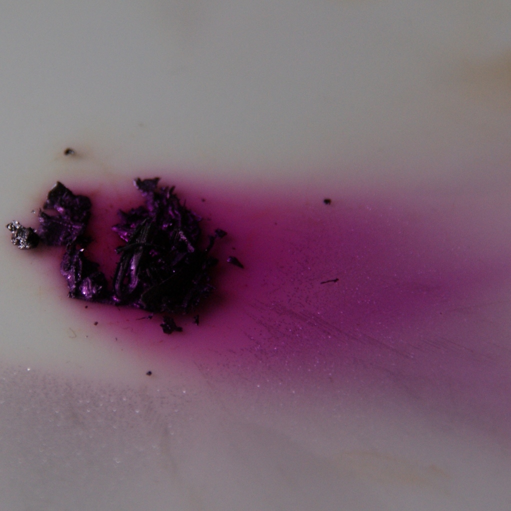
Iodine is a chemical element; it has symbol I and atomic number 53. The heaviest of the stable halogens, it exists at standard conditions as a semi-lustrous, non-metallic solid that melts to form a deep violet liquid at , and boils to a violet gas at . The element was discovered by the French chemist Bernard Courtois in 1811 and was named two years later by Joseph Louis Gay-Lussac, after the Ancient Greek , meaning 'violet'. Iodine occurs in many oxidation states, including iodide (I−), iodate (), and the various periodate anions. As the heaviest essential mineral nutrient, iodine is required for the synthesis of thyroid hormones. Iodine deficiency affects about two billion people and is the leading preventable cause of intellectual disabilities. The dominant producers of iodine today are Chile and Japan. Due to its high atomic number and ease of attachment to organic compounds, it has also found favour as a non-toxic radiocontrast material. Because of the spec ... [...More Info...] [...Related Items...] OR: [Wikipedia] [Google] [Baidu] |
Staining
Staining is a technique used to enhance contrast in samples, generally at the Microscope, microscopic level. Stains and dyes are frequently used in histology (microscopic study of biological tissue (biology), tissues), in cytology (microscopic study of cell (biology), cells), and in the medical fields of histopathology, hematology, and cytopathology that focus on the study and diagnoses of diseases at the microscopic level. Stains may be used to define biological tissues (highlighting, for example, muscle fibers or connective tissue), cell (biology), cell populations (classifying different blood cells), or organelles within individual cells. In biochemistry, it involves adding a class-specific (DNA, proteins, lipids, carbohydrates) dye to a substrate to qualify or quantify the presence of a specific compound. Staining and fluorescent tagging can serve similar purposes. Biological staining is also used to mark cells in flow cytometry, and to flag proteins or nucleic acids in gel ... [...More Info...] [...Related Items...] OR: [Wikipedia] [Google] [Baidu] |
Ascus
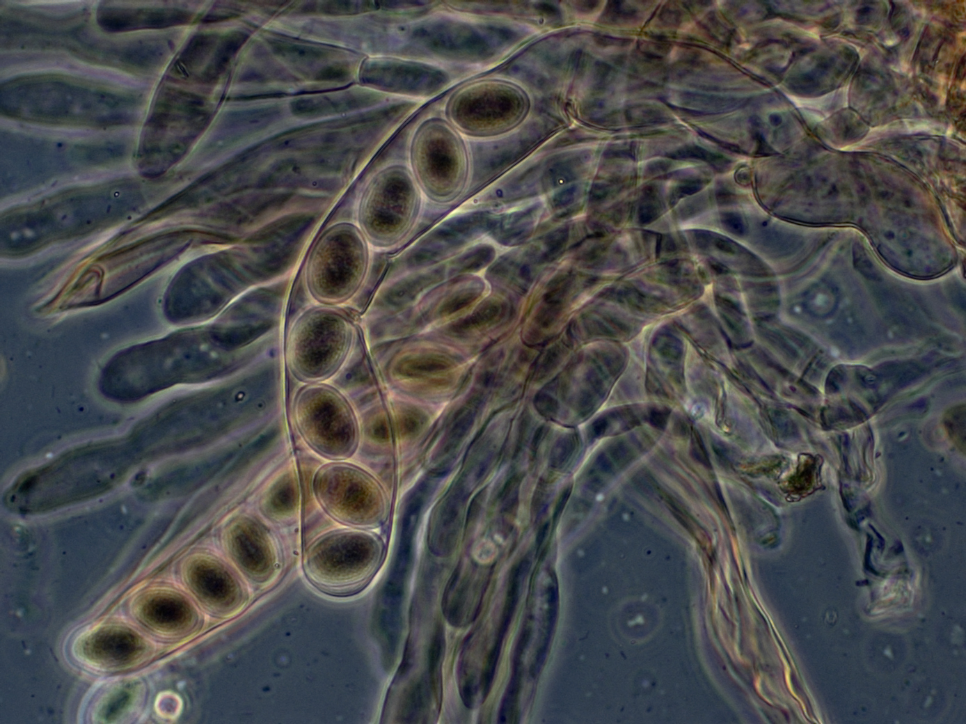
An ascus (; : asci) is the sexual spore-bearing cell produced in ascomycete fungi. Each ascus usually contains eight ascospores (or octad), produced by meiosis followed, in most species, by a mitotic cell division. However, asci in some genera or species can occur in numbers of one (e.g. '' Monosporascus cannonballus''), two, four, or multiples of four. In a few cases, the ascospores can bud off conidia that may fill the asci (e.g. '' Tympanis'') with hundreds of conidia, or the ascospores may fragment, e.g. some '' Cordyceps'', also filling the asci with smaller cells. Ascospores are nonmotile, usually single celled, but not infrequently may be coenocytic (lacking a septum), and in some cases coenocytic in multiple planes. Mitotic divisions within the developing spores populate each resulting cell in septate ascospores with nuclei. The term ocular chamber, or oculus, refers to the epiplasm (the portion of cytoplasm not used in ascospore formation) that is surrounded by the ... [...More Info...] [...Related Items...] OR: [Wikipedia] [Google] [Baidu] |
Green Algae
The green algae (: green alga) are a group of chlorophyll-containing autotrophic eukaryotes consisting of the phylum Prasinodermophyta and its unnamed sister group that contains the Chlorophyta and Charophyta/ Streptophyta. The land plants ( Embryophytes) have emerged deep within the charophytes as a sister of the Zygnematophyceae. Since the realization that the Embryophytes emerged within the green algae, some authors are starting to include them. The completed clade that includes both green algae and embryophytes is monophyletic and is referred to as the clade Viridiplantae and as the kingdom Plantae. The green algae include unicellular and colonial flagellates, most with two flagella per cell, as well as various colonial, coccoid (spherical), and filamentous forms, and macroscopic, multicellular seaweeds. There are about 22,000 species of green algae, many of which live most of their lives as single cells, while other species form coenobia (colonies), long filaments ... [...More Info...] [...Related Items...] OR: [Wikipedia] [Google] [Baidu] |
Trentepohlia (alga)
''Trentepohlia'' is a genus of filamentous chlorophyte green algae in the family Trentepohliaceae, living free on terrestrial supports such as tree trunks and wet rocks or symbiotically in lichens. The filaments of ''Trentepohlia'' often have a strong orange colour (photograph at right) caused by the presence of large quantities of carotenoid pigments which mask the green of the chlorophyll. Nomenclature Organisms belonging to the genus ''Trentepohlia'' were first described by Linnaeus in 1759; he named his species ''Byssus aureus'' (currently known as ''Trentepohlia aurea''). The genus was circumscribed by Carl Friedrich Philipp von Martius in Fl. Crypt. Erlang. on page 351 in 1817. The genus name of ''Trentepohlia'' is in honour of Johann Friedrich Trentepohl (1748–1806), who was a German clergyman and botanist. He worked as a lecturer and Pastor in various places in Wesermarsch. Martius' name was conserved in favor of the moss genus ''Trentepohlia'' and the Brassicaceae ... [...More Info...] [...Related Items...] OR: [Wikipedia] [Google] [Baidu] |
Thallus
Thallus (: thalli), from Latinized Greek (), meaning "a green shoot" or "twig", is the vegetative tissue of some organisms in diverse groups such as algae, fungi, some liverworts, lichens, and the Myxogastria. A thallus usually names the entire body of a multicellular non-moving organism in which there is no organization of the tissues into organs. Many of these organisms were previously known as the thallophytes, a polyphyletic group of distantly related organisms. An organism or structure resembling a thallus is called thalloid, thalloidal, thalliform, thalline, or thallose. Even though thalli do not have organized and distinct parts ( leaves, roots, and stems) as do the vascular plants, they may have analogous structures that resemble their vascular "equivalents". The analogous structures have similar function or macroscopic structure, but different microscopic structure; for example, no thallus has vascular tissue. In exceptional cases such as the Lemnoideae, where th ... [...More Info...] [...Related Items...] OR: [Wikipedia] [Google] [Baidu] |


