|
Inferior Salivary Nucleus
The salivatory nuclei are the superior salivatory nucleus, and the inferior salivatory nucleus that innervate the salivary glands. They are located in the pontine tegmentum in the brainstem. They both are examples of cranial nerve nuclei. The superior salivatory nucleus innervates the submandibular gland and the sublingual gland and is part of the facial nerve The inferior salivatory nucleus innervates the parotid gland by way of the otic ganglion and forms the parasympathetic component of the glossopharyngeal nerve. Superior salivatory nucleus The superior salivatory nucleus (or nucleus salivatorius superior) of the facial nerve is a visceromotor cranial nerve nucleus located in the pontine tegmentum. It is one of the salivatory nuclei. Parasympathetic efferent fibers of the facial nerve (preganglionic fibers) arise according to some authors from the small cells of the facial nucleus, or according to others from a special nucleus of cells scattered in the reticular formatio ... [...More Info...] [...Related Items...] OR: [Wikipedia] [Google] [Baidu] |
Salivary Gland
The salivary glands in mammals are exocrine glands that produce saliva through a system of ducts. Humans have three paired major salivary glands (parotid, submandibular, and sublingual), as well as hundreds of minor salivary glands. Salivary glands can be classified as serous, mucous, or seromucous (mixed). In serous secretions, the main type of protein secreted is alpha-amylase, an enzyme that breaks down starch into maltose and glucose, whereas in mucous secretions, the main protein secreted is mucin, which acts as a lubricant. In humans, 1200 to 1500 ml of saliva are produced every day. The secretion of saliva (salivation) is mediated by parasympathetic stimulation; acetylcholine is the active neurotransmitter and binds to muscarinic receptors in the glands, leading to increased salivation. The fourth pair of salivary glands, the tubarial glands discovered in 2020, are named for their location, being positioned in front and over the torus tubarius. However, this find ... [...More Info...] [...Related Items...] OR: [Wikipedia] [Google] [Baidu] |
Postsynaptic
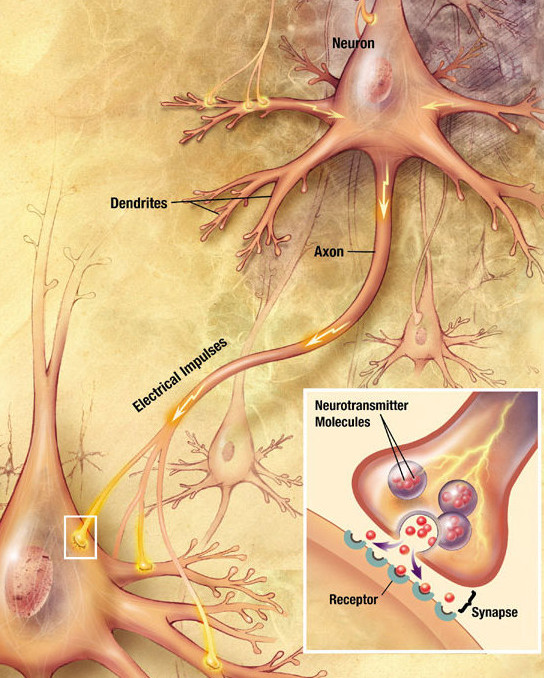
Chemical synapses are biological junctions through which neurons' signals can be sent to each other and to non-neuronal cells such as those in muscles or glands. Chemical synapses allow neurons to form circuits within the central nervous system. They are crucial to the biological computations that underlie perception and thought. They allow the nervous system to connect to and control other systems of the body. At a chemical synapse, one neuron releases neurotransmitter molecules into a small space (the synaptic cleft) that is adjacent to another neuron. The neurotransmitters are contained within small sacs called synaptic vesicles, and are released into the synaptic cleft by exocytosis. These molecules then bind to neurotransmitter receptors on the postsynaptic cell. Finally, the neurotransmitters are cleared from the synapse through one of several potential mechanisms including enzymatic degradation or re-uptake by specific transporters either on the presynaptic cel ... [...More Info...] [...Related Items...] OR: [Wikipedia] [Google] [Baidu] |
Dorsal Nucleus Of The Vagus Nerve
The dorsal nucleus of vagus nerve (or posterior nucleus of vagus nerve or dorsal vagal nucleus or nucleus dorsalis nervi vagi or nucleus posterior nervi vagi) is a cranial nerve nucleus for the vagus nerve in the medulla that lies ventral to the floor of the fourth ventricle. It mostly serves parasympathetic vagal functions in the gastrointestinal tract, lungs, and other thoracic and abdominal vagal innervations. These functions include, among others, bronchoconstriction and gland secretion. The cell bodies for the preganglionic parasympathetic vagal neurons that innervate the heart reside in the nucleus ambiguus. Additional cell bodies are found in the nucleus ambiguus, which give rise to the branchial efferent motor fibers of the vagus nerve (CN X) terminating in the laryngeal, pharyngeal muscles, and musculus uvulae. Additional images File:Gray694.png, Section of the medulla oblongata at about the middle of the olive. File:Gray696.png, The cranial nerve nuclei schematically ... [...More Info...] [...Related Items...] OR: [Wikipedia] [Google] [Baidu] |
Superior Salivatory Nucleus
The salivatory nuclei are the superior salivatory nucleus, and the inferior salivatory nucleus that innervate the salivary glands. They are located in the pontine tegmentum in the brainstem. They both are examples of cranial nerve nuclei. The superior salivatory nucleus innervates the submandibular gland and the sublingual gland and is part of the facial nerve The inferior salivatory nucleus innervates the parotid gland by way of the otic ganglion and forms the parasympathetic component of the glossopharyngeal nerve. Superior salivatory nucleus The superior salivatory nucleus (or nucleus salivatorius superior) of the facial nerve is a visceromotor cranial nerve nucleus located in the pontine tegmentum. It is one of the salivatory nuclei. Parasympathetic efferent fibers of the facial nerve (preganglionic fibers) arise according to some authors from the small cells of the facial nucleus, or according to others from a special nucleus of cells scattered in the reticular formation ... [...More Info...] [...Related Items...] OR: [Wikipedia] [Google] [Baidu] |
Saliva
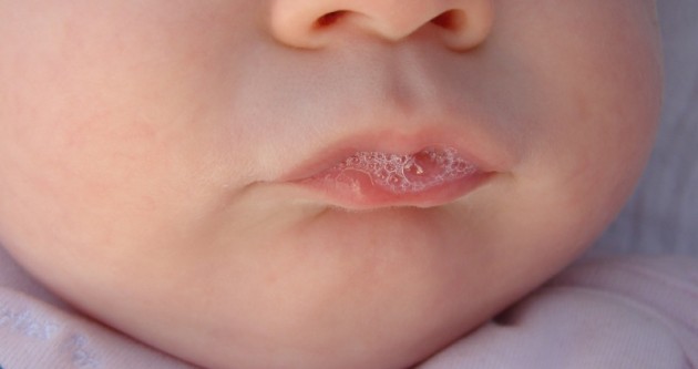
Saliva (commonly referred to as spit) is an extracellular fluid produced and secreted by salivary glands in the mouth. In humans, saliva is around 99% water, plus electrolytes, mucus, white blood cells, epithelial cells (from which DNA can be extracted), enzymes (such as lipase and amylase), antimicrobial agents (such as secretory IgA, and lysozymes). The enzymes found in saliva are essential in beginning the process of digestion of dietary starches and fats. These enzymes also play a role in breaking down food particles entrapped within dental crevices, thus protecting teeth from bacterial decay. Saliva also performs a lubricating function, wetting food and permitting the initiation of swallowing, and protecting the oral mucosa from drying out. Various animal species have special uses for saliva that go beyond predigestion. Some swifts use their gummy saliva to build nests. ''Aerodramus'' nests form the basis of bird's nest soup. Cobras, vipers, and certain other membe ... [...More Info...] [...Related Items...] OR: [Wikipedia] [Google] [Baidu] |
Parasympathetic Nervous System
The parasympathetic nervous system (PSNS) is one of the three divisions of the autonomic nervous system, the others being the sympathetic nervous system and the enteric nervous system. The enteric nervous system is sometimes considered part of the autonomic nervous system, and sometimes considered an independent system. The autonomic nervous system is responsible for regulating the body's unconscious actions. The parasympathetic system is responsible for stimulation of "rest-and-digest" or "feed and breed" activities that occur when the body is at rest, especially after eating, including sexual arousal, salivation, lacrimation (tears), urination, digestion, and defecation. Its action is described as being complementary to that of the sympathetic nervous system, which is responsible for stimulating activities associated with the fight-or-flight response. Nerve fibres of the parasympathetic nervous system arise from the central nervous system. Specific nerves include several ... [...More Info...] [...Related Items...] OR: [Wikipedia] [Google] [Baidu] |
General Visceral Efferent Fibers
General visceral efferent fibers (GVE) or visceral efferents or autonomic efferents, are the efferent nerve fibers of the autonomic nervous system (also known as the ''visceral efferent nervous system'' that provide motor innervation to smooth muscle, cardiac muscle, and glands (contrast with special visceral efferent (SVE) fibers) through postganglionic varicosities. GVE fibers may be either sympathetic or parasympathetic. The cranial nerves containing GVE fibers include the oculomotor nerve (CN III), the facial nerve (CN VII), the glossopharyngeal nerve (CN IX) and the vagus nerve (CN X).Mehta, Samir et al. Step-Up: A High-Yield, Systems-Based Review for the USMLE Step 1. Baltimore, MD: LWW, 2003. Additional images File:Gray840.png, Sympathetic connections of the ciliary and superior cervical ganglia. See also * Nerve fiber * Preganglionic fibers * Efferent nerve Efferent nerve fibers refer to axonal projections that ''exit'' a particular region; as opposed to Af ... [...More Info...] [...Related Items...] OR: [Wikipedia] [Google] [Baidu] |
Medulla Oblongata
The medulla oblongata or simply medulla is a long stem-like structure which makes up the lower part of the brainstem. It is anterior and partially inferior to the cerebellum. It is a cone-shaped neuronal mass responsible for autonomic (involuntary) functions, ranging from vomiting to sneezing. The medulla contains the cardiac, respiratory, vomiting and vasomotor centers, and therefore deals with the autonomic functions of breathing, heart rate and blood pressure as well as the sleep–wake cycle. During embryonic development, the medulla oblongata develops from the myelencephalon. The myelencephalon is a secondary vesicle which forms during the maturation of the rhombencephalon, also referred to as the hindbrain. The bulb is an archaic term for the medulla oblongata. In modern clinical usage, the word bulbar (as in bulbar palsy) is retained for terms that relate to the medulla oblongata, particularly in reference to medical conditions. The word bulbar can refer to the nerves ... [...More Info...] [...Related Items...] OR: [Wikipedia] [Google] [Baidu] |
Pons
The pons (from Latin , "bridge") is part of the brainstem that in humans and other bipeds lies inferior to the midbrain, superior to the medulla oblongata and anterior to the cerebellum. The pons is also called the pons Varolii ("bridge of Varolius"), after the Italian anatomist and surgeon Costanzo Varolio (1543–75). This region of the brainstem includes neural pathways and tracts that conduct signals from the brain down to the cerebellum and medulla, and tracts that carry the sensory signals up into the thalamus.Saladin Kenneth S.(2007) Anatomy & physiology the unity of form and function. Dubuque, IA: McGraw-Hill Structure The pons is in the brainstem situated between the midbrain and the medulla oblongata, and in front of the cerebellum. A separating groove between the pons and the medulla is the inferior pontine sulcus. The superior pontine sulcus separates the pons from the midbrain. The pons can be broadly divided into two parts: the basilar part of the pons (ventral ... [...More Info...] [...Related Items...] OR: [Wikipedia] [Google] [Baidu] |
Neuron
A neuron, neurone, or nerve cell is an electrically excitable cell that communicates with other cells via specialized connections called synapses. The neuron is the main component of nervous tissue in all animals except sponges and placozoa. Non-animals like plants and fungi do not have nerve cells. Neurons are typically classified into three types based on their function. Sensory neurons respond to stimuli such as touch, sound, or light that affect the cells of the sensory organs, and they send signals to the spinal cord or brain. Motor neurons receive signals from the brain and spinal cord to control everything from muscle contractions to glandular output. Interneurons connect neurons to other neurons within the same region of the brain or spinal cord. When multiple neurons are connected together, they form what is called a neural circuit. A typical neuron consists of a cell body (soma), dendrites, and a single axon. The soma is a compact structure, and the axon and dend ... [...More Info...] [...Related Items...] OR: [Wikipedia] [Google] [Baidu] |
Salivary Glands
The salivary glands in mammals are exocrine glands that produce saliva through a system of ducts. Humans have three paired major salivary glands (parotid, submandibular, and sublingual), as well as hundreds of minor salivary glands. Salivary glands can be classified as serous, mucous, or seromucous (mixed). In serous secretions, the main type of protein secreted is alpha-amylase, an enzyme that breaks down starch into maltose and glucose, whereas in mucous secretions, the main protein secreted is mucin, which acts as a lubricant. In humans, 1200 to 1500 ml of saliva are produced every day. The secretion of saliva (salivation) is mediated by parasympathetic stimulation; acetylcholine is the active neurotransmitter and binds to muscarinic receptors in the glands, leading to increased salivation. The fourth pair of salivary glands, the tubarial glands discovered in 2020, are named for their location, being positioned in front and over the torus tubarius. However, this finding ... [...More Info...] [...Related Items...] OR: [Wikipedia] [Google] [Baidu] |
Submandibular Ganglion
The submandibular ganglion (or submaxillary ganglion in older texts) is part of the human autonomic nervous system. It is one of four parasympathetic ganglia of the head and neck. (The others are the otic ganglion, pterygopalatine ganglion, and ciliary ganglion). Location and relations The submandibular ganglion is small and fusiform in shape. It is situated above the deep portion of the submandibular gland, on the hyoglossus muscle, near the posterior border of the mylohyoid muscle. The ganglion 'hangs' by two nerve filaments from the lower border of the lingual nerve (itself a branch of the mandibular nerve, CN V3). It is suspended from the lingual nerve by two filaments, one anterior and one posterior. Through the posterior of these it receives a branch from the chorda tympani nerve which runs in the sheath of the lingual nerve. Fibers Like other parasympathetic ganglia of the head and neck, the submandibular ganglion is the site of synapse for parasympathetic fibers and car ... [...More Info...] [...Related Items...] OR: [Wikipedia] [Google] [Baidu] |


