|
Inferior Gluteal Artery
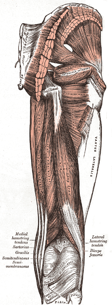
The inferior gluteal artery (sciatic artery), the smaller of the two terminal branches of the anterior trunk of the internal iliac artery, is distributed chiefly to the buttock and back of the thigh. It passes down on the sacral plexus of nerves and the piriformis muscle, behind the internal pudendal artery. It passes through the lower part of the greater sciatic foramen. It escapes from the pelvis between piriformis muscle and coccygeus muscle. It then descends in the interval between the greater trochanter of the femur and tuberosity of the ischium. It is accompanied by the sciatic nerve and the posterior femoral cutaneous nerves, and covered by the gluteus maximus. It continues down the back of the thigh, supplying the skin, and anastomosing with branches of the perforating arteries. Additional images File:Gray544.png, The arteries of the gluteal and posterior femoral regions. File:Gray829.png, Dissection of side wall of pelvis showing sacral and pudendal plexuses. See a ... [...More Info...] [...Related Items...] OR: [Wikipedia] [Google] [Baidu] |
Sciatic Nerve
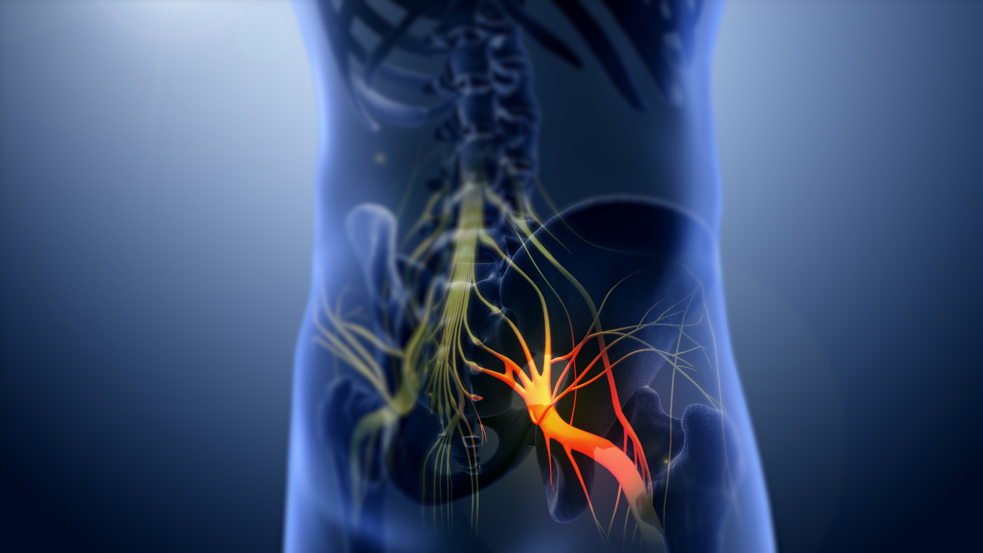
The sciatic nerve, also called the ischiadic nerve, is a large nerve in humans and other vertebrate animals which is the largest branch of the sacral plexus and runs alongside the hip joint and down the lower limb. It is the longest and widest single nerve in the human body, going from the top of the leg to the foot on the posterior aspect. The sciatic nerve has no cutaneous branches for the thigh. This nerve provides the connection to the nervous system for the skin of the lateral leg and the whole foot, the muscles of the back of the thigh, and those of the leg and foot. It is derived from spinal nerves L4 to S3. It contains fibers from both the anterior and posterior divisions of the lumbosacral plexus. Structure In humans, the sciatic nerve is formed from the L4 to S3 segments of the sacral plexus, a collection of nerve fibres that emerge from the sacral part of the spinal cord. The lumbosacral trunk from the L4 and L5 roots descends between the sacral promontory and ala and ... [...More Info...] [...Related Items...] OR: [Wikipedia] [Google] [Baidu] |
Internal Pudendal Artery
The internal pudendal artery is one of the three pudendal arteries. It branches off the internal iliac artery, and provides blood to the external genitalia. Structure The internal pudendal artery is the terminal branch of the anterior trunk of the internal iliac artery. It is smaller in the female than in the male. Path It arises from the anterior division of internal iliac artery. It runs on the lateral pelvic wall. It exits the pelvic cavity through the greater sciatic foramen, inferior to the piriformis muscle, to enter the gluteal region. It then curves around the sacrospinous ligament to enter the perineum through the lesser sciatic foramen. It travels through the pudendal canal with the internal pudendal veins and the pudendal nerve. Branches The internal pudendal artery gives off the following branches: The deep artery of clitoris is a branch of the internal pudendal artery and supplies the clitoral crura. Another branch of the internal pudendal artery is the dorsal ... [...More Info...] [...Related Items...] OR: [Wikipedia] [Google] [Baidu] |
Perforating Arteries
The perforating arteries, usually three in number, are so named because they perforate the tendon of the Adductor magnus to reach the back of the thigh. They pass backward close to the linea aspera of the femur under cover of small tendinous arches in the muscle. The first is given off above the Adductor brevis, the second in front of that muscle, and the third immediately below it. First The ''first perforating artery'' (a. perforans prima) passes posteriorly between the Pectineus and Adductor brevis (sometimes it perforates the latter); it then pierces the Adductor magnus close to the linea aspera. It gives branches to the Adductores brevis and magnus, Biceps femoris, and Gluteus maximus, and anastomoses with the inferior gluteal, medial and lateral femoral circumflex and second perforating arteries. Second The ''second perforating artery'' (a. perforans secunda), larger than the first, pierces the tendons of the Adductores brevis and magnus, and divides into ascending and de ... [...More Info...] [...Related Items...] OR: [Wikipedia] [Google] [Baidu] |
Gluteus Maximus
The gluteus maximus is the main extensor muscle of the hip. It is the largest and outermost of the three gluteal muscles and makes up a large part of the shape and appearance of each side of the hips. It is the single largest muscle in the human body. Its thick fleshy mass, in a quadrilateral shape, forms the prominence of the buttocks. The other gluteal muscles are the medius and minimus, and sometimes informally these are collectively referred to as the glutes. Its large size is one of the most characteristic features of the muscular system in humans,Norman Eizenberg et al., ''General Anatomy: Principles and Applications'' (2008), p. 17. connected as it is with the power of maintaining the trunk in the erect posture. Other primates have much flatter hips and cannot sustain standing erectly. The muscle is made up of muscle fascicles lying parallel with one another, and are collected together into larger bundles separated by fibrous septa. Structure The gluteus maximus is the ... [...More Info...] [...Related Items...] OR: [Wikipedia] [Google] [Baidu] |
Posterior Femoral Cutaneous Nerves
The posterior cutaneous nerve of the thigh (also called the posterior femoral cutaneous nerve) is a sensory nerve in the thigh. It supplies the skin of the posterior surface of the thigh, leg, buttock, and also the perineum. Structure The posterior cutaneous nerve of the thigh is a nerve from the sacral plexus. It arises partly from the dorsal divisions of the S1 and S2, and from the ventral divisions of S2 and S3 sacral spinal nerves. It leaves the pelvis through the greater sciatic foramen below the piriformis muscle. It then descends beneath the gluteus maximus muscle with the inferior gluteal artery, and runs down the back of the thigh beneath the fascia lata. It runs over the long head of the biceps femoris to the back of the knee. It then pierces the deep fascia, and accompanies the small saphenous vein to about the middle of the back of the leg. Its terminal branches communicate with the sural nerve. Branches Its branches are all cutaneous, and are distributed t ... [...More Info...] [...Related Items...] OR: [Wikipedia] [Google] [Baidu] |
Ischium
The ischium () forms the lower and back region of the (''os coxae''). Situated below the ilium and behind the pubis, it is one of three regions whose fusion creates the . The superior portion of this region forms approximately one-third of the acetabulum. |
Femur
The femur (; ), or thigh bone, is the proximal bone of the hindlimb in tetrapod vertebrates. The head of the femur articulates with the acetabulum in the pelvic bone forming the hip joint, while the distal part of the femur articulates with the tibia (shinbone) and patella (kneecap), forming the knee joint. By most measures the two (left and right) femurs are the strongest bones of the body, and in humans, the largest and thickest. Structure The femur is the only bone in the upper leg. The two femurs converge medially toward the knees, where they articulate with the proximal ends of the tibiae. The angle of convergence of the femora is a major factor in determining the femoral-tibial angle. Human females have thicker pelvic bones, causing their femora to converge more than in males. In the condition ''genu valgum'' (knock knee) the femurs converge so much that the knees touch one another. The opposite extreme is ''genu varum'' (bow-leggedness). In the general populatio ... [...More Info...] [...Related Items...] OR: [Wikipedia] [Google] [Baidu] |
Greater Trochanter
The greater trochanter of the femur is a large, irregular, quadrilateral eminence and a part of the skeletal system. It is directed lateral and medially and slightly posterior. In the adult it is about 2–4 cm lower than the femoral head.Standring, Susan, editor. ''Gray’s Anatomy: The Anatomical Basis of Clinical Practice''. Forty-First edition, Elsevier Limited, 2016, p. 1327. Because the pelvic outlet in the female is larger than in the male, there is a greater distance between the greater trochanters in the female. It has two surfaces and four borders. It is a traction epiphysis. Surfaces The ''lateral surface'', quadrilateral in form, is broad, rough, convex, and marked by a diagonal impression, which extends from the postero-superior to the antero-inferior angle, and serves for the insertion of the tendon of the gluteus medius. Above the impression is a triangular surface, sometimes rough for part of the tendon of the same muscle, sometimes smooth for the interposi ... [...More Info...] [...Related Items...] OR: [Wikipedia] [Google] [Baidu] |
Coccygeus Muscle
The coccygeus muscle or ischiococcygeus is a muscle of the pelvic floor, located posterior to levator ani and anterior to the sacrospinous ligament. Structure The coccygeus muscle is posterior to levator ani and anterior to the sacrospinous ligament in the pelvic floor. It is a triangular plane of muscular and tendinous fibers. It arises by its apex from the spine of the ischium and sacrospinous ligament. It is inserted by its base into the margin of the coccyx and into the side of the lowest piece of the sacrum. In combination with the levator ani, it forms the pelvic diaphragm. The pudendal nerve runs between the coccygeus muscle and the piriformis muscle, superficial to the coccygeus muscle. Nerve supply The coccygeus muscle is innervated by the pudendal nerve, which runs between it and the piriformis muscle. Function The coccygeus muscle assists the levator ani and piriformis muscle in closing in the back part of the outlet of the pelvis. This helps to support the ... [...More Info...] [...Related Items...] OR: [Wikipedia] [Google] [Baidu] |
Greater Sciatic Foramen
The greater sciatic foramen is an opening (foramen) in the posterior human pelvis. It is formed by the sacrotuberous and sacrospinous ligaments. The piriformis muscle passes through the foramen and occupies most of its volume. The greater sciatic foramen is wider in women than in men. Structure It is bounded as follows: * anterolaterally by the greater sciatic notch of the ilium. * posteromedially by the sacrotuberous ligament. * inferiorly by the sacrospinous ligament and the ischial spine. * superiorly by the anterior sacroiliac ligament. Function The piriformis, which exits the pelvis through the foramen, occupies most of its volume. The following structures also exit the pelvis through the greater sciatic foramen: See also *Lesser sciatic foramen The lesser sciatic foramen is an opening (foramen) between the pelvis and the back of the thigh. The foramen is formed by the sacrotuberous ligament which runs between the sacrum and the ischial tuberosity and the sacrospino ... [...More Info...] [...Related Items...] OR: [Wikipedia] [Google] [Baidu] |
Saunders (imprint)
Saunders is an American academic publisher based in the United States. It is currently an imprint of Elsevier. Formerly independent, the W. B. Saunders company was acquired by CBS in 1968, who added it to their publishing division Holt, Rinehart & Winston. When CBS left the publishing field in 1986, it sold the academic publishing units to Harcourt Brace Jovanovich. Harcourt was acquired by Reed Elsevier in 2001. . . Retrieved May 2, 2015. W. B. Saunders published the Kinsey Reports and |
Piriformis Muscle
The piriformis muscle () is a flat, pyramidally-shaped muscle in the gluteal region of the lower limbs. It is one of the six muscles in the lateral rotator group. The piriformis muscle has its origin upon the front surface of the sacrum, and inserts onto the greater trochanter of the femur. Depending upon the given position of the leg, it acts either as external (lateral) rotator of the thigh or as abductor of the thigh. It is innervated by the piriformis nerve. Structure The piriformis is a flat muscle, and is pyramidal in shape. Origin The piriformis muscle originates from the anterior (front) surface of the sacrum by three fleshy digitations attached to the second, third, and fourth sacral vertebra. It also arises from the superior margin of the greater sciatic notch, the gluteal surface of the ilium (near the posterior inferior iliac spine), the sacroiliac joint capsule, and (sometimes) the sacrotuberous ligament (more specifically, the superior part of the pelvic sur ... [...More Info...] [...Related Items...] OR: [Wikipedia] [Google] [Baidu] |


