|
Iliac Blade
The crest of the ilium (or iliac crest) is the superior border of the wing of ilium and the superiolateral margin of the greater pelvis. Structure The iliac crest stretches posteriorly from the anterior superior iliac spine (ASIS) to the posterior superior iliac spine (PSIS). Behind the ASIS, it divides into an outer and inner lip separated by the intermediate zone. The outer lip bulges laterally into the iliac tubercle. Platzer (2004), p 186 Palpable in its entire length, the crest is convex superiorly but is sinuously curved, being concave inward in front, concave outward behind. Palastanga (2006), p 243 It is thinner at the center than at the extremities. Development The iliac crest is derived from endochondral bone. Function To the external lip are attached the ''Tensor fasciae latae'', '' Obliquus externus abdominis'', and ''Latissimus dorsi'', and along its whole length the ''fascia lata''; to the intermediate line, the ''Obliquus internus abdominis''. To the int ... [...More Info...] [...Related Items...] OR: [Wikipedia] [Google] [Baidu] |
Wing Of Ilium
The wing (ala) of ilium is the large expanded portion of the ilium, the bone which bounds the greater pelvis laterally. It presents for examination two surfaces—an external and an internal—a crest, and two borders—an anterior and a posterior. External surface The external surface, known as the dorsum ossis ilium, is directed backward and lateralward behind, and downward and lateralward in front. It is smooth, convex in front, deeply concave behind; bounded above by the crest, below by the upper border of the acetabulum, in front and behind by the anterior and posterior borders. This surface is crossed in an arched direction by three lines—the posterior, anterior, and inferior gluteal lines. The ''posterior gluteal line'' (superior curved line), the shortest of the three, begins at the crest, about 5 cm in front of its posterior extremity; it is at first distinctly marked, but as it passes downward to the upper part of the greater sciatic notch, where it ends, ... [...More Info...] [...Related Items...] OR: [Wikipedia] [Google] [Baidu] |
Abdominal External Oblique Muscle
The abdominal external oblique muscle (also external oblique muscle, or exterior oblique) is the largest and outermost of the three flat abdominal muscles of the lateral anterior abdomen. Structure The external oblique is situated on the lateral and anterior parts of the abdomen. It is broad, thin, and irregularly quadrilateral, its muscular portion occupying the side, its aponeurosis the anterior wall of the abdomen. In most humans (especially females), the oblique is not visible, due to subcutaneous fat deposits and the small size of the muscle. It arises from eight fleshy digitations, each from the external surfaces and inferior borders of the fifth to twelfth ribs (lower eight ribs). These digitations are arranged in an oblique line which runs inferiorly and anteriorly, with the upper digitations being attached close to the cartilages of the corresponding ribs, the lowest to the apex of the cartilage of the last rib, the intermediate ones to the ribs at some distance from ... [...More Info...] [...Related Items...] OR: [Wikipedia] [Google] [Baidu] |
Bone Marrow Transplantation
Hematopoietic stem-cell transplantation (HSCT) is the transplantation of multipotent hematopoietic stem cells, usually derived from bone marrow, peripheral blood, or umbilical cord blood in order to replicate inside of a patient and to produce additional normal blood cells. It may be autologous (the patient's own stem cells are used), allogeneic (the stem cells come from a donor) or syngeneic (from an identical twin). It is most often performed for patients with certain cancers of the blood or bone marrow, such as multiple myeloma or leukemia. In these cases, the recipient's immune system is usually destroyed with radiation or chemotherapy before the transplantation. Infection and graft-versus-host disease are major complications of allogeneic HSCT. HSCT remains a dangerous procedure with many possible complications; it is reserved for patients with life-threatening diseases. As survival following the procedure has increased, its use has expanded beyond cancer to autoimmune ... [...More Info...] [...Related Items...] OR: [Wikipedia] [Google] [Baidu] |
Stem Cells
In multicellular organisms, stem cells are undifferentiated or partially differentiated cells that can differentiate into various types of cells and proliferate indefinitely to produce more of the same stem cell. They are the earliest type of cell in a cell lineage. They are found in both embryonic and adult organisms, but they have slightly different properties in each. They are usually distinguished from progenitor cells, which cannot divide indefinitely, and precursor or blast cells, which are usually committed to differentiating into one cell type. In mammals, roughly 50–150 cells make up the inner cell mass during the blastocyst stage of embryonic development, around days 5–14. These have stem-cell capability. ''In vivo'', they eventually differentiate into all of the body's cell types (making them pluripotent). This process starts with the differentiation into the three germ layers – the ectoderm, mesoderm and endoderm – at the gastrulation stage. However, whe ... [...More Info...] [...Related Items...] OR: [Wikipedia] [Google] [Baidu] |
Bone Marrow
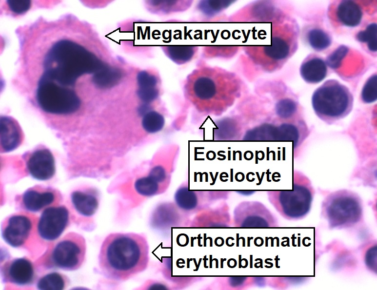
Bone marrow is a semi-solid biological tissue, tissue found within the Spongy bone, spongy (also known as cancellous) portions of bones. In birds and mammals, bone marrow is the primary site of new blood cell production (or haematopoiesis). It is composed of Blood cell, hematopoietic cells, marrow adipose tissue, and supportive stromal cells. In adult humans, bone marrow is primarily located in the Rib cage, ribs, vertebrae, sternum, and Pelvis, bones of the pelvis. Bone marrow comprises approximately 5% of total body mass in healthy adult humans, such that a man weighing 73 kg (161 lbs) will have around 3.7 kg (8 lbs) of bone marrow. Human marrow produces approximately 500 billion blood cells per day, which join the Circulatory system, systemic circulation via permeable vasculature sinusoids within the medullary cavity. All types of hematopoietic cells, including both Myeloid tissue, myeloid and Lymphocyte, lymphoid lineages, are created in bone marrow; howev ... [...More Info...] [...Related Items...] OR: [Wikipedia] [Google] [Baidu] |
Transverse Fascia
The transversalis fascia (or transverse fascia) is a thin aponeurotic membrane of the abdomen. It lies between the inner surface of the transverse abdominal muscle and the parietal peritoneum. It forms part of the general layer of fascia lining the abdominal parietes. It is directly continuous with the iliac fascia, the internal spermatic fascia, and pelvic fasciae. Structure In the inguinal region, the transversalis fascia is thick and dense. It is joined by fibers from the aponeurosis of the transverse abdominal muscle. It becomes thin as it ascends to the diaphragm and blends with the fascia covering the under surface of this muscle. It is directly continuous with the iliac fascia, the internal spermatic fascia, and pelvic fasciae. Borders Behind, it is lost in the fat which covers the posterior surfaces of the kidneys. Below, it has the following attachments: posteriorly, to the whole length of the iliac crest, between the attachments of the transverse abdominal ... [...More Info...] [...Related Items...] OR: [Wikipedia] [Google] [Baidu] |
Iliac Fascia
The iliac fascia, or Abernethy's fascia, is a fascia in the region of the ilium of the pelvis. It has the following connections: * ''laterally'', to the whole length of the inner lip of the iliac crest. * ''medially'', to the linea terminalis of the lesser pelvis, where it is continuous with the periosteum. At the iliopectineal eminence it receives the tendon of insertion of the Psoas minor, when that muscle exists. Lateral to the femoral vessels it is intimately connected to the posterior margin of the inguinal ligament, and is continuous with the transversalis fascia. Immediately lateral to the femoral vessels the iliac fascia is prolonged backward and medialward from the inguinal ligament as a band, the iliopectineal fascia, which is attached to the iliopectineal eminence. This fascia divides the space between the inguinal ligament and the hip bone into two lacunæ or compartments: * the ''medial'' vascular lacuna transmits the femoral vessels. * the ''lateral'' muscula ... [...More Info...] [...Related Items...] OR: [Wikipedia] [Google] [Baidu] |
Fascia Lata
The fascia lata is the deep fascia of the thigh. It encloses the thigh muscles and forms the outer limit of the fascial compartments of thigh, which are internally separated by the medial intermuscular septum and the lateral intermuscular septum. The fascia lata is thickened at its lateral side where it forms the iliotibial tract, a structure that runs to the tibia and serves as a site of muscle attachment. Structure The fascia lata is an investment for the whole of the thigh, but varies in thickness in different parts. It is thicker in the upper and lateral part of the thigh, where it receives a fibrous expansion from the gluteus maximus, and where the tensor fasciae latae is inserted between its layers; it is very thin behind and at the upper and medial part, where it covers the adductor muscles, and again becomes stronger around the knee, receiving fibrous expansions from the tendon of the biceps femoris laterally, from the sartorius medially, and from the quadriceps f ... [...More Info...] [...Related Items...] OR: [Wikipedia] [Google] [Baidu] |
Tensor Fasciae Latae Muscle
The tensor fasciae latae (or tensor fasciæ latæ or, formerly, tensor vaginae femoris) is a muscle of the thigh. Together with the gluteus maximus, it acts on the iliotibial band and is continuous with the iliotibial tract, which attaches to the tibia. The muscle assists in keeping the balance of the pelvis while standing, walking, or running. Structure It arises from the anterior part of the outer lip of the iliac crest; from the outer surface of the anterior superior iliac spine, and part of the outer border of the notch below it, between the gluteus medius and sartorius; and from the deep surface of the fascia lata. It is inserted between the two layers of the iliotibial tract of the fascia lata about the junction of the middle and upper thirds of the thigh. The tensor fasciae latae tautens the iliotibial tract and braces the knee, especially when the opposite foot is lifted.Saladin, Kenneth. Anatomy and Physiology. 6th ed. Mc-Graw Hill. 2010. The terminal insertion poin ... [...More Info...] [...Related Items...] OR: [Wikipedia] [Google] [Baidu] |
Longissimus
The longissimus ( la, the longest one) is the muscle lateral to the semispinalis muscles. It is the longest subdivision of the erector spinae muscles that extends forward into the transverse processes of the posterior cervical vertebrae. Structure Longissimus thoracis et lumborum The longissimus thoracis et lumborum is the intermediate and largest of the continuations of the erector spinae. In the lumbar region (longissimus lumborum), where it is as yet blended with the iliocostalis, some of its fibers are attached to the whole length of the posterior surfaces of the transverse processes and the accessory processes of the lumbar vertebrae, and to the anterior layer of the lumbodorsal fascia. In the thoracic region (longissimus thoracis), it is inserted, by rounded tendons, into the tips of the transverse processes of all the thoracic vertebrae, and by fleshy processes into the lower nine or ten ribs between their tubercles and angles. Longissimus cervicis The longissimu ... [...More Info...] [...Related Items...] OR: [Wikipedia] [Google] [Baidu] |
Iliocostalis
Iliocostalis muscle is the muscle immediately lateral to the longissimus that is the nearest to the furrow that separates the epaxial muscles from the hypaxial. It lies very deep to the fleshy portion of the serratus posterior muscle. It laterally flexes the vertebral column to the same side. Structure Iliocostalis muscle has a common origin from the iliac crest, the sacrum, the thoracolumbar fascia, and the spinous processes of the vertebrae from T11 to L5. Iliocostalis cervicis (cervicalis ascendens) arises from the angles of the third, fourth, fifth, and sixth ribs, and is inserted into the posterior tubercles of the transverse processes of the fourth, fifth, and sixth cervical vertebrae. Iliocostalis thoracis (musculus accessorius; iliocostalis thoracis) arises by flattened tendons from the upper borders of the angles of the lower six ribs medial to the tendons of insertion of the iliocostalis lumborum; these become muscular, and are inserted into the upper borders o ... [...More Info...] [...Related Items...] OR: [Wikipedia] [Google] [Baidu] |


