|
Hip Examination
In medicine, physiotherapy, chiropractic, and osteopathy the hip examination, or hip exam, is undertaken when a patient has a complaint of hip pain and/or signs and/or symptoms suggestive of hip joint pathology. It is a physical examination maneuver. Examination steps The hip examination, like all examinations of the joints, is typically divided into the following sections: * Position/lighting/draping * Inspection * Palpation * Motion * Special maneuvers The middle three steps are often remembered with the saying ''look, feel, move''. Position/lighting/draping Position – for most of the exam the patient should be supine and the bed or examination table should be flat. The patient's hands should remain at their sides with the head resting on a pillow. The knees and hips should be in the anatomical position (knee extended, hip neither flexed nor extended). Lighting – adjusted so that it is ideal. Draping – both of the patient's hips should be exposed so that the quadrice ... [...More Info...] [...Related Items...] OR: [Wikipedia] [Google] [Baidu] |
Medicine
Medicine is the science and practice of caring for a patient, managing the diagnosis, prognosis, prevention, treatment, palliation of their injury or disease, and promoting their health. Medicine encompasses a variety of health care practices evolved to maintain and restore health by the prevention and treatment of illness. Contemporary medicine applies biomedical sciences, biomedical research, genetics, and medical technology to diagnose, treat, and prevent injury and disease, typically through pharmaceuticals or surgery, but also through therapies as diverse as psychotherapy, external splints and traction, medical devices, biologics, and ionizing radiation, amongst others. Medicine has been practiced since prehistoric times, and for most of this time it was an art (an area of skill and knowledge), frequently having connections to the religious and philosophical beliefs of local culture. For example, a medicine man would apply herbs and say prayers for healing, o ... [...More Info...] [...Related Items...] OR: [Wikipedia] [Google] [Baidu] |
Proprioception
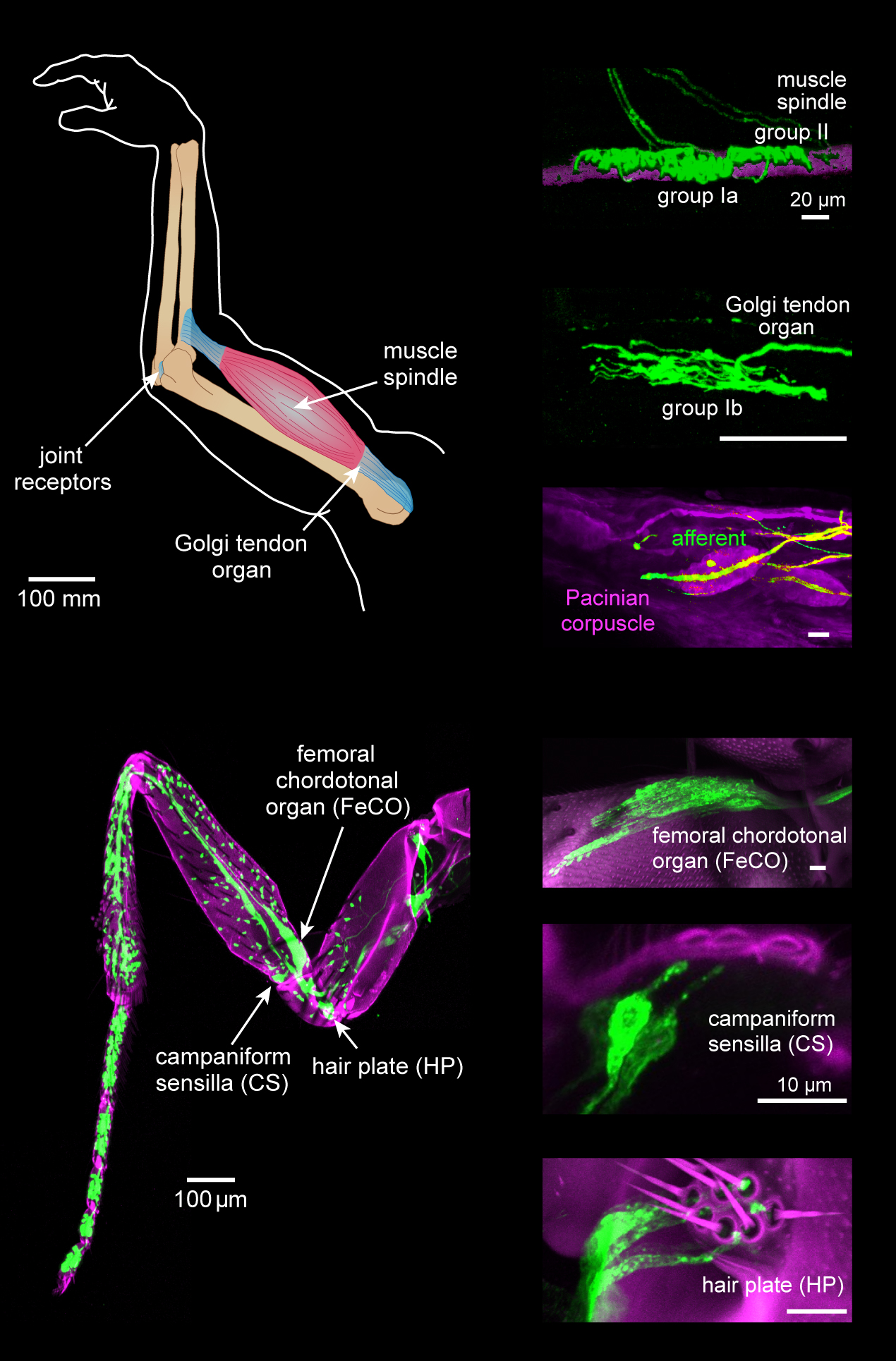
Proprioception ( ), also referred to as kinaesthesia (or kinesthesia), is the sense of self-movement, force, and body position. It is sometimes described as the "sixth sense". Proprioception is mediated by proprioceptors, mechanosensory neurons located within muscles, tendons, and joints. Most animals possess multiple subtypes of proprioceptors, which detect distinct kinematic parameters, such as joint position, movement, and load. Although all mobile animals possess proprioceptors, the structure of the sensory organs can vary across species. Proprioceptive signals are transmitted to the central nervous system, where they are integrated with information from other sensory systems, such as the visual system and the vestibular system, to create an overall representation of body position, movement, and acceleration. In many animals, sensory feedback from proprioceptors is essential for stabilizing body posture and coordinating body movement. System overview In vertebrates, limb ve ... [...More Info...] [...Related Items...] OR: [Wikipedia] [Google] [Baidu] |
Flexion Contracture
Motion, the process of movement, is described using specific anatomical terms. Motion includes movement of organs, joints, limbs, and specific sections of the body. The terminology used describes this motion according to its direction relative to the anatomical position of the body parts involved. Anatomists and others use a unified set of terms to describe most of the movements, although other, more specialized terms are necessary for describing unique movements such as those of the hands, feet, and eyes. In general, motion is classified according to the anatomical plane it occurs in. ''Flexion'' and ''extension'' are examples of ''angular'' motions, in which two axes of a joint are brought closer together or moved further apart. ''Rotational'' motion may occur at other joints, for example the shoulder, and are described as ''internal'' or ''external''. Other terms, such as ''elevation'' and ''depression'', describe movement above or below the horizontal plane. Many anatomica ... [...More Info...] [...Related Items...] OR: [Wikipedia] [Google] [Baidu] |
Anterior Superior Iliac Spine
The anterior superior iliac spine (abbreviated: ASIS) is a bony projection of the iliac bone, and an important landmark of surface anatomy. It refers to the anterior extremity of the iliac crest of the pelvis. It provides attachment for the inguinal ligament, and the sartorius muscle. The tensor fasciae latae muscle attaches to the lateral aspect of the superior anterior iliac spine, and also about 5 cm away at the iliac tubercle. Structure The anterior superior iliac spine refers to the anterior extremity of the iliac crest of the pelvis. This is a key surface landmark, and easily palpated. It provides attachment for the inguinal ligament, the sartorius muscle, and the tensor fasciae latae muscle. A variety of structures lie close to the anterior superior iliac spine, including the subcostal nerve, the femoral artery (which passes between it and the pubic symphysis), and the iliohypogastric nerve. Clinical significance The anterior superior iliac spine provides a c ... [...More Info...] [...Related Items...] OR: [Wikipedia] [Google] [Baidu] |
Posterior Superior Iliac Spine
The posterior border of the ala, shorter than the anterior, also presents two projections separated by a notch, the posterior superior iliac spine and the posterior inferior iliac spine. The posterior superior iliac spine serves for the attachment of the oblique portion of the posterior sacroiliac ligaments and the multifidus. See also * Dimples of Venus The dimples of Venus (also known as back dimples, Duffy Dimples, butt dimples or Veneral dimples) are sagittally symmetrical indentations sometimes visible on the human lower back, just superior to the gluteal cleft. They are directly superfici ... References External links * – "Posterior view of the skeleton of the trunk." * – "The Female Pelvis - bones" * – "The Sacral and Coccygeal Vertebrae, Posterior View" * * * https://web.archive.org/web/20170917131455/http://healthandspine.com/ Bones of the pelvis Ilium (bone) {{musculoskeletal-stub ... [...More Info...] [...Related Items...] OR: [Wikipedia] [Google] [Baidu] |
Gaenslen's Test
Gaenslen's test, also known as Gaenslen's maneuver, is a medical test used to detect musculoskeletal abnormalities and primary-chronic inflammation of the lumbar vertebrae and sacroiliac joint. This test is often used to test for spondyloarthritis, sciatica, or other forms of rheumatism, and is often performed during checkup visits in patients who have been diagnosed with one of the former disorders. It is named after Frederick Julius Gaenslen, the orthopedic surgeon who invented the test. This test is often performed alongside Patrick's test and Yeoman's test. To perform Gaenslen's test, the hip joint is flexed maximally on one side and the opposite hip joint is extended, stressing both sacroiliac joints simultaneously. This is often done by having the patient lying on his or her back, lifting the knee to push towards the patient's chest while the other leg is allowed to fall over the side of an examination table, and is pushed toward the floor, flexing both sacroiliac joints. Th ... [...More Info...] [...Related Items...] OR: [Wikipedia] [Google] [Baidu] |
FABER Test
Patrick's test or FABER test is performed to evaluate pathology of the hip joint or the sacroiliac joint. The test is performed by having the tested leg flexed and the thigh abducted and externally rotated. If pain is elicited on the ipsilateral side anteriorly, it is suggestive of a hip joint disorder on the same side. If pain is elicited on the contralateral side posteriorly around the sacroiliac joint, it is suggestive of pain mediated by dysfunction in that joint. History Patrick's test is named after the American neurologist Hugh Talbot Patrick. See also * Gaenslen's test * Physical medicine and rehabilitation Physical medicine and rehabilitation, also known as physiatry, is a branch of medicine that aims to enhance and restore functional ability and quality of life to people with physical impairments or disabilities. This can include conditions su ... References Orthopedic surgical procedures {{Med-sign-stub ... [...More Info...] [...Related Items...] OR: [Wikipedia] [Google] [Baidu] |
Patrick's Test
Patrick's test or FABER test is performed to evaluate pathology of the hip joint or the sacroiliac joint. The test is performed by having the tested leg flexed and the thigh abducted and externally rotated. If pain is elicited on the ipsilateral side anteriorly, it is suggestive of a hip joint disorder on the same side. If pain is elicited on the contralateral side posteriorly around the sacroiliac joint, it is suggestive of pain mediated by dysfunction in that joint. History Patrick's test is named after the American neurologist Hugh Talbot Patrick. See also * Gaenslen's test * Physical medicine and rehabilitation Physical medicine and rehabilitation, also known as physiatry, is a branch of medicine that aims to enhance and restore functional ability and quality of life to people with physical impairments or disabilities. This can include conditions su ... References Orthopedic surgical procedures {{Med-sign-stub ... [...More Info...] [...Related Items...] OR: [Wikipedia] [Google] [Baidu] |
Inferior Pubic Ramus
In vertebrates, the pubic region ( la, pubis) is the most forward-facing (ventral and anterior) of the three main regions making up the coxal bone. The left and right pubic regions are each made up of three sections, a superior ramus, inferior ramus, and a body. Structure The pubic region is made up of a ''body'', ''superior ramus'', and ''inferior ramus'' (). The left and right coxal bones join at the pubic symphysis. It is covered by a layer of fat, which is covered by the mons pubis. The pubis is the lower limit of the suprapubic region. In the female, the pubic region is anterior to the urethral sponge. Body The body forms the wide, strong, middle and flat part of the pubic region. The bodies of the left and right pubic regions join at the pubic symphysis. The rough upper edge is the pubic crest, ending laterally in the pubic tubercle. This tubercle, found roughly 3 cm from the pubic symphysis, is a distinctive feature on the lower part of the abdominal wall; important ... [...More Info...] [...Related Items...] OR: [Wikipedia] [Google] [Baidu] |
Superior Pubic Ramus
In vertebrates, the pubic region ( la, pubis) is the most forward-facing (ventral and anterior) of the three main regions making up the coxal bone. The left and right pubic regions are each made up of three sections, a superior ramus, inferior ramus, and a body. Structure The pubic region is made up of a ''body'', ''superior ramus'', and ''inferior ramus'' (). The left and right coxal bones join at the pubic symphysis. It is covered by a layer of fat, which is covered by the mons pubis. The pubis is the lower limit of the suprapubic region. In the female, the pubic region is anterior to the urethral sponge. Body The body forms the wide, strong, middle and flat part of the pubic region. The bodies of the left and right pubic regions join at the pubic symphysis. The rough upper edge is the pubic crest, ending laterally in the pubic tubercle. This tubercle, found roughly 3 cm from the pubic symphysis, is a distinctive feature on the lower part of the abdominal wall; important ... [...More Info...] [...Related Items...] OR: [Wikipedia] [Google] [Baidu] |
Iliac Spine
The ilium is a bone of the pelvic girdle with four bony projections, each serving as attachment points for muscles and ligaments: * Anterior superior iliac spine * Anterior inferior iliac spine * Posterior superior iliac spine * Posterior inferior iliac spine The posterior inferior iliac spine (Sweeney's Tubercle) is an anatomical landmark that describes a bony "spine", or projection, at the posterior and inferior surface of the iliac bone. It is one of two such spines on the posterior surface, the o ... {{set index Pelvis Skeletal system Ilium (bone) ... [...More Info...] [...Related Items...] OR: [Wikipedia] [Google] [Baidu] |
Xiphisternum
The xiphoid process , or xiphisternum or metasternum, is a small cartilaginous process (extension) of the inferior (lower) part of the sternum, which is usually ossified in the adult human. It may also be referred to as the ensiform process. Both the Greek-derived ''xiphoid'' and its Latin equivalent ''ensiform'' mean 'swordlike' or 'sword-shaped' Structure The xiphoid process is considered to be at the level of the 9th thoracic vertebra and the T7 dermatome. Development In newborns and young (especially small) infants, the tip of the xiphoid process may be both seen and felt as a lump just below the sternal notch. At 15 to 29 years old, the xiphoid usually fuses to the body of the sternum with a fibrous joint. Unlike the synovial articulation of major joints, this is non-movable. Ossification of the xiphoid process occurs around age 40. Variation The xiphoid process can be naturally bifurcated or sometimes perforated (xiphoidal foramen). These variances in morphology are inhe ... [...More Info...] [...Related Items...] OR: [Wikipedia] [Google] [Baidu] |


