|
Hilgenreiner's Line
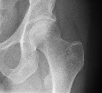
Hilgenreiner's line is a horizontal line drawn on an AP radiograph of the pelvis running between the inferior aspects of both triradiate cartilages of the acetabulums. It is named for Heinrich Hilgenreiner. Clinical Use Used in conjunction with Perkin's line Perkin's line is a line drawn on an AP radiograph of the pelvis perpendicular to Hilgenreiner's line at the lateral aspects of the triradiate cartilage of the acetabulum. Clinical use Used in conjunction with Hilgenreiner's line, Perkin's line ... or the acetabular angle, Hilgenreiner's line is useful in the diagnosis of developmental dysplasia of the hip. References {{Orthopedics-stub Musculoskeletal radiographic signs ... [...More Info...] [...Related Items...] OR: [Wikipedia] [Google] [Baidu] |
Anteroposterior
Standard anatomical terms of location are used to unambiguously describe the anatomy of animals, including humans. The terms, typically derived from Latin or Greek roots, describe something in its standard anatomical position. This position provides a definition of what is at the front ("anterior"), behind ("posterior") and so on. As part of defining and describing terms, the body is described through the use of anatomical planes and anatomical axes. The meaning of terms that are used can change depending on whether an organism is bipedal or quadrupedal. Additionally, for some animals such as invertebrates, some terms may not have any meaning at all; for example, an animal that is radially symmetrical will have no anterior surface, but can still have a description that a part is close to the middle ("proximal") or further from the middle ("distal"). International organisations have determined vocabularies that are often used as standard vocabularies for subdisciplines of anatom ... [...More Info...] [...Related Items...] OR: [Wikipedia] [Google] [Baidu] |
Medical Radiography
Radiography is an imaging technique using X-rays, gamma rays, or similar ionizing radiation and non-ionizing radiation to view the internal form of an object. Applications of radiography include medical radiography ("diagnostic" and "therapeutic") and industrial radiography. Similar techniques are used in airport security (where "body scanners" generally use backscatter X-ray). To create an image in conventional radiography, a beam of X-rays is produced by an X-ray generator and is projected toward the object. A certain amount of the X-rays or other radiation is absorbed by the object, dependent on the object's density and structural composition. The X-rays that pass through the object are captured behind the object by a detector (either photographic film or a digital detector). The generation of flat two dimensional images by this technique is called projectional radiography. In computed tomography (CT scanning) an X-ray source and its associated detectors rotate around the su ... [...More Info...] [...Related Items...] OR: [Wikipedia] [Google] [Baidu] |
Human Pelvis
The pelvis (plural pelves or pelvises) is the lower part of the Trunk (anatomy), trunk, between the human abdomen, abdomen and the thighs (sometimes also called pelvic region), together with its embedded skeleton (sometimes also called bony pelvis, or pelvic skeleton). The pelvic region of the trunk includes the bony pelvis, the pelvic cavity (the space enclosed by the bony pelvis), the pelvic floor, below the pelvic cavity, and the perineum, below the pelvic floor. The pelvic skeleton is formed in the area of the back, by the sacrum and the coccyx and anteriorly and to the left and right sides, by a pair of hip bones. The two hip bones connect the spine with the lower limbs. They are attached to the sacrum posteriorly, connected to each other anteriorly, and joined with the two femurs at the hip joints. The gap enclosed by the bony pelvis, called the pelvic cavity, is the section of the body underneath the abdomen and mainly consists of the reproductive organs (sex organs) and ... [...More Info...] [...Related Items...] OR: [Wikipedia] [Google] [Baidu] |
Triradiate Cartilage
The triradiate cartilage (in Latin cartilago ypsiloformis) is the 'Y'-shaped epiphyseal plate between the ilium, ischium and pubis to form the acetabulum of the os coxae. Human development In children, the triradiate cartilage closes at an approximate bone age of 12 years for girls and 14 years for boys. Clinical use Evaluating the position of the triradiate cartilage on an AP radiograph of the pelvis with both Perkin's line and Hilgenreiner's line Hilgenreiner's line is a horizontal line drawn on an AP radiograph of the pelvis running between the inferior aspects of both triradiate cartilages of the acetabulums. It is named for Heinrich Hilgenreiner. Clinical Use Used in conjunction wi ... can help establish a diagnosis of developmental dysplasia of the hip. References See also {{Pelvis Pelvis ... [...More Info...] [...Related Items...] OR: [Wikipedia] [Google] [Baidu] |
Acetabulum
The acetabulum (), also called the cotyloid cavity, is a concave surface of the pelvis. The head of the femur meets with the pelvis at the acetabulum, forming the hip joint. Structure There are three bones of the ''os coxae'' (hip bone) that come together to form the ''acetabulum''. Contributing a little more than two-fifths of the structure is the ischium, which provides lower and side boundaries to the acetabulum. The ilium forms the upper boundary, providing a little less than two-fifths of the structure of the acetabulum. The rest is formed by the pubis, near the midline. It is bounded by a prominent uneven rim, which is thick and strong above, and serves for the attachment of the acetabular labrum, which reduces its opening, and deepens the surface for formation of the hip joint. At the lower part of the ''acetabulum'' is the acetabular notch, which is continuous with a circular depression, the acetabular fossa, at the bottom of the cavity of the ''acetabulum''. The re ... [...More Info...] [...Related Items...] OR: [Wikipedia] [Google] [Baidu] |
Heinrich Hilgenreiner
Heinrich Hilgenreiner (3 November 1870 – 24 October 1954) German surgeon and orthopedist. Biography Born in Prague, and raised in a German family in Bohemia (which at the time was part of the Habsburg monarchy), he served as a medical officer in the First World War. After the war, he became a professor of the German Charles-Ferdinand University in Prague and director of the ''Kinderklinik'' (children's clinic). In 1946 he was forced to leave Czechoslovakia for Austria, where he lived until his death. He was the younger brother of Karl Hilgenreiner, a theologian and politician, also professor at Charles University. He is the grandfather of the Austrian artist Gerhard Gleich. He died in 1954 in Spillern, Austria. Work As a professor at the Karls-Universität in Prague, he became a specialist on the diagnosis and cure of congenital luxation of the hip joint in infants and young children. "Hilgenreiner's line Hilgenreiner's line is a horizontal line drawn on an AP radiograph of ... [...More Info...] [...Related Items...] OR: [Wikipedia] [Google] [Baidu] |
Perkin's Line
Perkin's line is a line drawn on an AP radiograph of the pelvis perpendicular to Hilgenreiner's line at the lateral aspects of the triradiate cartilage of the acetabulum. Clinical use Used in conjunction with Hilgenreiner's line, Perkin's line is useful in the diagnosis of developmental dysplasia of the hip; the upper femoral epiphysis The epiphysis () is the rounded end of a long bone, at its joint with adjacent bone(s). Between the epiphysis and diaphysis (the long midsection of the long bone) lies the metaphysis, including the epiphyseal plate (growth plate). At the join ... should be in the inferomedial quadrant on a normal radiograph. Lateral displacement relative to Perkin's line is indicative of DDH. References External links Wheeless Online Musculoskeletal radiographic signs {{Orthopedics-stub ... [...More Info...] [...Related Items...] OR: [Wikipedia] [Google] [Baidu] |
Acetabular Angle
In vertebrate anatomy, hip (or "coxa"Latin ''coxa'' was used by Celsus in the sense "hip", but by Pliny the Elder in the sense "hip bone" (Diab, p 77) in medical terminology) refers to either an anatomical region or a joint. The hip region is located lateral and anterior to the gluteal region, inferior to the iliac crest, and overlying the greater trochanter of the femur, or "thigh bone". In adults, three of the bones of the pelvis have fused into the hip bone or acetabulum which forms part of the hip region. The hip joint, scientifically referred to as the acetabulofemoral joint (''art. coxae''), is the joint between the head of the femur and acetabulum of the pelvis and its primary function is to support the weight of the body in both static (e.g., standing) and dynamic (e.g., walking or running) postures. The hip joints have very important roles in retaining balance, and for maintaining the pelvic inclination angle. Pain of the hip may be the result of numerous causes, i ... [...More Info...] [...Related Items...] OR: [Wikipedia] [Google] [Baidu] |
Hip Dysplasia (human)
Hip dysplasia is an abnormality of the hip joint where the socket portion does not fully cover the ball portion, resulting in an increased risk for joint dislocation. Hip dysplasia may occur at birth or develop in early life. Regardless, it does not typically produce symptoms in babies less than a year old. Occasionally one leg may be shorter than the other. The left hip is more often affected than the right. Complications without treatment can include arthritis, limping, and low back pain. Females are affected more often than males. Hip dysplasia was described at least as early as the 300s BC by Hippocrates. Risk factors for hip dysplasia include female sex, family history, certain swaddling practices, and breech presentation whether an infant is delivered vaginally or by cesarean section. If one identical twin is affected, there is a 40% risk the other will also be affected. Screening all babies for the condition by physical examination is recommended. Ultrasonography may ... [...More Info...] [...Related Items...] OR: [Wikipedia] [Google] [Baidu] |


