|
Hepatization Of Lungs
Hepatization is conversion into a substance resembling the liver; a state of the lungs when gorged with effuse matter, so that they are no longer pervious to the air. Red hepatization is when there are red blood cells, neutrophils, and fibrin in the pulmonary alveolus/ alveoli; it precedes gray hepatization, where the red cells have been broken down leaving a fibrinosuppurative exudate. The main cause is lobar pneumonia Lobar pneumonia is a form of pneumonia characterized by inflammatory exudate within the intra-alveolar space resulting in consolidation that affects a large and continuous area of the lobe of a lung. It is one of three anatomic classifications o .... Transformation from Red hepatization to gray hepatization is an example for acute inflammation turning into a chronic inflammation. References Further readingLectures on the diseases of the lungs and heartby Thomas DaviesMedical Times 1841 [...More Info...] [...Related Items...] OR: [Wikipedia] [Google] [Baidu] |
Liver
The liver is a major Organ (anatomy), organ only found in vertebrates which performs many essential biological functions such as detoxification of the organism, and the Protein biosynthesis, synthesis of proteins and biochemicals necessary for digestion and growth. In humans, it is located in the quadrant (anatomy), right upper quadrant of the abdomen, below the thoracic diaphragm, diaphragm. Its other roles in metabolism include the regulation of Glycogen, glycogen storage, decomposition of red blood cells, and the production of hormones. The liver is an accessory digestive organ that produces bile, an alkaline fluid containing cholesterol and bile acids, which helps the fatty acid degradation, breakdown of fat. The gallbladder, a small pouch that sits just under the liver, stores bile produced by the liver which is later moved to the small intestine to complete digestion. The liver's highly specialized biological tissue, tissue, consisting mostly of hepatocytes, regulates a w ... [...More Info...] [...Related Items...] OR: [Wikipedia] [Google] [Baidu] |
Lung
The lungs are the primary organs of the respiratory system in humans and most other animals, including some snails and a small number of fish. In mammals and most other vertebrates, two lungs are located near the backbone on either side of the heart. Their function in the respiratory system is to extract oxygen from the air and transfer it into the bloodstream, and to release carbon dioxide from the bloodstream into the atmosphere, in a process of gas exchange. Respiration is driven by different muscular systems in different species. Mammals, reptiles and birds use their different muscles to support and foster breathing. In earlier tetrapods, air was driven into the lungs by the pharyngeal muscles via buccal pumping, a mechanism still seen in amphibians. In humans, the main muscle of respiration that drives breathing is the diaphragm. The lungs also provide airflow that makes vocal sounds including human speech possible. Humans have two lungs, one on the left and on ... [...More Info...] [...Related Items...] OR: [Wikipedia] [Google] [Baidu] |
Red Blood Cells
Red blood cells (RBCs), also referred to as red cells, red blood corpuscles (in humans or other animals not having nucleus in red blood cells), haematids, erythroid cells or erythrocytes (from Greek language, Greek ''erythros'' for "red" and ''kytos'' for "hollow vessel", with ''-cyte'' translated as "cell" in modern usage), are the most common type of blood cell and the vertebrate's principal means of delivering oxygen (O2) to the body tissue (biology), tissues—via blood flow through the circulatory system. RBCs take up oxygen in the lungs, or in fish the gills, and release it into tissues while squeezing through the body's capillary, capillaries. The cytoplasm of a red blood cell is rich in hemoglobin, an iron-containing biomolecule that can bind oxygen and is responsible for the red color of the cells and the blood. Each human red blood cell contains approximately 270 million hemoglobin molecules. The cell membrane is composed of proteins and lipids, and this structure ... [...More Info...] [...Related Items...] OR: [Wikipedia] [Google] [Baidu] |
Neutrophil
Neutrophils (also known as neutrocytes or heterophils) are the most abundant type of granulocytes and make up 40% to 70% of all white blood cells in humans. They form an essential part of the innate immune system, with their functions varying in different animals. They are formed from stem cells in the bone marrow and Cellular differentiation, differentiated into #Subpopulations, subpopulations of neutrophil-killers and neutrophil-cagers. They are short-lived and highly mobile, as they can enter parts of tissue where other cells/molecules cannot. Neutrophils may be subdivided into segmented neutrophils and banded neutrophils (or Band cell, bands). They form part of the polymorphonuclear cells family (PMNs) together with basophils and eosinophils. The name ''neutrophil'' derives from staining characteristics on hematoxylin and eosin (H&E stain, H&E) histology, histological or cell biology, cytological preparations. Whereas basophilic white blood cells stain dark blue and eosinoph ... [...More Info...] [...Related Items...] OR: [Wikipedia] [Google] [Baidu] |
Fibrin
Fibrin (also called Factor Ia) is a fibrous, non-globular protein involved in the clotting of blood. It is formed by the action of the protease thrombin on fibrinogen, which causes it to polymerize. The polymerized fibrin, together with platelets, forms a hemostatic plug or clot over a wound site. When the lining of a blood vessel is broken, platelets are attracted, forming a platelet plug. These platelets have thrombin receptors on their surfaces that bind serum thrombin molecules, which in turn convert soluble fibrinogen in the serum into fibrin at the wound site. Fibrin forms long strands of tough insoluble protein that are bound to the platelets. Factor XIII completes the cross-linking of fibrin so that it hardens and contracts. The cross-linked fibrin forms a mesh atop the platelet plug that completes the clot. Fibrin was discovered by Marcello Malpighi in 1666. Role in disease Excessive generation of fibrin due to activation of the coagulation cascade leads to thrombos ... [...More Info...] [...Related Items...] OR: [Wikipedia] [Google] [Baidu] |
Pulmonary Alveolus
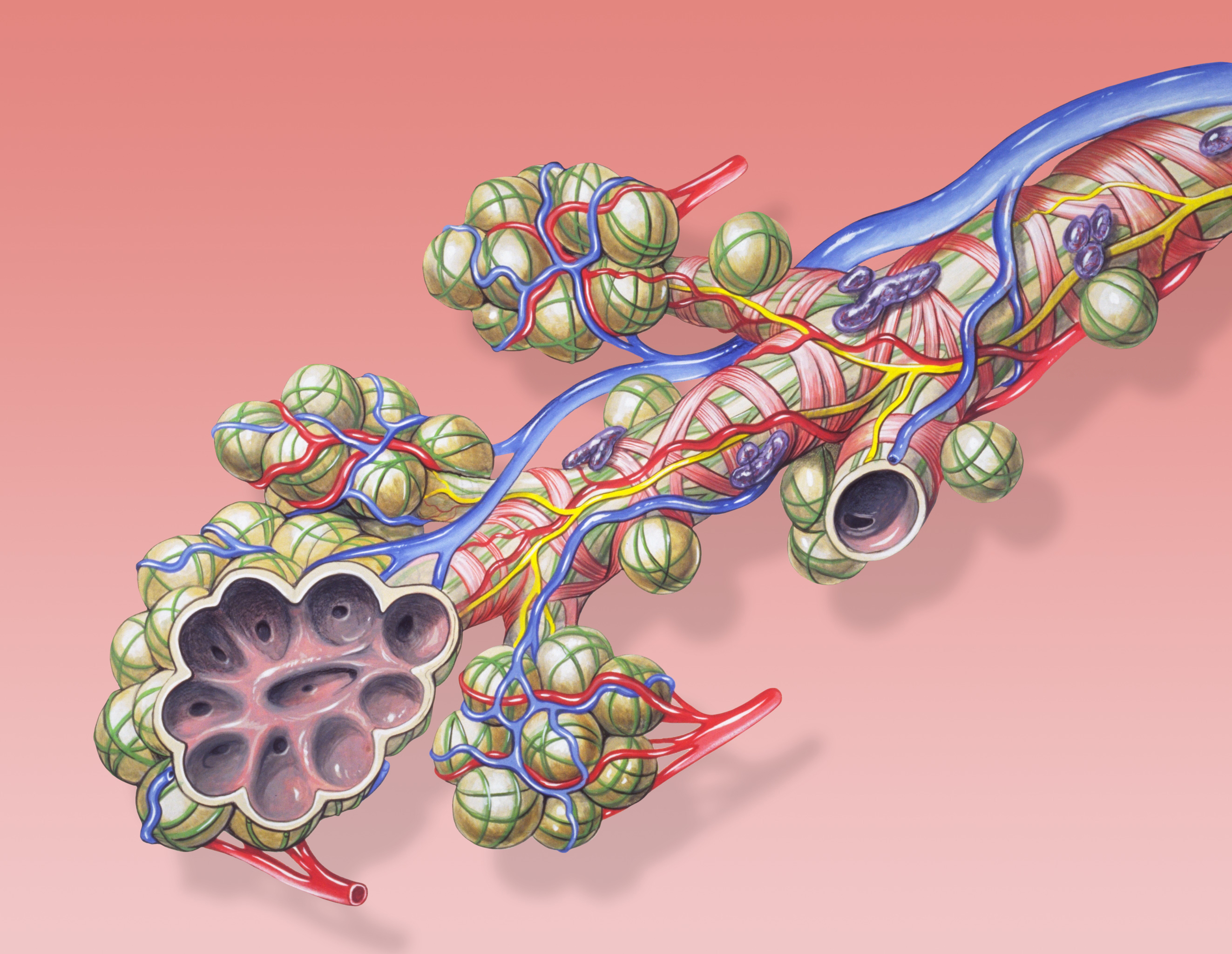
A pulmonary alveolus (plural: alveoli, from Latin ''alveolus'', "little cavity"), also known as an air sac or air space, is one of millions of hollow, distensible cup-shaped cavities in the lungs where oxygen is exchanged for carbon dioxide. Alveoli make up the functional tissue of the mammalian lungs known as the lung parenchyma, which takes up 90 percent of the total lung volume. Alveoli are first located in the respiratory bronchioles that mark the beginning of the respiratory zone. They are located sparsely in these bronchioles, line the walls of the alveolar ducts, and are more numerous in the blind-ended alveolar sacs. The acini are the basic units of respiration, with gas exchange taking place in all the alveoli present. The alveolar membrane is the gas exchange surface, surrounded by a network of capillaries. Across the membrane oxygen is diffused into the capillaries and carbon dioxide released from the capillaries into the alveoli to be breathed out. Alveoli are pa ... [...More Info...] [...Related Items...] OR: [Wikipedia] [Google] [Baidu] |
Lobar Pneumonia
Lobar pneumonia is a form of pneumonia characterized by inflammatory exudate within the intra-alveolar space resulting in consolidation that affects a large and continuous area of the lobe of a lung. It is one of three anatomic classifications of pneumonia (the other being bronchopneumonia and atypical pneumonia). In children round pneumonia develops instead because the pores of Kohn which allow the lobar spread of infection are underdeveloped. Mechanism The invading organism starts multiplying, thereby releasing toxins that cause inflammation and edema of the lung parenchyma. This leads to the accumulation of cellular debris within the lungs. This leads to consolidation or solidification, which is a term that is used for macroscopic or radiologic appearance of the lungs affected by pneumonia. Bacterial pneumonia is mainly classified into lobar and diffuse depending on the degree of lung irritation or damage. Stages Lobar pneumonia usually has an acute progression. Classica ... [...More Info...] [...Related Items...] OR: [Wikipedia] [Google] [Baidu] |
Davies, Thomas (1792-1839) (DNB00)
*Thomas Davies (bishop) (c. 1511–1573), Welsh clergyman, Bishop of St Asaph, 1561–1573 * Thomas Davies (physician) (died 1615) English physician, see Lumleian Lectures *Thomas Davies (bookseller) (c. 1713–1785), London-based Scottish bookseller * Thomas Davies (brewer) (?–1869), see Don Brewery *Thomas Davies (British Army officer) (c. 1737–1812), Royal Artillery officer, artist, and naturalist * Thomas Alfred Davies (1809–1899), American Civil War general * Thomas Davies (Conservative politician) (1858–1939), British Member of Parliament for Cirencester and Tewkesbury, 1918–1929 *Thomas Davies (Australian politician) (1881–1942), Australian politician *Thomas F. Davis (1804–1871), fifth Episcopal Bishop of South Carolina * Thomas Frederick Davies Sr. (1831–1905), third bishop of the Episcopal Diocese of Michigan, 1889–1905 *Thomas Frederick Davies Jr. (1872–1936), second bishop of the Episcopal Diocese of Western Massachusetts, 1911–1936 *Thomas Henry Has ... [...More Info...] [...Related Items...] OR: [Wikipedia] [Google] [Baidu] |


