|
Henri Chaput
Victor Alexandre Henri Chaput (17 November 1857, Tonnerre – 1919) was a French surgeon who specialized in intestinal surgery. Biography He studied medicine in Paris, where he received his doctorate in 1885. In 1888 he became a hospital surgeon, subsequently performing surgery at the Hôpitaux Broussais, Boucicaut, and Lariboisièr during his career.Henri Victor Chaput at Who Named It He was at the forefront of surgical asepsis, advocating the use of sterile rubber ( caoutchouc) gloves during operations. He was known for his preference of [...More Info...] [...Related Items...] OR: [Wikipedia] [Google] [Baidu] |
Tibia
The tibia (; ), also known as the shinbone or shankbone, is the larger, stronger, and anterior (frontal) of the two bones in the leg below the knee in vertebrates (the other being the fibula, behind and to the outside of the tibia); it connects the knee with the ankle. The tibia is found on the medial side of the leg next to the fibula and closer to the median plane. The tibia is connected to the fibula by the interosseous membrane of leg, forming a type of fibrous joint called a syndesmosis with very little movement. The tibia is named for the flute ''tibia''. It is the second largest bone in the human body, after the femur. The leg bones are the strongest long bones as they support the rest of the body. Structure In human anatomy, the tibia is the second largest bone next to the femur. As in other vertebrates the tibia is one of two bones in the lower leg, the other being the fibula, and is a component of the knee and ankle joints. The ossification or formation of the bone ... [...More Info...] [...Related Items...] OR: [Wikipedia] [Google] [Baidu] |
1919 Deaths
Events January * January 1 ** The Czechoslovak Legions occupy much of the self-proclaimed "free city" of Pressburg (now Bratislava), enforcing its incorporation into the new republic of Czechoslovakia. ** HMY ''Iolaire'' sinks off the coast of the Hebrides; 201 people, mostly servicemen returning home to Lewis and Harris, are killed. * January 2– 22 – Russian Civil War: The Red Army's Caspian-Caucasian Front begins the Northern Caucasus Operation against the White Army, but fails to make progress. * January 3 – The Faisal–Weizmann Agreement is signed by Emir Faisal (representing the Arab Kingdom of Hejaz) and Zionist leader Chaim Weizmann, for Arab–Jewish cooperation in the development of a Jewish homeland in Palestine, and an Arab nation in a large part of the Middle East. * January 5 – In Germany: ** Spartacist uprising in Berlin: The Marxist Spartacus League, with the newly formed Communist Party of Germany and the Independent Social Democ ... [...More Info...] [...Related Items...] OR: [Wikipedia] [Google] [Baidu] |
1857 Births
Events January–March * January 1 – The biggest Estonian newspaper, ''Postimees'', is established by Johann Voldemar Jannsen. * January 7 – The partly French-owned London General Omnibus Company begins operating. * January 9 – The 7.9 Fort Tejon earthquake shakes Central and Southern California, with a maximum Mercalli intensity of IX (''Violent''). * January 24 – The University of Calcutta is established in Calcutta, as the first multidisciplinary modern university in South Asia. The University of Bombay is also established in Bombay, British India, this year. * February 3 – The National Deaf Mute College (later renamed Gallaudet University) is established in Washington, D.C., becoming the first school for the advanced education of the deaf. * February 5 – The Federal Constitution of the United Mexican States is promulgated. * March – The Austrian garrison leaves Bucharest. * March 3 ** France and the United Kingdom for ... [...More Info...] [...Related Items...] OR: [Wikipedia] [Google] [Baidu] |
Malleolus
A malleolus is the bony prominence on each side of the human ankle. Each leg is supported by two bones, the tibia on the inner side (medial) of the leg and the fibula on the outer side (lateral) of the leg. The medial malleolus is the prominence on the inner side of the ankle, formed by the lower end of the tibia. The lateral malleolus is the prominence on the outer side of the ankle, formed by the lower end of the fibula. The word ''malleolus'' (), plural ''malleoli'' (), comes from Latin and means "small hammer". (It is cognate with ''mallet''.) Medial malleolus The medial malleolus is found at the foot end of the tibia. The medial surface of the lower extremity of tibia is prolonged downward to form a strong pyramidal process, flattened from without inward - the medial malleolus. * The ''medial surface'' of this process is convex and subcutaneous. * The ''lateral'' or ''articular surface'' is smooth and slightly concave, and articulates with the talus. * The ''anterior bo ... [...More Info...] [...Related Items...] OR: [Wikipedia] [Google] [Baidu] |
Peritoneum
The peritoneum is the serous membrane forming the lining of the abdominal cavity or coelom in amniotes and some invertebrates, such as annelids. It covers most of the intra-abdominal (or coelomic) organs, and is composed of a layer of mesothelium supported by a thin layer of connective tissue. This peritoneal lining of the cavity supports many of the abdominal organs and serves as a conduit for their blood vessels, lymphatic vessels, and nerves. The abdominal cavity (the space bounded by the vertebrae, abdominal muscles, diaphragm, and pelvic floor) is different from the intraperitoneal space (located within the abdominal cavity but wrapped in peritoneum). The structures within the intraperitoneal space are called "intraperitoneal" (e.g., the stomach and intestines), the structures in the abdominal cavity that are located behind the intraperitoneal space are called "retroperitoneal" (e.g., the kidneys), and those structures below the intraperitoneal space are called "subp ... [...More Info...] [...Related Items...] OR: [Wikipedia] [Google] [Baidu] |
Rectum
The rectum is the final straight portion of the large intestine in humans and some other mammals, and the Gastrointestinal tract, gut in others. The adult human rectum is about long, and begins at the rectosigmoid junction (the end of the sigmoid colon) at the level of the third sacral vertebra or the sacral promontory depending upon what definition is used. Its diameter is similar to that of the sigmoid colon at its commencement, but it is dilated near its termination, forming the rectal ampulla. It terminates at the level of the anorectal ring (the level of the puborectalis sling) or the dentate line, again depending upon which definition is used. In humans, the rectum is followed by the anal canal which is about long, before the gastrointestinal tract terminates at the anal verge. The word rectum comes from the Latin ''Wikt:rectum, rectum Wikt:intestinum, intestinum'', meaning ''straight intestine''. Structure The rectum is a part of the lower gastrointestinal tract ... [...More Info...] [...Related Items...] OR: [Wikipedia] [Google] [Baidu] |
Bile Ducts
A bile duct is any of a number of long tube-like structures that carry bile, and is present in most vertebrates. Bile is required for the digestion of food and is secreted by the liver into passages that carry bile toward the hepatic duct. It joins the cystic duct (carrying bile to and from the gallbladder) to form the common bile duct which then opens into the intestine. Structure The top half of the common bile duct is associated with the liver, while the bottom half of the common bile duct is associated with the pancreas, through which it passes on its way to the intestine. It opens into the part of the intestine called the duodenum via the ampulla of Vater. Segments The biliary tree (see below) is the whole network of various sized ducts branching through the liver. The path is as follows: Bile canaliculi → Canals of Hering → interlobular bile ducts → intrahepatic bile ducts → left and right hepatic ducts ''merge to form'' → common hepatic duct ''exits liver a ... [...More Info...] [...Related Items...] OR: [Wikipedia] [Google] [Baidu] |
Kneecap
The patella, also known as the kneecap, is a flat, rounded triangular bone which articulates with the femur (thigh bone) and covers and protects the anterior articular surface of the knee joint. The patella is found in many tetrapods, such as mice, cats, birds and dogs, but not in whales, or most reptiles. In humans, the patella is the largest sesamoid bone (i.e., embedded within a tendon or a muscle) in the body. Babies are born with a patella of soft cartilage which begins to ossify into bone at about four years of age. Structure The patella is a sesamoid bone roughly triangular in shape, with the apex of the patella facing downwards. The apex is the most inferior (lowest) part of the patella. It is pointed in shape, and gives attachment to the patellar ligament. The front and back surfaces are joined by a thin margin and towards centre by a thicker margin. The tendon of the quadriceps femoris muscle attaches to the base of the patella., with the vastus intermedius muscle ... [...More Info...] [...Related Items...] OR: [Wikipedia] [Google] [Baidu] |
Octave Terrillon
Octave Roch Simon Terrillon (17 May 1844, Oigny-sur-Seine – 22 December 1895, Paris) was a French physician and surgeon, known as a pioneer of aseptic surgery. From 1868 he worked as a hospital interne in Paris, where in 1873 he received his medical doctorate. In 1876 he qualified as a hospital surgeon, and eventually became associated with the Salpêtrière Hospital. In 1878 he became an associate professor at the faculty of medicine in Paris. On April 13, 1957, a French postage stamp featuring a portrait of Dr. Terrillon was issued. Included on the stamp were images of a microscope, an autoclave and some surgical instruments. Somewhere around 1882 he advocated the procedure of using boiling water, a heat sterilisation technique for disinfecting surgical instruments. Selected works * ''Leçons de clinique chirurgicale professées à la Salpêtrière'', 1889 – Lessons taught in the surgical clinic at the Salpêtrière. * ''Traité des maladies du testicule et de ... [...More Info...] [...Related Items...] OR: [Wikipedia] [Google] [Baidu] |
Paul Jules Tillaux
Paul Jules Tillaux (8 December 1834 – 20 October 1904) was a French physician who was a native of Aunay-sur-Odon, département Calvados. Tillaux was a surgeon and professor of surgery in Paris, and in 1879 became a member of the ''Académie de Médecine''. He was director of the Amphitheatre d'Anatomie des Hopitaux de Paris from 1868 to 1890. In 1892 Tillaux was the first physician to describe an uncommon Salter Harris Type III fracture of the tibia. He performed experiments on cadavers and discovered that stress to the anterior inferior tibiofibular ligament could lead to this type of avulsion fracture. This fracture is unique because it occurs during a certain period of adolescence, when there is a differential rate of growth of the epiphysis. In honor of his discovery, a fracture of the anteriolateral tibial epiphysis is now called a Tillaux fracture — often misdiagnosed as a simple sprain. A similar fracture to the posterolateral tibia was later identified by surge ... [...More Info...] [...Related Items...] OR: [Wikipedia] [Google] [Baidu] |
Epiphysis
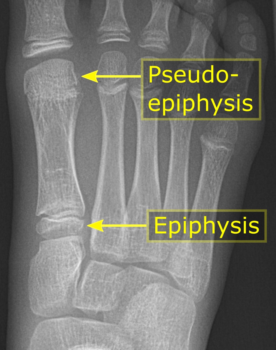
The epiphysis () is the rounded end of a long bone, at its joint with adjacent bone(s). Between the epiphysis and diaphysis (the long midsection of the long bone) lies the metaphysis, including the epiphyseal plate (growth plate). At the joint, the epiphysis is covered with articular cartilage; below that covering is a zone similar to the epiphyseal plate, known as subchondral bone. The epiphysis is filled with red bone marrow, which produces erythrocytes (red blood cells). Structure There are four types of epiphysis: # Pressure epiphysis: The region of the long bone that forms the joint is a pressure epiphysis (e.g. the head of the femur, part of the hip joint complex). Pressure epiphyses assist in transmitting the weight of the human body and are the regions of the bone that are under pressure during movement or locomotion. Another example of a pressure epiphysis is the head of the humerus which is part of the shoulder complex. condyles of femur and tibia also comes under ... [...More Info...] [...Related Items...] OR: [Wikipedia] [Google] [Baidu] |


