|
Hemothorax
A hemothorax (derived from hemo- lood+ thorax hest plural ''hemothoraces'') is an accumulation of blood within the pleural cavity. The symptoms of a hemothorax may include chest pain and difficulty breathing, while the clinical signs may include reduced breath sounds on the affected side and a rapid heart rate. Hemothoraces are usually caused by an injury, but they may occur spontaneously due to cancer invading the pleural cavity, as a result of a blood clotting disorder, as an unusual manifestation of endometriosis, in response to a collapsed lung, or rarely in association with other conditions. Hemothoraces are usually diagnosed using a chest X-ray, but they can be identified using other forms of imaging including ultrasound, a CT scan, or an MRI. They can be differentiated from other forms of fluid within the pleural cavity by analysing a sample of the fluid, and are defined as having a hematocrit of greater than 50% that of the person's blood. Hemothoraces may be tre ... [...More Info...] [...Related Items...] OR: [Wikipedia] [Google] [Baidu] |
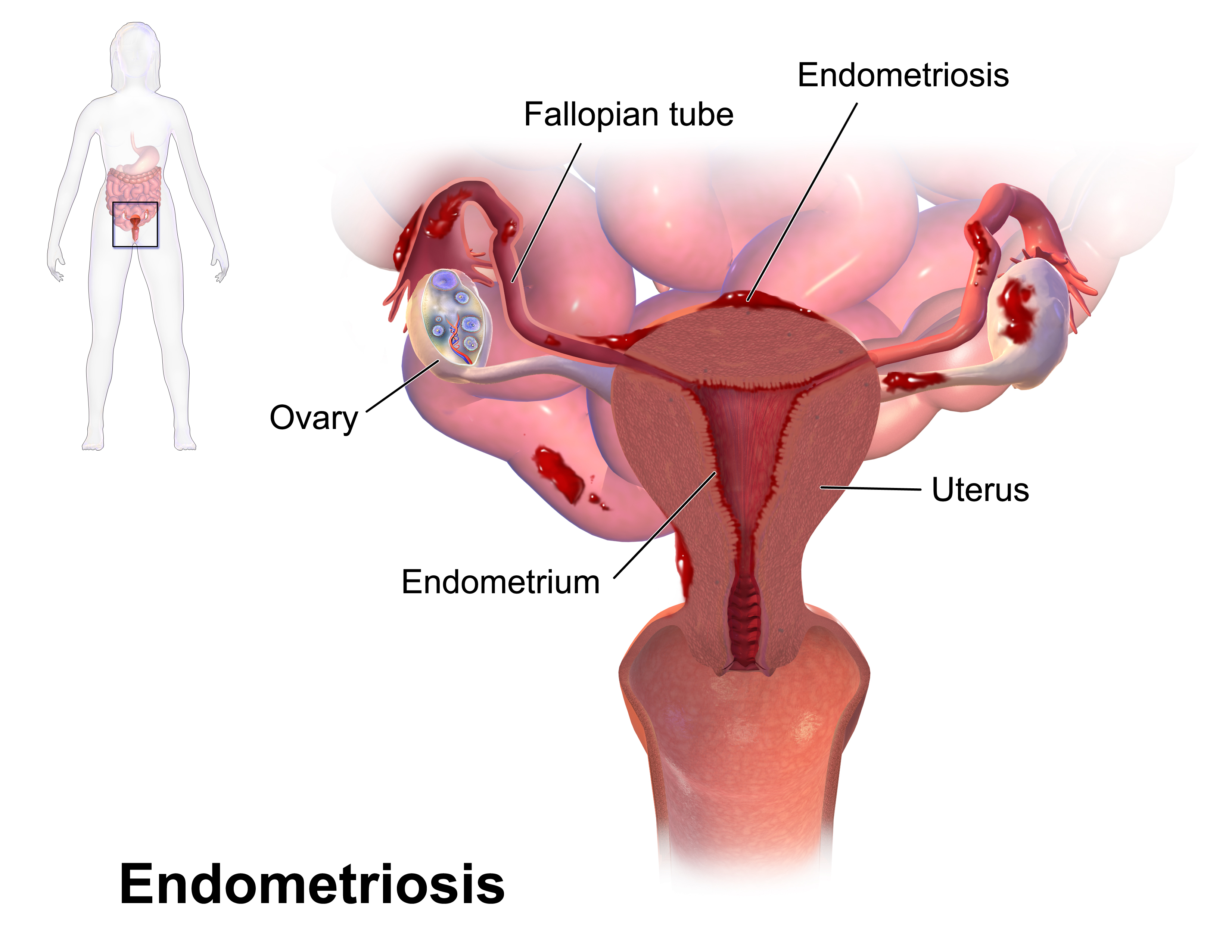
Endometriosis
Endometriosis is a disease of the female reproductive system in which cells similar to those in the endometrium, the layer of tissue that normally covers the inside of the uterus, grow outside the uterus. Most often this is on the ovaries, fallopian tubes, and tissue around the uterus and ovaries; in rare cases it may also occur in other parts of the body. Some symptoms include pelvic pain, heavy periods, pain with bowel movements, and infertility. Nearly half of those affected have chronic pelvic pain, while in 70% pain occurs during menstruation. Pain during sexual intercourse is also common. Infertility occurs in up to half of affected individuals. About 25% of individuals have no symptoms and 85% of those seen with infertility in a tertiary center have no pain. Endometriosis can have both social and psychological effects. The cause is not entirely clear. Risk factors include having a family history of the condition. The areas of endometriosis bleed each month (menstrua ... [...More Info...] [...Related Items...] OR: [Wikipedia] [Google] [Baidu] |
Pleural Effusion
A pleural effusion is accumulation of excessive fluid in the pleural space, the potential space that surrounds each lung. Under normal conditions, pleural fluid is secreted by the parietal pleural capillaries at a rate of 0.6 millilitre per kilogram weight per hour, and is cleared by lymphatic absorption leaving behind only 5–15 millilitres of fluid, which helps to maintain a functional vacuum between the parietal and visceral pleurae. Excess fluid within the pleural space can impair inspiration by upsetting the functional vacuum and hydrostatically increasing the resistance against lung expansion, resulting in a fully or partially collapsed lung. Various kinds of fluid can accumulate in the pleural space, such as serous fluid (hydrothorax), blood (hemothorax), pus (pyothorax, more commonly known as pleural empyema), chyle ( chylothorax), or very rarely urine (urinothorax). When unspecified, the term "pleural effusion" normally refers to hydrothorax. A pleural effusion can a ... [...More Info...] [...Related Items...] OR: [Wikipedia] [Google] [Baidu] |
Tube Thoracostomy
A chest tube (also chest drain, thoracic catheter, tube thoracostomy or intercostal drain) is a surgical drain that is inserted through the chest wall and into the pleural space or the mediastinum in order to remove clinically undesired substances such as air (pneumothorax), excess fluid (pleural effusion or hydrothorax), blood (hemothorax), chyle (chylothorax) or pus (empyema) from the intrathoracic space. An intrapleural chest tube is also known as a Bülau drain or an intercostal catheter (ICC), and can either be a thin, flexible silicone tube (known as a "pigtail" drain), or a larger, semi-rigid, fenestrated plastic tube, which often involves a flutter valve or underwater seal. The concept of chest drainage was first advocated by Hippocrates when he described the treatment of empyema by means of incision, cautery and insertion of metal tubes. However, the technique was not widely used until the influenza epidemic of 1918 to evacuate post-pneumonic empyema, which was first do ... [...More Info...] [...Related Items...] OR: [Wikipedia] [Google] [Baidu] |
Chest Tube
A chest tube (also chest drain, thoracic catheter, tube thoracostomy or intercostal drain) is a surgical drain that is inserted through the chest wall and into the pleural space or the mediastinum in order to remove clinically undesired substances such as air (pneumothorax), excess fluid (pleural effusion or hydrothorax), blood (hemothorax), chyle ( chylothorax) or pus (empyema) from the intrathoracic space. An intrapleural chest tube is also known as a Bülau drain or an intercostal catheter (ICC), and can either be a thin, flexible silicone tube (known as a "pigtail" drain), or a larger, semi-rigid, fenestrated plastic tube, which often involves a flutter valve or underwater seal. The concept of chest drainage was first advocated by Hippocrates when he described the treatment of empyema by means of incision, cautery and insertion of metal tubes. However, the technique was not widely used until the influenza epidemic of 1918 to evacuate post-pneumonic empyema, which was first ... [...More Info...] [...Related Items...] OR: [Wikipedia] [Google] [Baidu] |
Thoracotomy
A thoracotomy is a surgical procedure to gain access into the pleural space of the chest. It is performed by surgeons (emergency physicians or paramedics under certain circumstances) to gain access to the thoracic organs, most commonly the heart, the lungs, or the esophagus, or for access to the thoracic aorta or the anterior spine (the latter may be necessary to access tumors in the spine). A thoracotomy is the first step in thoracic surgeries including lobectomy or pneumonectomy for lung cancer or to gain thoracic access in major trauma. Approaches There are many different surgical approaches to performing a thoracotomy. Some common forms of thoracotomies include: * Median sternotomy provides wide access to the mediastinum and is the incision of choice for most open-heart surgery and access to the anterior mediastinum * Posterolateral thoracotomy is an incision through an intercostal space on the back, and is often widened with rib spreaders. It is a very common approach ... [...More Info...] [...Related Items...] OR: [Wikipedia] [Google] [Baidu] |
Pneumothorax
A pneumothorax is an abnormal collection of air in the pleural space between the lung and the chest wall. Symptoms typically include sudden onset of sharp, one-sided chest pain and shortness of breath. In a minority of cases, a one-way valve is formed by an area of damaged tissue, and the amount of air in the space between chest wall and lungs increases; this is called a tension pneumothorax. This can cause a steadily worsening oxygen shortage and low blood pressure. This leads to a type of shock called obstructive shock, which can be fatal unless reversed. Very rarely, both lungs may be affected by a pneumothorax. It is often called a "collapsed lung", although that term may also refer to atelectasis. A primary spontaneous pneumothorax is one that occurs without an apparent cause and in the absence of significant lung disease. A secondary spontaneous pneumothorax occurs in the presence of existing lung disease. Smoking increases the risk of primary spontaneous pneumothora ... [...More Info...] [...Related Items...] OR: [Wikipedia] [Google] [Baidu] |
Thoracentesis
Thoracentesis , also known as thoracocentesis (from Greek ''thōrax'' 'chest, thorax'—GEN ''thōrakos''—and ''kentēsis'' 'pricking, puncture'), pleural tap, needle thoracostomy, or needle decompression (often used term), is an invasive medical procedure to remove fluid or air from the pleural space for diagnostic or therapeutic purposes. A cannula, or hollow needle, is carefully introduced into the thorax, generally after administration of local anesthesia. The procedure was first performed by Morrill Wyman in 1850 and then described by Henry Ingersoll Bowditch in 1852. The recommended location varies depending upon the source. Some sources recommend the midaxillary line, in the eighth, ninth, or tenth intercostal space. Whenever possible, the procedure should be performed under ultrasound guidance, which has shown to reduce complications. Tension pneumothorax is a medical emergency that requires emergent needle decompression before a chest tube is placed. Indications 48 ... [...More Info...] [...Related Items...] OR: [Wikipedia] [Google] [Baidu] |
Hydrothorax
Hydrothorax is a type of pleural effusion in which transudate accumulates in the pleural cavity. This condition is most likely to develop secondary to congestive heart failure, following an increase in hydrostatic pressure within the lungs. More rarely, hydrothorax can develop in 10% of patients with ascites which is called hepatic hydrothorax. It is often difficult to manage in end-stage liver failure and often fails to respond to therapy. Pleural effusions may also develop following the accumulation of other fluids within the pleural cavity; if the fluid is blood it is known as hemothorax (as in major chest injuries), if the fluid is pus it is known as pyothorax (resulting from chest infections), and if the fluid is lymph it is known as chylothorax (resulting from rupture of the thoracic duct). Treatment Treatment of hydrothorax is difficult for several reasons. The underlying condition needs to be corrected; however, often the source of the hydrothorax is end stage liver dise ... [...More Info...] [...Related Items...] OR: [Wikipedia] [Google] [Baidu] |
Fibrinolytic Therapy
Thrombolysis, also called fibrinolytic therapy, is the breakdown (lysis) of blood clots formed in blood vessels, using medication. It is used in ST elevation myocardial infarction, stroke, and in cases of severe venous thromboembolism (massive pulmonary embolism or extensive deep vein thrombosis). The main complication is bleeding (which can be dangerous), and in some situations thrombolysis may therefore be unsuitable. Thrombolysis can also play an important part in reperfusion therapy that deals specifically with blocked arteries. Medical uses Diseases where thrombolysis is used: * ST elevation myocardial infarction: Large trials have shown that mortality can be reduced using thrombolysis (particularly fibrinolysis) in treating heart attacks. It works by stimulating secondary fibrinolysis by plasmin through infusion of analogs of tissue plasminogen activator (tPA), the protein that normally activates plasmin. * Stroke: Thrombolysis reduces major disability or death when given ... [...More Info...] [...Related Items...] OR: [Wikipedia] [Google] [Baidu] |
Fibrothorax
Fibrothorax is a medical condition characterised by severe scarring (fibrosis) and fusion of the layers of the pleural space surrounding the lungs resulting in decreased movement of the lung and ribcage. The main symptom of fibrothorax is shortness of breath. There also may be recurrent fluid collections surrounding the lungs. Fibrothorax may occur as a complication of many diseases, including infection of the pleural space known as an empyema or bleeding into the pleural space known as a haemothorax. Fibrosis in the pleura may be produced intentionally using a technique called pleurodesis to prevent recurrent punctured lung (pneumothorax), and the usually limited fibrosis that this produces can rarely be extensive enough to lead to fibrothorax. The condition is most often diagnosed using an X-ray or CT scan, the latter more readily detecting mild cases. Fibrothorax is often treated conservatively with watchful waiting but may require surgery. The outlook is usually good as long ... [...More Info...] [...Related Items...] OR: [Wikipedia] [Google] [Baidu] |
Streptokinase
Streptokinase (SK) is a thrombolytic medication activating plasminogen by nonenzymatic mechanism. As a medication it is used to break down clots in some cases of myocardial infarction (heart attack), pulmonary embolism, and arterial thromboembolism. The type of heart attack it is used in is an ST elevation myocardial infarction (STEMI). It is given by injection into a vein. Side effects include nausea, bleeding, low blood pressure, and allergic reactions. A second use in a person's lifetime is not recommended. While no harm has been found with use in pregnancy, it has not been well studied in this group. Streptokinase is in the antithrombotic family of medications and works by turning on the fibrinolytic system. Streptokinase was discovered in 1933 from beta-hemolytic streptococci. It is on the World Health Organization's List of Essential Medicines. It is no longer commercially available in the United States. Medical uses If percutaneous coronary intervention (PCI) is not ... [...More Info...] [...Related Items...] OR: [Wikipedia] [Google] [Baidu] |
Serous Fluid
In physiology, serous fluid or serosal fluid (originating from the Medieval Latin word ''serosus'', from Latin ''serum'') is any of various body fluids resembling Serum (blood), serum, that are typically pale yellow or transparent and of a benign nature. The fluid fills the inside of body cavity, body cavities. Serous fluid originates from serous glands, with secretions enriched with proteins and water. Serous fluid may also originate from mixed glands, which contain both mucous cell, mucous and serous cells. A common trait of serous fluids is their role in assisting digestion, excretion, and respiratory system, respiration. In medical fields, especially cytopathology, serous fluid is a synonym for effusion fluids from various body cavities. Examples of effusion fluid are pleural effusion and pericardial effusion. There are many causes of effusions which include involvement of the cavity by cancer. Cancer in a serous cavity is called a serous carcinoma. Cytopathology evaluation is ... [...More Info...] [...Related Items...] OR: [Wikipedia] [Google] [Baidu] |
.jpg)


.png)