|
Hematoma Development
A hematoma, also spelled haematoma, or blood suffusion is a localized bleeding outside of blood vessels, due to either disease or trauma including injury or surgery and may involve blood continuing to seep from broken capillaries. A hematoma is benign and is initially in liquid form spread among the tissues including in sacs between tissues where it may coagulate and solidify before blood is reabsorbed into blood vessels. An ecchymosis is a hematoma of the skin larger than 10 mm. They may occur among and or within many areas such as skin and other organs, connective tissues, bone, joints and muscle. A collection of blood (or even a hemorrhage) may be aggravated by anticoagulant medication (blood thinner). Blood seepage and collection of blood may occur if heparin is given via an intramuscular route; to avoid this, heparin must be given intravenously or subcutaneously. Signs and symptoms Some hematomas are visible under the surface of the skin (commonly called bruises) or p ... [...More Info...] [...Related Items...] OR: [Wikipedia] [Google] [Baidu] |
Contusion
A bruise, also known as a contusion, is a type of hematoma of tissue, the most common cause being capillaries damaged by trauma, causing localized bleeding that extravasates into the surrounding interstitial tissues. Most bruises occur close enough to the epidermis such that the bleeding causes a visible discoloration. The bruise then remains visible until the blood is either absorbed by tissues or cleared by immune system action. Bruises which do not blanch under pressure can involve capillaries at the level of skin, subcutaneous tissue, muscle, or bone. Bruises are not to be confused with other similar-looking lesions. (Such lesions include petechia (less than , resulting from numerous and diverse etiologies such as adverse reactions from medications such as warfarin, straining, asphyxiation, platelet disorders and diseases such as '' cytomegalovirus''), purpura (, classified as palpable purpura or non-palpable purpura and indicates various pathologic conditions such as t ... [...More Info...] [...Related Items...] OR: [Wikipedia] [Google] [Baidu] |
Hematoma At Backside
A hematoma, also spelled haematoma, or blood suffusion is a localized bleeding outside of blood vessels, due to either disease or trauma including injury or surgery and may involve blood continuing to seep from broken capillaries. A hematoma is benign and is initially in liquid form spread among the tissues including in sacs between tissues where it may coagulate and solidify before blood is reabsorbed into blood vessels. An ecchymosis is a hematoma of the skin larger than 10 mm. They may occur among and or within many areas such as skin and other organs, connective tissues, bone, joints and muscle. A collection of blood (or even a hemorrhage) may be aggravated by anticoagulant medication (blood thinner). Blood seepage and collection of blood may occur if heparin is given via an intramuscular route; to avoid this, heparin must be given intravenously or subcutaneously. Signs and symptoms Some hematomas are visible under the surface of the skin (commonly called bruises) or p ... [...More Info...] [...Related Items...] OR: [Wikipedia] [Google] [Baidu] |
Subarachnoid Hematoma
Subarachnoid hemorrhage (SAH) is bleeding into the subarachnoid space—the area between the arachnoid membrane and the pia mater surrounding the brain. Symptoms may include a severe headache of rapid onset, vomiting, decreased level of consciousness, fever, and sometimes seizures. Neck stiffness or neck pain are also relatively common. In about a quarter of people a small bleed with resolving symptoms occurs within a month of a larger bleed. SAH may occur as a result of a head injury or spontaneously, usually from a ruptured cerebral aneurysm. Risk factors for spontaneous cases include high blood pressure, smoking, family history, alcoholism, and cocaine use. Generally, the diagnosis can be determined by a CT scan of the head if done within six hours of symptom onset. Occasionally, a lumbar puncture is also required. After confirmation further tests are usually performed to determine the underlying cause. Treatment is by prompt neurosurgery or endovascular coiling. Medicatio ... [...More Info...] [...Related Items...] OR: [Wikipedia] [Google] [Baidu] |
Arachnoid Mater
The arachnoid mater (or simply arachnoid) is one of the three meninges, the protective membranes that cover the brain and spinal cord. It is so named because of its resemblance to a spider web. The arachnoid mater is a derivative of the neural crest mesoectoderm in the embryo. Structure It is interposed between the two other meninges, the more superficial and much thicker dura mater and the deeper pia mater, from which it is separated by the subarachnoid space. The delicate arachnoid layer is not attached to the inside of the dura but against it and surrounds the brain and spinal cord. It does not line the brain down into its sulci (folds), as does the pia mater, with the exception of the longitudinal fissure, which divides the left and right cerebral hemispheres. Cerebrospinal fluid (CSF) flows under the arachnoid in the subarachnoid space, within a meshwork of trabeculae which span between the arachnoid and the pia. The arachnoid mater makes arachnoid villi, small protrusions ... [...More Info...] [...Related Items...] OR: [Wikipedia] [Google] [Baidu] |
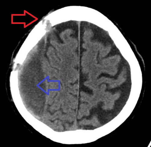
Subdural Hematoma
A subdural hematoma (SDH) is a type of bleeding in which a Hematoma, collection of blood—usually but not always associated with a traumatic brain injury—gathers between the inner layer of the dura mater and the arachnoid mater of the meninges surrounding the brain. It usually results from tears in bridging veins that cross the subdural space. Subdural hematomas may cause an increase in the intracranial pressure, pressure inside the skull, which in turn can cause compression of and damage to delicate brain tissue. Acute subdural hematomas are often life-threatening. Chronic subdural hematomas have a better prognosis if properly managed. In contrast, epidural hematomas are usually caused by tears in arteries, resulting in a build-up of blood between the dura mater and the skull. The third type of brain hemorrhage, known as a subarachnoid hemorrhage, causes bleeding into the subarachnoid space between the arachnoid mater and the pia mater. __TOC__ Signs and symptoms The sympt ... [...More Info...] [...Related Items...] OR: [Wikipedia] [Google] [Baidu] |
Dura Mater
In neuroanatomy, dura mater is a thick membrane made of dense irregular connective tissue that surrounds the brain and spinal cord. It is the outermost of the three layers of membrane called the meninges that protect the central nervous system. The other two meningeal layers are the arachnoid mater and the pia mater. It envelops the arachnoid mater, which is responsible for keeping in the cerebrospinal fluid. It is derived primarily from the neural crest cell population, with postnatal contributions of the paraxial mesoderm. Structure The dura mater has several functions and layers. The dura mater is a membrane that envelops the arachnoid mater. It surrounds and supports the dural sinuses (also called dural venous sinuses, cerebral sinuses, or cranial sinuses) and carries blood from the brain toward the heart. Cranial dura mater has two layers called ''lamellae'', a superficial layer (also called the periosteal layer), which serves as the skull's inner periosteum, called the ... [...More Info...] [...Related Items...] OR: [Wikipedia] [Google] [Baidu] |
Epidural Hematoma
Epidural hematoma is when bleeding occurs between the tough outer membrane covering the brain (dura mater) and the skull. Often there is loss of consciousness following a head injury, a brief regaining of consciousness, and then loss of consciousness again. Other symptoms may include headache, confusion, vomiting, and an inability to move parts of the body. Complications may include seizures. The cause is typically head injury that results in a break of the temporal bone and bleeding from the middle meningeal artery. Occasionally it can occur as a result of a bleeding disorder or blood vessel malformation. Diagnosis is typically by a CT scan or MRI. When this condition occurs in the spine it is known as a spinal epidural hematoma. Treatment is generally by urgent surgery in the form of a craniotomy or burr hole. Without treatment, death typically results. The condition occurs in one to four percent of head injuries. Typically it occurs in young adults. Males are more often ... [...More Info...] [...Related Items...] OR: [Wikipedia] [Google] [Baidu] |
Skull
The skull is a bone protective cavity for the brain. The skull is composed of four types of bone i.e., cranial bones, facial bones, ear ossicles and hyoid bone. However two parts are more prominent: the cranium and the mandible. In humans, these two parts are the neurocranium and the viscerocranium ( facial skeleton) that includes the mandible as its largest bone. The skull forms the anterior-most portion of the skeleton and is a product of cephalisation—housing the brain, and several sensory structures such as the eyes, ears, nose, and mouth. In humans these sensory structures are part of the facial skeleton. Functions of the skull include protection of the brain, fixing the distance between the eyes to allow stereoscopic vision, and fixing the position of the ears to enable sound localisation of the direction and distance of sounds. In some animals, such as horned ungulates (mammals with hooves), the skull also has a defensive function by providing the mount (on the front ... [...More Info...] [...Related Items...] OR: [Wikipedia] [Google] [Baidu] |
Cephalohematoma
A cephalohaematoma is a hemorrhage of blood between the skull and the periosteum of any age human, including a newborn baby secondary to rupture of blood vessels crossing the periosteum. Because the swelling is subperiosteal, its boundaries are limited by the individual bones, in contrast to a caput succedaneum. Symptoms and signs Swelling appears after 2-3 days after birth. If severe the child may develop jaundice, anemia or hypotension. In some cases it may be an indication of a linear skull fracture or be at risk of an infection leading to osteomyelitis or meningitis. The swelling of a cephalohematoma takes weeks to resolve as the blood clot is slowly absorbed from the periphery towards the centre. In time the swelling hardens (calcification) leaving a relatively softer centre so that it appears as a 'depressed fracture'. Cephalohematoma should be distinguished from another scalp bleeding called subgaleal hemorrhage (also called subaponeurotic hemorrhage), which is blood betwe ... [...More Info...] [...Related Items...] OR: [Wikipedia] [Google] [Baidu] |
Periosteum
The periosteum is a membrane that covers the outer surface of all bones, except at the articular surfaces (i.e. the parts within a joint space) of long bones. Endosteum lines the inner surface of the medullary cavity of all long bones. Structure The periosteum consists of an outer fibrous layer, and an inner cambium layer (or osteogenic layer). The fibrous layer is of dense irregular connective tissue, containing fibroblasts, while the cambium layer is highly cellular containing progenitor cells that develop into osteoblasts. These osteoblasts are responsible for increasing the width of a long bone and the overall size of the other bone types. After a bone fracture, the progenitor cells develop into osteoblasts and chondroblasts, which are essential to the healing process. The outer fibrous layer and the inner cambium layer is differentiated under electron micrography. As opposed to osseous tissue, the periosteum has nociceptors, sensory neurons that make it very sensitive to ... [...More Info...] [...Related Items...] OR: [Wikipedia] [Google] [Baidu] |
Galea Aponeurosis
The epicranial aponeurosis (aponeurosis epicranialis, galea aponeurotica) is an aponeurosis (a tough layer of dense fibrous tissue). It covers the upper part of the skull in humans and many other animals. Structure In humans, the epicranial aponeurosis originates from the external occipital protuberance and highest nuchal lines of the occipital bone. It merges with the occipitofrontalis muscle. In front, it forms a short and narrow prolongation between its union with the frontalis muscle (the frontal part of the occipitofrontalis muscle). On either side, the epicranial aponeurosis attaches to the anterior auricular muscles and the superior auricular muscles. Here it is less aponeurotic, and is continued over the temporal fascia to the zygomatic arch as a layer of laminated areolar tissue. It is closely connected to the integument by the firm, dense, fibro-fatty layer which forms the superficial fascia of the scalp. It is attached to the pericranium by loose cellular tissue, ... [...More Info...] [...Related Items...] OR: [Wikipedia] [Google] [Baidu] |
Subgaleal Hematoma
Subgaleal hemorrhage, also known as subgaleal hematoma, is bleeding in the potential space between the skull periosteum and the scalp galea aponeurosis. Symptoms The diagnosis is generally clinical, with a fluctuant boggy mass developing over the scalp (especially over the occiput) with superficial skin bruising. The swelling develops gradually 12–72 hours after delivery, although it may be noted immediately after delivery in severe cases. Subgaleal hematoma growth is insidious, as it spreads across the whole calvaria and may not be recognized for hours to days. If enough blood accumulates, a visible fluid wave may be seen. Patients may develop periorbital ecchymosis ("raccoon eyes"). Patients with subgaleal hematoma may present with hemorrhagic shock given the volume of blood that can be lost into the potential space between the skull periosteum and the scalp galea aponeurosis, which has been found to be as high as 20-40% of the neonatal blood volume in some studies. The swel ... [...More Info...] [...Related Items...] OR: [Wikipedia] [Google] [Baidu] |


