|
Hassall–Henle Bodies
Hassall–Henle bodies are small transparent growths on the posterior surface of Descemet's membrane at the periphery of the cornea. These bodies contain collagenous matter in which numerous cracks and fissures are filled with extrusions of the corneal endothelium. The condition is usually associated with the aging process. Hassall–Henle bodies are named after British physician Arthur Hill Hassall (1817–1894) and German anatomist Friedrich Gustav Jakob Henle Friedrich Gustav Jakob Henle (; 9 July 1809 – 13 May 1885) was a German physician, pathologist, and anatomist. He is credited with the discovery of the loop of Henle in the kidney. His essay, "On Miasma and Contagia," was an early argument for ... (1809–1885). They are sometimes referred to as Hassall–Henle warts or Henle's warts. References * {{DEFAULTSORT:Hassall-Henle bodies Ophthalmology ... [...More Info...] [...Related Items...] OR: [Wikipedia] [Google] [Baidu] |
Descemet's Membrane
Descemet's membrane ( or the Descemet membrane) is the basement membrane that lies between the corneal proper substance, also called stroma, and the endothelial layer of the cornea. It is composed of different kinds of collagen (Type IV and VIII) than the stroma. The endothelial layer is located at the posterior of the cornea. Descemet's membrane, as the basement membrane for the endothelial layer, is secreted by the single layer of squamous epithelial cells that compose the endothelial layer of the cornea. Structure Its thickness ranges from 3 μm at birth to 8–10 μm in adults.Johnson DH, Bourne WM, Campbell RJ: The ultrastructure of Descemet's membrane. I. Changes with age in normal cornea. Arch Ophthalmol 100:1942, 1982 The corneal endothelium is a single layer of squamous cells covering the surface of the cornea that faces the anterior chamber. Clinical significance Significant damage to the membrane may require a corneal transplant. Damage caused by the hereditary co ... [...More Info...] [...Related Items...] OR: [Wikipedia] [Google] [Baidu] |
Cornea
The cornea is the transparent front part of the eye that covers the iris, pupil, and anterior chamber. Along with the anterior chamber and lens, the cornea refracts light, accounting for approximately two-thirds of the eye's total optical power. In humans, the refractive power of the cornea is approximately 43 dioptres. The cornea can be reshaped by surgical procedures such as LASIK. While the cornea contributes most of the eye's focusing power, its focus is fixed. Accommodation (the refocusing of light to better view near objects) is accomplished by changing the geometry of the lens. Medical terms related to the cornea often start with the prefix "'' kerat-''" from the Greek word κέρας, ''horn''. Structure The cornea has unmyelinated nerve endings sensitive to touch, temperature and chemicals; a touch of the cornea causes an involuntary reflex to close the eyelid. Because transparency is of prime importance, the healthy cornea does not have or need blood vessels with ... [...More Info...] [...Related Items...] OR: [Wikipedia] [Google] [Baidu] |
Endothelium
The endothelium is a single layer of squamous endothelial cells that line the interior surface of blood vessels and lymphatic vessels. The endothelium forms an interface between circulating blood or lymph in the lumen and the rest of the vessel wall. Endothelial cells form the barrier between vessels and tissue and control the flow of substances and fluid into and out of a tissue. Endothelial cells in direct contact with blood are called vascular endothelial cells whereas those in direct contact with lymph are known as lymphatic endothelial cells. Vascular endothelial cells line the entire circulatory system, from the heart to the smallest capillaries. These cells have unique functions that include fluid filtration, such as in the glomerulus of the kidney, blood vessel tone, hemostasis, neutrophil recruitment, and hormone trafficking. Endothelium of the interior surfaces of the heart chambers is called endocardium. An impaired function can lead to serious health issues throug ... [...More Info...] [...Related Items...] OR: [Wikipedia] [Google] [Baidu] |
Arthur Hill Hassall
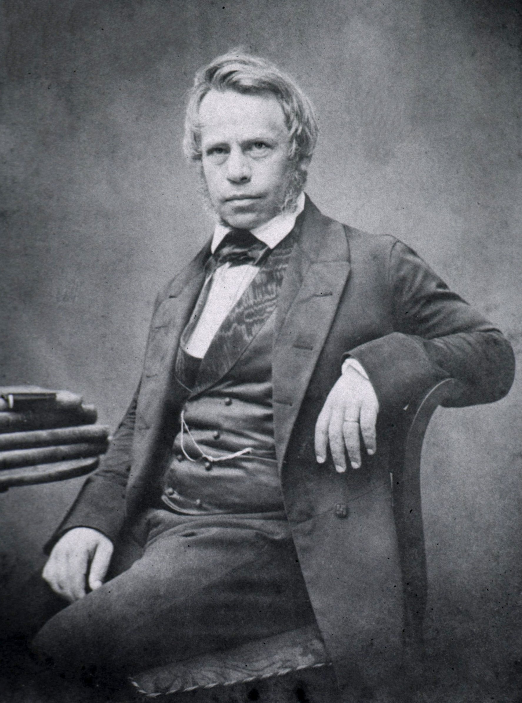
Arthur Hill Hassall (13 December 1817, Teddington – 9 April 1894, San Remo) was a British physician, chemist and microscopist who is primarily known for his work in public health and food safety. Biography Hassall was born in Middlesex as the youngest son of five children in a house of a surgeon. His father was Thomas Hassall (1771–1844) and his mother, née Ann Sherrock ( 1778–1817). He spent his school years in Richmond. He entered medicine through apprenticeship in 1834 to his uncle Sir James Murray (1788–1871), spending his early career in Dublin, where he also studied botany and the seashore. In 1846 he published a two-volume study, ''The Microscopic Anatomy of the Human Body in Health and Disease'', the first English textbook on the subject. After further studying botany at Kew and publishing on botanical topics, particularly freshwater algae, he came to public attention with his 1850 book ''A microscopical examination of the water supplied to the inhabitants of ... [...More Info...] [...Related Items...] OR: [Wikipedia] [Google] [Baidu] |
Friedrich Gustav Jakob Henle
Friedrich Gustav Jakob Henle (; 9 July 1809 – 13 May 1885) was a German physician, pathologist, and anatomist. He is credited with the discovery of the loop of Henle in the kidney. His essay, "On Miasma and Contagia," was an early argument for the germ theory of disease. He was an important figure in the development of modern medicine. Biography Henle was born in Fürth, Bavaria, to Simon and Rachel Diesbach Henle (Hähnlein). He was Jewish. After studying medicine at Heidelberg and at Bonn, where he took his doctor's degree in 1832, he became prosector in anatomy to Johannes Müller at Berlin. During the six years he spent in that position he published a large amount of work, including three anatomical monographs on new species of animals and papers on the structure of the lymphatic system, the distribution of epithelium in the human body, the structure and development of the hair, and the formation of mucus and pus. In 1840, he accepted the chair of anatomy at Zürich an ... [...More Info...] [...Related Items...] OR: [Wikipedia] [Google] [Baidu] |


