|
Hamp Cook
Hepcidin is a protein that in humans is encoded by the ''HAMP'' gene. Hepcidin is a key regulator of the entry of iron into the circulation in mammals. During conditions in which the hepcidin level is abnormally high, such as inflammation, serum iron falls due to iron trapping within macrophages and liver cells and decreased gut iron absorption. This typically leads to anemia due to an inadequate amount of serum iron being available for developing red blood cells. When the hepcidin level is abnormally low such as in hemochromatosis, iron overload occurs due to increased ferroportin mediated iron efflux from storage and increased gut iron absorption. Structure Hepcidin exists as a preprohormone (84 amino acids), prohormone (60 amino acids), and hormone (25 amino acids). Twenty- and 22-amino acid metabolites of hepcidin also exist in the urine. Deletion of 5 ''N''-terminal amino acids results in loss of function. The conversion of prohepcidin to hepcidin is mediated by the ... [...More Info...] [...Related Items...] OR: [Wikipedia] [Google] [Baidu] |
Protein Nuclear Magnetic Resonance Spectroscopy
Nuclear magnetic resonance spectroscopy of proteins (usually abbreviated protein NMR) is a field of structural biology in which NMR spectroscopy is used to obtain information about the structure and dynamics of proteins, and also nucleic acids, and their complexes. The field was pioneered by Richard R. Ernst and Kurt Wüthrich at the ETH, and by Ad Bax, Marius Clore, Angela Gronenborn at the NIH, and Gerhard Wagner at Harvard University, among others. Structure determination by NMR spectroscopy usually consists of several phases, each using a separate set of highly specialized techniques. The sample is prepared, measurements are made, interpretive approaches are applied, and a structure is calculated and validated. NMR involves the quantum-mechanical properties of the central core ("nucleus") of the atom. These properties depend on the local molecular environment, and their measurement provides a map of how the atoms are linked chemically, how close they are in space, and how rapid ... [...More Info...] [...Related Items...] OR: [Wikipedia] [Google] [Baidu] |
Disulfide
In biochemistry, a disulfide (or disulphide in British English) refers to a functional group with the structure . The linkage is also called an SS-bond or sometimes a disulfide bridge and is usually derived by the coupling of two thiol groups. In biology, disulfide bridges formed between thiol groups in two cysteine residues are an important component of the secondary and tertiary structure of proteins. ''Persulfide'' usually refers to compounds. In inorganic chemistry disulfide usually refers to the corresponding anion (−S−S−). Organic disulfides Symmetrical disulfides are compounds of the formula . Most disulfides encountered in organo sulfur chemistry are symmetrical disulfides. Unsymmetrical disulfides (also called heterodisulfides) are compounds of the formula . They are less common in organic chemistry, but most disulfides in nature are unsymmetrical. Properties The disulfide bonds are strong, with a typical bond dissociation energy of 60 kcal/mol (251&nbs ... [...More Info...] [...Related Items...] OR: [Wikipedia] [Google] [Baidu] |
Staphylococcus Aureus
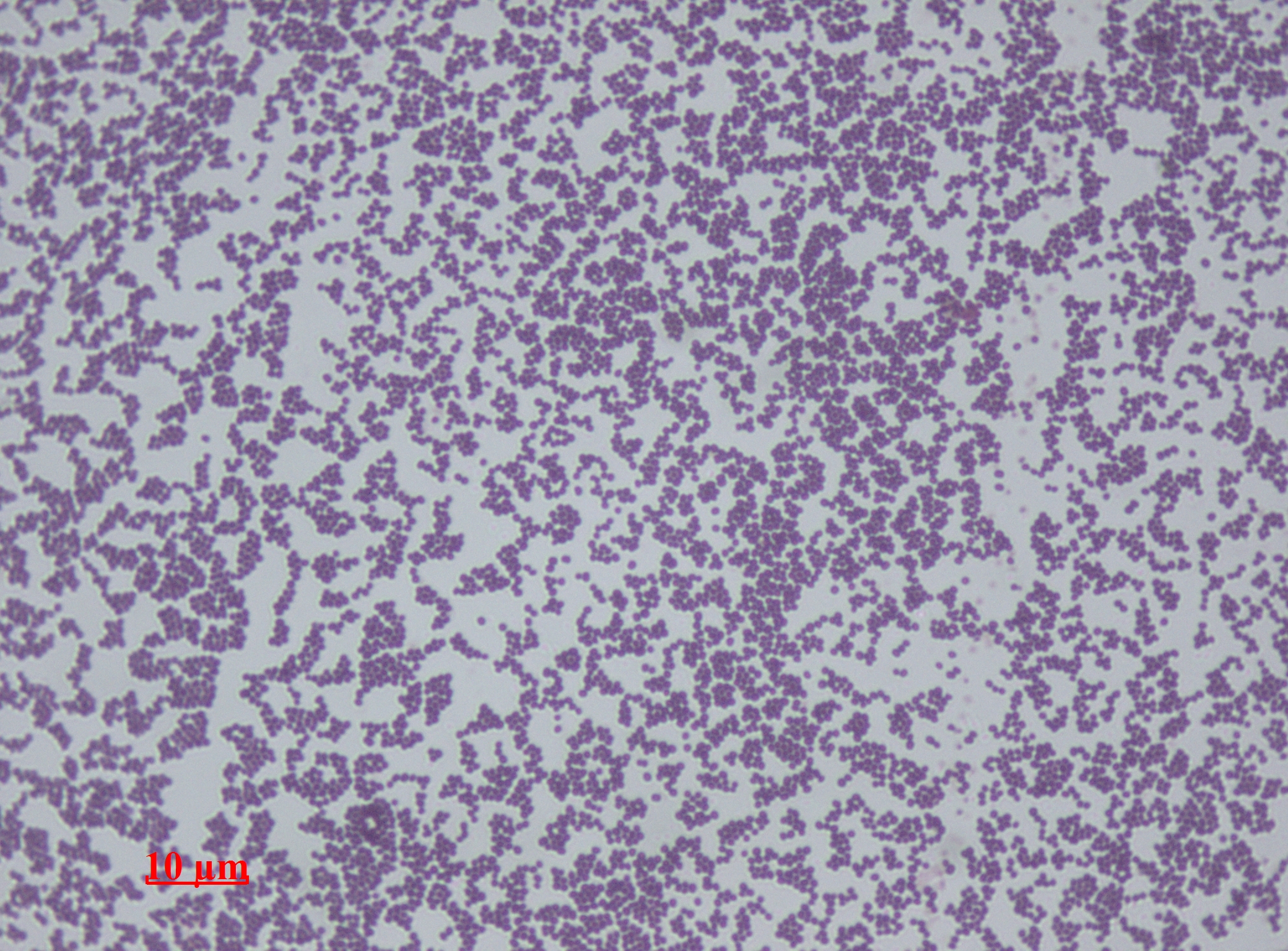
''Staphylococcus aureus'' is a Gram-positive spherically shaped bacterium, a member of the Bacillota, and is a usual member of the microbiota of the body, frequently found in the upper respiratory tract and on the skin. It is often positive for catalase and nitrate reduction and is a facultative anaerobe that can grow without the need for oxygen. Although ''S. aureus'' usually acts as a commensal of the human microbiota, it can also become an opportunistic pathogen, being a common cause of skin infections including abscesses, respiratory infections such as sinusitis, and food poisoning. Pathogenic strains often promote infections by producing virulence factors such as potent protein toxins, and the expression of a cell-surface protein that binds and inactivates antibodies. ''S. aureus'' is one of the leading pathogens for deaths associated with antimicrobial resistance and the emergence of antibiotic-resistant strains, such as methicillin-resistant ''S. aureus'' (MRSA ... [...More Info...] [...Related Items...] OR: [Wikipedia] [Google] [Baidu] |
Staphylococcus Epidermidis
''Staphylococcus epidermidis'' is a Gram-positive bacterium, and one of over 40 species belonging to the genus '' Staphylococcus''. It is part of the normal human microbiota, typically the skin microbiota, and less commonly the mucosal microbiota and also found in marine sponges. It is a facultative anaerobic bacteria. Although ''S. epidermidis'' is not usually pathogenic, patients with compromised immune systems are at risk of developing infection. These infections are generally hospital-acquired. ''S. epidermidis'' is a particular concern for people with catheters or other surgical implants because it is known to form biofilms that grow on these devices. Being part of the normal skin microbiota, ''S. epidermidis'' is a frequent contaminant of specimens sent to the diagnostic laboratory. Some strains of ''S. epidermidis'' are highly salt tolerant and commonly found in marine environment. S.I. Paul et al. (2021) isolated and identified salt tolerant strains of ''S. epiderm ... [...More Info...] [...Related Items...] OR: [Wikipedia] [Google] [Baidu] |
Neisseria Cinerea
''Neisseria cinerea'' is a commensal species grouped with the Gram-negative, oxidase-positive, and catalase-positive diplococci. It was first classified as ''Micrococcus cinereus'' by Alexander von Lingelsheim in 1906. Using DNA hybridization, ''N. cinerea'' exhibits 50% similarity to '' Neisseria gonorrhoeae''. Classification ''Neisseria cinerea'' has been formerly classified as many other organisms, including ''Moraxella catarrhalis'' and ''Neisseria pseudocatarrhalis''. It is often mistaken for ''N. gonorrhoeae'' with which it shares morphological and biochemical traits. Both are isolated from similar infections, including endocervical and rectal infections, neonatal conjunctivitis, and lymphadenitis.''N. cinerea'' has even been isolated on gonococcal selective media specifically made to isolate ''N. gonorrhoeae''. Colony morphology One important difference between ''N. cinerea'' and ''N. gonorrhoeae'' is that ''N. cinerea'' is slightly more pigmented and exhibits a golden ... [...More Info...] [...Related Items...] OR: [Wikipedia] [Google] [Baidu] |
Escherichia Coli
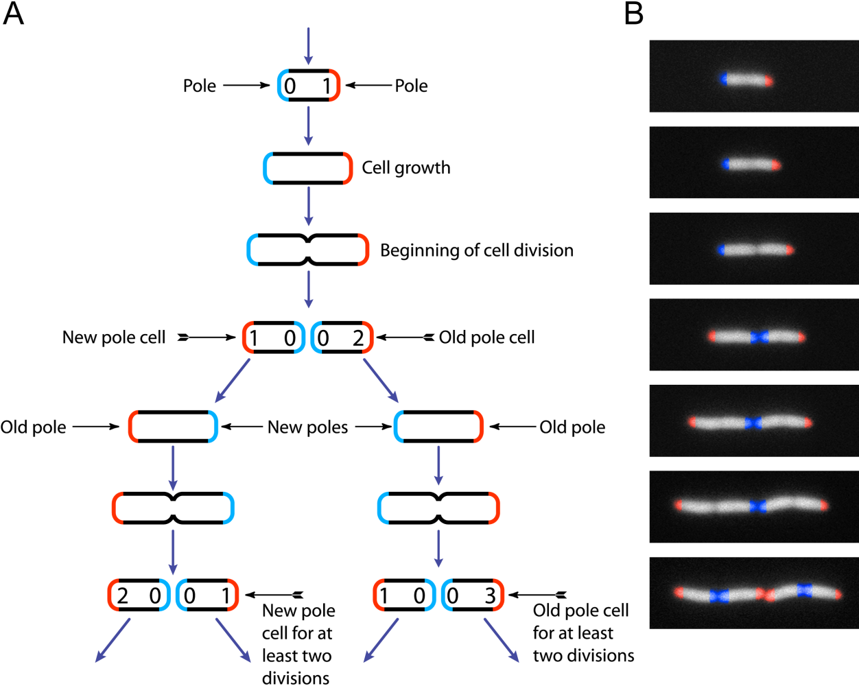
''Escherichia coli'' (),Wells, J. C. (2000) Longman Pronunciation Dictionary. Harlow ngland Pearson Education Ltd. also known as ''E. coli'' (), is a Gram-negative, facultative anaerobic, rod-shaped, coliform bacterium of the genus ''Escherichia'' that is commonly found in the lower intestine of warm-blooded organisms. Most ''E. coli'' strains are harmless, but some serotypes ( EPEC, ETEC etc.) can cause serious food poisoning in their hosts, and are occasionally responsible for food contamination incidents that prompt product recalls. Most strains do not cause disease in humans and are part of the normal microbiota of the gut; such strains are harmless or even beneficial to humans (although these strains tend to be less studied than the pathogenic ones). For example, some strains of ''E. coli'' benefit their hosts by producing vitamin K2 or by preventing the colonization of the intestine by pathogenic bacteria. These mutually beneficial relationships between ''E. col ... [...More Info...] [...Related Items...] OR: [Wikipedia] [Google] [Baidu] |
Hemojuvelin
Hemojuvelin (HJV), also known as repulsive guidance molecule C (RGMc) or hemochromatosis type 2 protein (HFE2), is a membrane-bound and soluble protein in mammals that is responsible for the iron overload condition known as juvenile hemochromatosis in humans, a severe form of hemochromatosis. In humans, the hemojuvelin protein is encoded by the ''HFE2'' gene. Hemojuvelin is a member of the repulsive guidance molecule family of proteins. Both RGMa and RGMb are found in the nervous system, while hemojuvelin is found in skeletal muscle and the liver. Function For many years the signal transduction pathways that regulate systemic iron homeostasis have been unknown. However it has been demonstrated that hemojuvelin interacts with bone morphogenetic protein (BMP), possibly as a co-receptor, and may signal via the SMAD pathway to regulate hepcidin expression. Associations with BMP2 and BMP4 have been described. Mouse HJV knock-out models confirmed that HJV is the gene responsib ... [...More Info...] [...Related Items...] OR: [Wikipedia] [Google] [Baidu] |
Juvenile Hemochromatosis
Juvenile hemochromatosis, also known as hemochromatosis type 2, is a rare form of hereditary hemochromatosis, which emerges in young individuals, typically between 15 and 30 years of age, but occasionally later. It is characterized by an inability to control how much iron is absorbed by the body, in turn leading to iron overload, where excess iron accumulates in many areas of the body and causes damage to the places it accumulates. It is a genetic disorder that can be caused by mutations in either the HJV (also called HFE2) or HAMP genes, and is inherited in an autosomal recessive fashion. Depending on which of these genes is affected, the disease can be further subdivided into types 2A and 2B. Signs and Symptoms The most common symptoms of juvenile hemochromatosis are as follows: * Weakness * Lethargy * Hyperpigmentation (darkening of the skin) * Arthropathy (joint disease) * Diabetes * Heart disease (dilated cardiomyopathy). Complications of heart disease are the main cause ... [...More Info...] [...Related Items...] OR: [Wikipedia] [Google] [Baidu] |
Kidney Failure
Kidney failure, also known as end-stage kidney disease, is a medical condition in which the kidneys can no longer adequately filter waste products from the blood, functioning at less than 15% of normal levels. Kidney failure is classified as either acute kidney failure, which develops rapidly and may resolve; and chronic kidney failure, which develops slowly and can often be irreversible. Symptoms may include leg swelling, feeling tired, vomiting, loss of appetite, and confusion. Complications of acute and chronic failure include uremia, high blood potassium, and volume overload. Complications of chronic failure also include heart disease, high blood pressure, and anemia. Causes of acute kidney failure include low blood pressure, blockage of the urinary tract, certain medications, muscle breakdown, and hemolytic uremic syndrome. Causes of chronic kidney failure include diabetes, high blood pressure, nephrotic syndrome, and polycystic kidney disease. Diagnosis of acute failure ... [...More Info...] [...Related Items...] OR: [Wikipedia] [Google] [Baidu] |
Hepatic Portal System
In human anatomy, the hepatic portal system is the system of veins comprising the hepatic portal vein and its tributaries. It is also called the portal venous system (although it is not the only example of a portal venous system) and splanchnic veins, which is ''not'' synonymous with ''hepatic portal system'' and is imprecise (as it means ''visceral veins'' and not necessarily the ''veins of the abdominal viscera'').Splanchnic circulation. Online Medical Dictionary. URLhttp://cancerweb.ncl.ac.uk/cgi-bin/omd?splanchnic+circulation Accessed on: October 22, 2008. Structure Large veins that are considered part of the ''portal venous system'' are the: *Hepatic portal vein * Splenic vein * Superior mesenteric vein *Inferior mesenteric vein The superior mesenteric vein and the splenic vein come together to form the actual hepatic portal vein. The inferior mesenteric vein connects in the majority of people on the splenic vein, but in some people, it is known to connect on the p ... [...More Info...] [...Related Items...] OR: [Wikipedia] [Google] [Baidu] |
Lysosome
A lysosome () is a membrane-bound organelle found in many animal cells. They are spherical vesicles that contain hydrolytic enzymes that can break down many kinds of biomolecules. A lysosome has a specific composition, of both its membrane proteins, and its lumenal proteins. The lumen's pH (~4.5–5.0) is optimal for the enzymes involved in hydrolysis, analogous to the activity of the stomach. Besides degradation of polymers, the lysosome is involved in various cell processes, including secretion, plasma membrane repair, apoptosis, cell signaling, and energy metabolism. Lysosomes act as the waste disposal system of the cell by digesting used materials in the cytoplasm, from both inside and outside the cell. Material from outside the cell is taken up through endocytosis, while material from the inside of the cell is digested through autophagy. The sizes of the organelles vary greatly—the larger ones can be more than 10 times the size of the smaller ones. They were discov ... [...More Info...] [...Related Items...] OR: [Wikipedia] [Google] [Baidu] |
Enterocytes
Enterocytes, or intestinal absorptive cells, are simple columnar epithelial cells which line the inner surface of the small and large intestines. A glycocalyx surface coat contains digestive enzymes. Microvilli on the apical surface increase its surface area. This facilitates transport of numerous small molecules into the enterocyte from the intestinal lumen. These include broken down proteins, fats, and sugars, as well as water, electrolytes, vitamins, and bile salts. Enterocytes also have an endocrine role, secreting hormones such as leptin. Function The major functions of enterocytes include: *Ion uptake, including sodium, calcium, magnesium, iron, zinc, and copper. This typically occurs through active transport. *Water uptake. This follows the osmotic gradient established by Na+/K+ ATPase on the basolateral surface. This can occur transcellularly or paracellularly. *Sugar uptake. Polysaccharides and disaccharidases in the glycocalyx break down large sugar molecules, which a ... [...More Info...] [...Related Items...] OR: [Wikipedia] [Google] [Baidu] |


