|
HLA-E
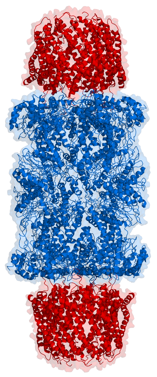
HLA class I histocompatibility antigen, alpha chain E (HLA-E) also known as MHC class I antigen E is a protein that in humans is encoded by the ''HLA-E'' gene. The human HLA-E is a non-classical MHC class I molecule that is characterized by a limited polymorphism and a lower cell surface expression than its classical paralogues. The functional homolog in mice is called Qa-1b, officially known as H2-T23. Structure Like other MHC class I molecules, HLA-E is a heterodimer consisting of an α heavy chain and a light chain ( β-2 microglobulin). The heavy chain is approximately 45 kDa and anchored in the membrane. The HLA-E gene contains 8 exons. Exon one encodes the signal peptide, exons 2 and 3 encode the α1 and α2 domains, which both bind the peptide, exon 4 encodes the α3 domain, exon 5 encodes the transmembrane domain, and exons 6 and 7 encode the cytoplasmic tail. Function HLA-E has a very specialized role in cell recognition by natural killer cells (NK cells). HLA-E bind ... [...More Info...] [...Related Items...] OR: [Wikipedia] [Google] [Baidu] |
MHC Class I
MHC class I molecules are one of two primary classes of major histocompatibility complex (MHC) molecules (the other being MHC class II) and are found on the cell surface of all nucleated cells in the bodies of vertebrates. They also occur on platelets, but not on red blood cells. Their function is to display peptide fragments of proteins from within the cell to cytotoxic T cells; this will trigger an immediate response from the immune system against a particular non-self antigen displayed with the help of an MHC class I protein. Because MHC class I molecules present peptides derived from cytosolic proteins, the pathway of MHC class I presentation is often called ''cytosolic'' or ''endogenous pathway''. In humans, the HLAs corresponding to MHC class I are HLA-A, HLA-B, and HLA-C. Function Class I MHC molecules bind peptides generated mainly from degradation of cytosolic proteins by the proteasome. The MHC I:peptide complex is then inserted via endoplasmic reticulum into the ext ... [...More Info...] [...Related Items...] OR: [Wikipedia] [Google] [Baidu] |
Qa-1b
Within molecular and cell biology, Qa-1b is a MHC class I molecule and is the functional homolog of HLA-E in humans. Qa-1b is characterised by its limited polymorphisms and small peptide repertoire. Qa-1b binds to peptides derived from signal peptides of MHC class Ia molecule and interact with the CD94/NKG2 receptors on natural killer cell Natural killer cells, also known as NK cells or large granular lymphocytes (LGL), are a type of cytotoxic lymphocyte critical to the innate immune system that belong to the rapidly expanding family of known innate lymphoid cells (ILC) and repres ...s. The Qa-1b-peptide complex signals natural killer cells not to engage in cell lysis. Despite its homology with HLA-E, it seems that Qa-1b evolved a similar function to HLA-E coincidentally. References {{Surface antigens Biomolecules ... [...More Info...] [...Related Items...] OR: [Wikipedia] [Google] [Baidu] |
CD94
CD94 (Cluster of Differentiation 94), also known as killer cell lectin-like receptor subfamily D, member 1 (KLRD1) is a human gene. The protein encoded by CD94 gene is a lectin, cluster of differentiation and a receptor that is involved in cell signaling and is expressed on the surface of natural killer cells in the innate immune system. CD94 pairs with the NKG2 molecule as a heterodimer. The CD94/NKG2 complex, on the surface of natural killer cells interacts with HLA-E, Human Leukocyte Antigen (HLA)-E on target cells. Function Natural killer (NK) cells are a distinct lineage of lymphocytes that mediate cytotoxic activity and secrete cytokines upon immune stimulation. Several genes of the C-type lectin superfamily, including members of the NKG2 family, are expressed by NK cells and may be involved in the regulation of NK cell function. KLRD1 (CD94) is an antigen preferentially expressed on NK cells and is classified as a type II membrane protein because it has an external C te ... [...More Info...] [...Related Items...] OR: [Wikipedia] [Google] [Baidu] |
Natural Killer Cells
Natural killer cells, also known as NK cells or large granular lymphocytes (LGL), are a type of cytotoxic lymphocyte critical to the innate immune system that belong to the rapidly expanding family of known innate lymphoid cells (ILC) and represent 5–20% of all circulating lymphocytes in humans. The role of NK cells is analogous to that of cytotoxic T cells in the vertebrate adaptive immune response. NK cells provide rapid responses to virus-infected cell and other intracellular pathogens acting at around 3 days after infection, and respond to tumor formation. Typically, immune cells detect the major histocompatibility complex (MHC) presented on infected cell surfaces, triggering cytokine release, causing the death of the infected cell by lysis or apoptosis. NK cells are unique, however, as they have the ability to recognize and kill stressed cells in the absence of antibodies and MHC, allowing for a much faster immune reaction. They were named "natural killers" because of the n ... [...More Info...] [...Related Items...] OR: [Wikipedia] [Google] [Baidu] |
Signal Peptide Peptidase
In molecular biology, the Signal Peptide Peptidase (SPP) is a type of protein that specifically cleaves parts of other proteins. It is an intramembrane aspartyl protease with the conserved active site motifs 'YD' and 'GxGD' in adjacent transmembrane domains (TMDs). Its sequences is highly conserved in different vertebrate species. SPP cleaves remnant signal peptides left behind in membrane by the action of signal peptidase and also plays key roles in immune surveillance and the maturation of certain viral proteins. Biological function Physiologically SPP processes signal peptides of classical MHC class I preproteins. A nine amino acid-long cleavage fragment is then presented on HLA-E receptors and modulates the activity of natural killer cells. SPP also plays a pathophysiological role; it cleaves the structural nucleocapsid protein (also known as core protein) of the Hepatitis C virus and thus influences viral reproduction rate. In mice, a nonamer peptide originating from the ... [...More Info...] [...Related Items...] OR: [Wikipedia] [Google] [Baidu] |
NKG2
NKG2 also known as CD159 (Cluster of Differentiation 159) is a receptor for natural killer cells (NK cells). There are 7 NKG2 types: A, B, C, D, E, F and H. NKG2D is an activating receptor on the NK cell surface. NKG2A dimerizes with CD94 to make an inhibitory receptor (CD94/NKG2). IPH2201 is a monoclonal antibody targeted at NKG2A. Gene expression In both humans and mice, genes encoding the ''NKG2'' family are clustered – in human genome on chromosome 12, in mouse on chromosome 6. They are generally expressed on NK cells and a subset of CD8+ T cells, although the expression of ''NKG2D'' was also confirmed on γδ T cells, NKT cells, and even on some subsets of CD4+ T cells or myeloid cells. ''NKG2D'' expression can also be present on cancer cells and is proven to stimulate oncogenic bioenergetic metabolism, proliferation and metastases generation. On NK cells, ''NKG2'' genes are expressed through the ontogeny as well as in adulthood. As about 90% of fetal NK cells expre ... [...More Info...] [...Related Items...] OR: [Wikipedia] [Google] [Baidu] |
Protein
Proteins are large biomolecules and macromolecules that comprise one or more long chains of amino acid residues. Proteins perform a vast array of functions within organisms, including catalysing metabolic reactions, DNA replication, responding to stimuli, providing structure to cells and organisms, and transporting molecules from one location to another. Proteins differ from one another primarily in their sequence of amino acids, which is dictated by the nucleotide sequence of their genes, and which usually results in protein folding into a specific 3D structure that determines its activity. A linear chain of amino acid residues is called a polypeptide. A protein contains at least one long polypeptide. Short polypeptides, containing less than 20–30 residues, are rarely considered to be proteins and are commonly called peptides. The individual amino acid residues are bonded together by peptide bonds and adjacent amino acid residues. The sequence of amino acid residue ... [...More Info...] [...Related Items...] OR: [Wikipedia] [Google] [Baidu] |
Cell Membrane
The cell membrane (also known as the plasma membrane (PM) or cytoplasmic membrane, and historically referred to as the plasmalemma) is a biological membrane that separates and protects the interior of all cells from the outside environment (the extracellular space). The cell membrane consists of a lipid bilayer, made up of two layers of phospholipids with cholesterols (a lipid component) interspersed between them, maintaining appropriate membrane fluidity at various temperatures. The membrane also contains membrane proteins, including integral proteins that span the membrane and serve as membrane transporters, and peripheral proteins that loosely attach to the outer (peripheral) side of the cell membrane, acting as enzymes to facilitate interaction with the cell's environment. Glycolipids embedded in the outer lipid layer serve a similar purpose. The cell membrane controls the movement of substances in and out of cells and organelles, being selectively permeable to ions a ... [...More Info...] [...Related Items...] OR: [Wikipedia] [Google] [Baidu] |
Transporter Associated With Antigen Processing
Transporter associated with antigen processing (TAP) protein complex belongs to the ATP-binding-cassette transporter family. It delivers cytosolic peptides into the endoplasmic reticulum (ER), where they bind to nascent MHC class I molecules. The TAP structure is formed of two proteins: TAP-1 and TAP-2, which have one hydrophobic region and one ATP-binding region each. They assemble into a heterodimer, which results in a four-domain transporter. Function The TAP transporter is found in the ER lumen associated with the peptide-loading complex (PLC). This complex of β2 microglobulin, calreticulin, ERp57, TAP, tapasin, and MHC class I acts to keep hold of MHC molecules until they have been fully loaded with peptides. Peptide transport TAP-mediated peptide transport is a multistep process. The peptide-binding pocket is formed by TAP-1 and TAP-2. Association with TAP is an ATP-independent event, ‘in a fast bimolecular association step, peptide binds to TAP, followed by a sl ... [...More Info...] [...Related Items...] OR: [Wikipedia] [Google] [Baidu] |
Proteasome
Proteasomes are protein complexes which degrade unneeded or damaged proteins by proteolysis, a chemical reaction that breaks peptide bonds. Enzymes that help such reactions are called proteases. Proteasomes are part of a major mechanism by which cells regulate the concentration of particular proteins and degrade misfolded proteins. Proteins are tagged for degradation with a small protein called ubiquitin. The tagging reaction is catalyzed by enzymes called ubiquitin ligases. Once a protein is tagged with a single ubiquitin molecule, this is a signal to other ligases to attach additional ubiquitin molecules. The result is a ''polyubiquitin chain'' that is bound by the proteasome, allowing it to degrade the tagged protein. The degradation process yields peptides of about seven to eight amino acids long, which can then be further degraded into shorter amino acid sequences and used in synthesizing new proteins. Proteasomes are found inside all eukaryotes and archaea, and in so ... [...More Info...] [...Related Items...] OR: [Wikipedia] [Google] [Baidu] |
Signal Peptides
A signal peptide (sometimes referred to as signal sequence, targeting signal, localization signal, localization sequence, transit peptide, leader sequence or leader peptide) is a short peptide (usually 16-30 amino acids long) present at the N-terminus (or occasionally nonclassically at the C-terminus or internally) of most newly synthesized proteins that are destined toward the secretory pathway. These proteins include those that reside either inside certain organelles (the endoplasmic reticulum, Golgi or endosomes), secreted from the cell, or inserted into most cellular membranes. Although most type I membrane-bound proteins have signal peptides, the majority of type II and multi-spanning membrane-bound proteins are targeted to the secretory pathway by their first transmembrane domain, which biochemically resembles a signal sequence except that it is not cleaved. They are a kind of target peptide. Function (translocation) Signal peptides function to prompt a cell to transloc ... [...More Info...] [...Related Items...] OR: [Wikipedia] [Google] [Baidu] |


