|
HES1
Transcription factor HES1 (hairy and enhancer of split-1) is a protein that is encoded by the ''Hes1'' gene, and is the mammalian homolog of the hairy gene in ''Drosophila.'' HES1 is one of the seven members of the Hes gene family (HES1-7). Hes genes code nuclear proteins that suppress transcription. This protein belongs to the basic helix-loop-helix (bHLH) family of transcription factors. It is a transcriptional repressor of genes that require a bHLH protein for their transcription. The protein has a particular type of basic domain that contains a helix interrupting protein that binds to the N-box promoter region rather than the canonical enhancer box (E-box). As a member of the bHLH family, it is a transcriptional repressor that influences cell proliferation and differentiation in embryogenesis. HES1 regulates its own expression via a negative feedback loop, and oscillates with approximately 2-hour periodicity. Structure There are three conserved domains in Hes genes that ... [...More Info...] [...Related Items...] OR: [Wikipedia] [Google] [Baidu] |
HEY2
Hairy/enhancer-of-split related with YRPW motif protein 2 (HEY2) also known as cardiovascular helix-loop-helix factor 1 (CHF1) is a protein that in humans is encoded by the ''HEY2'' gene. This protein is a type of transcription factor that belongs to the hairy and enhancer of split-related (HESR) family of basic helix-loop-helix ( bHLH)-type transcription factors. It forms homo- or hetero-dimers that localize to the nucleus and interact with a histone deacetylase complex to repress transcription. During embryonic development, this mechanism is used to control the number of cells that develop into cardiac progenitor cells and myocardial cells. The relationship is inversely related, so as the number of cells that express the Hey2 gene increases, the more CHF1 is present to repress transcription and the number of cells that take on a myocardial fate decreases. Expression The expression of the Hey2 gene is induced by the Notch signaling pathway. In this mechanism, adjacent ... [...More Info...] [...Related Items...] OR: [Wikipedia] [Google] [Baidu] |
E-box
An E-box (enhancer box) is a DNA response element found in some eukaryotes that acts as a protein-binding site and has been found to regulate gene expression in neurons, muscles, and other tissues. Its specific DNA sequence, CANNTG (where N can be any nucleotide), with a palindromic canonical sequence of CACGTG, is recognized and bound by transcription factors to initiate gene transcription. Once the transcription factors bind to the promoters through the E-box, other enzymes can bind to the promoter and facilitate transcription from DNA to mRNA. Discovery The E-box was discovered in a collaboration between Susumu Tonegawa's and Walter Gilbert's laboratories in 1985 as a control element in immunoglobulin heavy-chain enhancer. They found that a region of 140 base pairs in the tissue-specific transcriptional enhancer element was sufficient for different levels of transcription enhancement in different tissues and sequences. They suggested that proteins made by specific tissues acted ... [...More Info...] [...Related Items...] OR: [Wikipedia] [Google] [Baidu] |
Sirtuin 1
Sirtuin 1, also known as NAD-dependent deacetylase sirtuin-1, is a protein that in humans is encoded by the SIRT1 gene. SIRT1 stands for sirtuin (silent mating type information regulation 2 homolog) 1 (''S. cerevisiae''), referring to the fact that its sirtuin homolog (biological equivalent across species) in yeast ''(Saccharomyces cerevisiae)'' is Sir2. SIRT1 is an enzyme located primarily in the cell nucleus that deacetylates transcription factors that contribute to cellular regulation (reaction to stressors, longevity). Function Sirtuin 1 is a member of the sirtuin family of proteins, homologs of the Sir2 gene in ''S. cerevisiae''. Members of the sirtuin family are characterized by a sirtuin core domain and grouped into four classes. The functions of human sirtuins have not yet been determined; however, yeast sirtuin proteins are known to regulate epigenetic gene silencing and suppress recombination of rDNA. Studies suggest that the human sirtuins may function as intracel ... [...More Info...] [...Related Items...] OR: [Wikipedia] [Google] [Baidu] |
TLE2
Transducin-like enhancer protein 2 is a protein that in humans is encoded by the ''TLE2'' gene. Interactions TLE2 has been shown to interact with TLE1 and HES1 Transcription factor HES1 (hairy and enhancer of split-1) is a protein that is encoded by the ''Hes1'' gene, and is the mammalian homolog of the hairy gene in ''Drosophila.'' HES1 is one of the seven members of the Hes gene family (HES1-7). Hes ge .... References Further reading * * * * * * * * * * * * * * * {{gene-19-stub ... [...More Info...] [...Related Items...] OR: [Wikipedia] [Google] [Baidu] |
ASCL1
Achaete-scute homolog 1 is a protein that in humans is encoded by the ''ASCL1'' gene. Because it was discovered subsequent to studies on its homolog in Drosophila, the Achaete-scute complex, it was originally named MASH-1 for mammalian achaete scute homolog-1. Function This gene encodes a member of the basic helix-loop-helix (BHLH) family of transcription factors. The protein activates transcription by binding to the E box (5'-CANNTG-3'). Dimerization with other BHLH proteins is required for efficient DNA binding. This protein plays a role in the neuronal commitment and differentiation and in the generation of olfactory and autonomic neurons. It is highly expressed in medullary thyroid cancer and Lung cancer#Small cell lung carcinoma .28SCLC.29, small cell lung cancer and may be a useful marker for these cancers. The presence of a CAG repeat in the gene suggests that it may also play a role in tumor formation. Role in neuronal commitment Development of the vertebrate nervous s ... [...More Info...] [...Related Items...] OR: [Wikipedia] [Google] [Baidu] |
Astrocyte
Astrocytes (from Ancient Greek , , "star" + , , "cavity", "cell"), also known collectively as astroglia, are characteristic star-shaped glial cells in the brain and spinal cord. They perform many functions, including biochemical control of endothelial cells that form the blood–brain barrier, provision of nutrients to the nervous tissue, maintenance of extracellular ion balance, regulation of cerebral blood flow, and a role in the repair and scarring process of the brain and spinal cord following infection and traumatic injuries. The proportion of astrocytes in the brain is not well defined; depending on the counting technique used, studies have found that the astrocyte proportion varies by region and ranges from 20% to 40% of all glia. Another study reports that astrocytes are the most numerous cell type in the brain. Astrocytes are the major source of cholesterol in the central nervous system. Apolipoprotein E transports cholesterol from astrocytes to neurons and other glial ... [...More Info...] [...Related Items...] OR: [Wikipedia] [Google] [Baidu] |
Pax6
Paired box protein Pax-6, also known as aniridia type II protein (AN2) or oculorhombin, is a protein that in humans is encoded by the ''PAX6'' gene. Function PAX6 is a member of the Pax gene family which is responsible for carrying the genetic information that will encode the Pax-6 protein. It acts as a "master control" gene for the development of eyes and other sensory organs, certain neural and epidermal tissues as well as other homologous structures, usually derived from ectodermal tissues. However, it has been recognized that a suite of genes is necessary for eye development, and therefore the term of "master control" gene may be inaccurate. Pax-6 is expressed as a transcription factor when neural ectoderm receives a combination of weak Sonic hedgehog (SHH) and strong TGF-Beta signaling gradients. Expression is first seen in the forebrain, hindbrain, head ectoderm and spinal cord followed by later expression in midbrain. This transcription factor is most noted for its u ... [...More Info...] [...Related Items...] OR: [Wikipedia] [Google] [Baidu] |
Knockout Mouse
A knockout mouse, or knock-out mouse, is a genetically modified mouse (''Mus musculus'') in which researchers have inactivated, or "knocked out", an existing gene by replacing it or disrupting it with an artificial piece of DNA. They are important animal models for studying the role of genes which have been sequenced but whose functions have not been determined. By causing a specific gene to be inactive in the mouse, and observing any differences from normal behaviour or physiology, researchers can infer its probable function. Mice are currently the laboratory animal species most closely related to humans for which the knockout technique can easily be applied. They are widely used in knockout experiments, especially those investigating genetic questions that relate to human physiology. Gene knockout in rats is much harder and has only been possible since 2003. The first recorded knockout mouse was created by Mario R. Capecchi, Martin Evans, and Oliver Smithies in 1989, for whi ... [...More Info...] [...Related Items...] OR: [Wikipedia] [Google] [Baidu] |
Subventricular Zone
The subventricular zone (SVZ) is a region situated on the outside wall of each lateral ventricle of the vertebrate brain. It is present in both the embryonic and adult brain. In embryonic life, the SVZ refers to a secondary proliferative zone containing neural progenitor cells, which divide to produce neurons in the process of neurogenesis. The primary neural stem cells of the brain and spinal cord, termed radial glial cells, instead reside in the ventricular zone (VZ) (so-called because the VZ lines the inside of the developing ventricles). In the developing cerebral cortex, which resides in the dorsal telencephalon, the SVZ and VZ are transient tissues that do not exist in the adult. However, the SVZ of the ventral telencephalon persists throughout life. The adult SVZ is composed of four distinct layers of variable thickness and cell density as well as cellular composition. Along with the dentate gyrus of the hippocampus, the SVZ is one of two places where neurogenesis has ... [...More Info...] [...Related Items...] OR: [Wikipedia] [Google] [Baidu] |
Radial Glial Cell
Radial glial cells, or radial glial progenitor cells (RGPs), are bipolar-shaped progenitor cells that are responsible for producing all of the neurons in the cerebral cortex. RGPs also produce certain lineages of glia, including astrocytes and oligodendrocytes. Their cell bodies (somata) reside in the embryonic ventricular zone, which lies next to the developing ventricular system. During development, newborn neurons use radial glia as scaffolds, traveling along the radial glial fibers in order to reach their final destinations. Despite the various possible fates of the radial glial population, it has been demonstrated through clonal analysis that most radial glia have restricted, unipotent or multipotent, fates. Radial glia can be found during the neurogenic phase in all vertebrates (studied to date). The term "radial glia" refers to the morphological characteristics of these cells that were first observed: namely, their radial processes and their similarity to astrocytes, an ... [...More Info...] [...Related Items...] OR: [Wikipedia] [Google] [Baidu] |
Neuroepithelial Cells
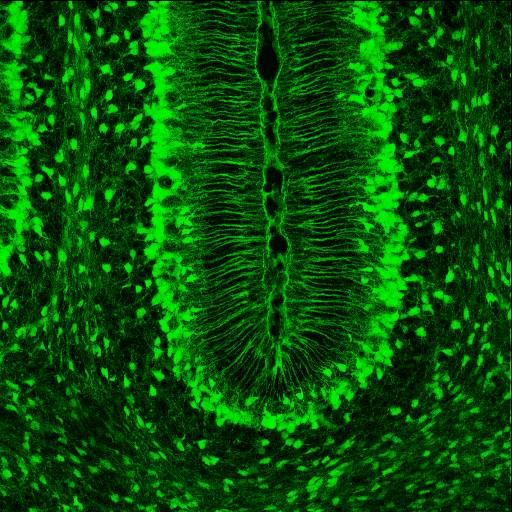
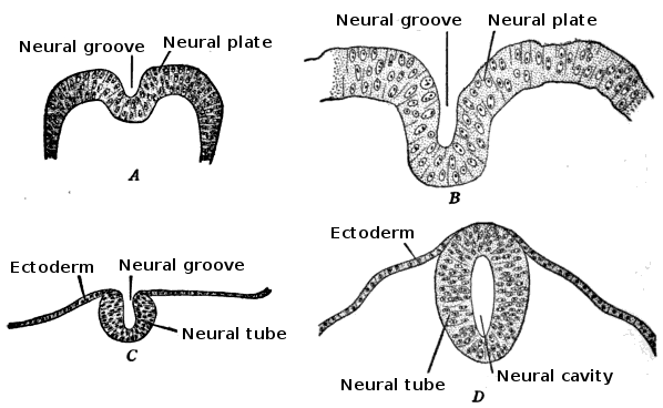
Neuroepithelial cells, or neuroectodermal cells, form the wall of the closed neural tube in early embryonic development. The neuroepithelial cells span the thickness of the tube's wall, connecting with the pial surface and with the ventricular or lumenal surface. They are joined at the lumen of the tube by junctional complexes, where they form a pseudostratified layer of epithelium called neuroepithelium. Neuroepithelial cells are the stem cells of the central nervous system, known as neural stem cells, and generate the intermediate progenitor cells known as radial glial cells, that differentiate into neurons and glia in the process of neurogenesis. Embryonic neural development Brain development During the third week of embryonic growth the brain begins to develop in the early fetus in a process called morphogenesis. Neuroepithelial cells of the ectoderm begin multiplying rapidly and fold in forming the neural plate, which invaginates during the fourth week of embryonic growth ... [...More Info...] [...Related Items...] OR: [Wikipedia] [Google] [Baidu] |


-_Drosophila_Model.jpg)


