|
Henneman's Size Principle
Henneman’s size principle describes relationships between properties of motor neurons and the muscle fibers they innervate and thus control, which together are called motor units. Motor neurons with large cell bodies tend to innervate fast-twitch, high-force, less fatigue-resistant muscle fibers, whereas motor neurons with small cell bodies tend to innervate slow-twitch, low-force, fatigue-resistant muscle fibers. In order to contract a particular muscle, motor neurons with small cell bodies are recruited (i.e. begin to fire action potentials) before motor neurons with large cell bodies. It was proposed by Elwood Henneman. History At the time of Henneman’s initial study of motor neuron recruitment, it was known that neurons varied greatly in size, that is in the diameter and extent of the dendritic arbor, size of the soma, and diameter of axon. However, the functional significance of neuron size was not yet known. In 1965, Henneman and colleagues published five papers de ... [...More Info...] [...Related Items...] OR: [Wikipedia] [Google] [Baidu] |
Motor Neuron
A motor neuron (or motoneuron), also known as efferent neuron is a neuron whose cell body is located in the motor cortex, brainstem or the spinal cord, and whose axon (fiber) projects to the spinal cord or outside of the spinal cord to directly or indirectly control effector organs, mainly muscles and glands. There are two types of motor neuron – upper motor neurons and lower motor neurons. Axons from upper motor neurons synapse onto interneurons in the spinal cord and occasionally directly onto lower motor neurons. The axons from the lower motor neurons are efferent nerve fibers that carry signals from the spinal cord to the effectors. Types of lower motor neurons are alpha motor neurons, beta motor neurons, and gamma motor neurons. A single motor neuron may innervate many muscle fibres and a muscle fibre can undergo many action potentials in the time taken for a single muscle twitch. Innervation takes place at a neuromuscular junction and twitches can become superimpo ... [...More Info...] [...Related Items...] OR: [Wikipedia] [Google] [Baidu] |
Myocyte
A muscle cell, also known as a myocyte, is a mature contractile Cell (biology), cell in the muscle of an animal. In humans and other vertebrates there are three types: skeletal muscle, skeletal, smooth muscle, smooth, and Cardiac muscle, cardiac (cardiomyocytes). A skeletal muscle cell is long and threadlike with multinucleated, many nuclei and is called a ''muscle fiber''. Muscle cells develop from embryonic precursor cells called myoblasts. Skeletal muscle cells form by cell fusion, fusion of myoblasts to produce multinucleated cells (syncytium, syncytia) in a process known as myogenesis. Skeletal muscle cells and cardiac muscle cells both contain myofibrils and sarcomeres and form a striated muscle tissue. Cardiac muscle cells form the cardiac muscle in the walls of the heart chambers, and have a single central Cell nucleus, nucleus. Cardiac muscle cells are joined to neighboring cells by intercalated discs, and when joined in a visible unit they are described as a ''cardiac m ... [...More Info...] [...Related Items...] OR: [Wikipedia] [Google] [Baidu] |
Motor Units
In biology, a motor unit is made up of a motor neuron and all of the skeletal muscle fibers innervated by the neuron's axon terminals, including the neuromuscular junctions between the neuron and the fibres. Groups of motor units often work together as a motor pool to coordinate the contractions of a single muscle. The concept was proposed by Charles Scott Sherrington. Usually muscle fibers in a motor unit are of the same fiber type. When a motor unit is activated, all of its fibers contract. In vertebrates, the force of a muscle contraction is controlled by the number of activated motor units. The number of muscle fibers within each unit can vary within a particular muscle and even more from muscle to muscle: the muscles that act on the largest body masses have motor units that contain more muscle fibers, whereas smaller muscles contain fewer muscle fibers in each motor unit. For instance, thigh muscles can have a thousand fibers in each unit, while extraocular muscles might ha ... [...More Info...] [...Related Items...] OR: [Wikipedia] [Google] [Baidu] |
Skeletal Striated Muscle
Skeletal muscle (commonly referred to as muscle) is one of the three types of vertebrate muscle tissue, the others being cardiac muscle and smooth muscle. They are part of the somatic nervous system, voluntary muscular system and typically are attached by tendons to bones of a skeleton. The skeletal muscle cells are much longer than in the other types of muscle tissue, and are also known as ''muscle fibers''. The tissue of a skeletal muscle is striated muscle tissue, striated – having a striped appearance due to the arrangement of the sarcomeres. A skeletal muscle contains multiple muscle fascicle, fascicles – bundles of muscle fibers. Each individual fiber and each muscle is surrounded by a type of connective tissue layer of fascia. Muscle fibers are formed from the cell fusion, fusion of developmental myoblasts in a process known as myogenesis resulting in long multinucleated cells. In these cells, the cell nucleus, nuclei, termed ''myonuclei'', are located along the inside ... [...More Info...] [...Related Items...] OR: [Wikipedia] [Google] [Baidu] |
Elwood Henneman
Elwood Henneman (1915 – 22 February 1996) was an American neurophysiologist who studied the properties of vertebrate motor neurons. Biography and Research Henneman received his bachelor's degree from Harvard College in Cambridge, Massachusetts in 1937. In 1943 he finished his medical studies at McGill University in Montreal. During a research fellowship at Johns Hopkins University in Baltimore, Maryland, Henneman and colleague, Vernon Mountcastle, showed that tactile information about the extremities is represented in an orderly map in the ventrolateral thalamus of the cat and monkey. Further research positions followed, including at the Royal Victorian Hospital and at the Illinois Neuropsychiatric Institute (NPI) in Chicago. At NPI, Henneman discovered that the drug Mephenesin (Myensin) inhibits interneurons in the spinal cord and thus causes muscle relaxation. This discovery helped lead to the development of muscle relaxant drugs. Of greater impact for the scientific communi ... [...More Info...] [...Related Items...] OR: [Wikipedia] [Google] [Baidu] |
Soleus Muscle
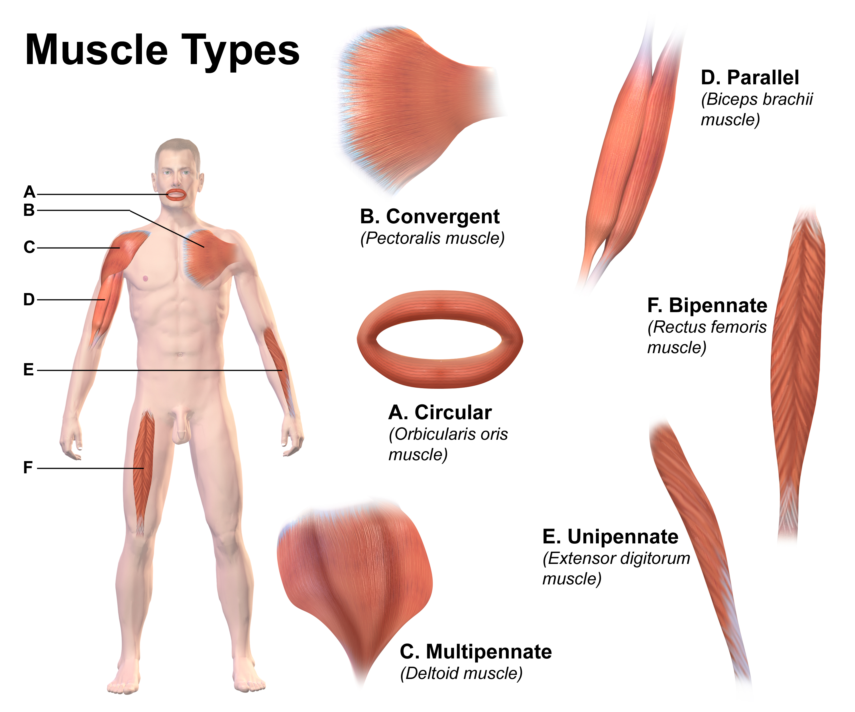
In humans and some other mammals, the soleus is a powerful muscle in the back part of the lower leg (the calf). It runs from just below the knee to the heel and is involved in standing and walking. It is closely connected to the gastrocnemius muscle, and some anatomists consider this combination to be a single muscle, the triceps surae. Its name is derived from the Latin word "solea", meaning "sandal". Structure The soleus is located in the superficial posterior compartment of the leg. The soleus exhibits significant morphological differences across species. It is unipennate in many species. In some animals, such as the rabbit, it is fused for much of its length with the gastrocnemius muscle. The soleus is a complex, multi-pennate muscle in humans, normally having a separate (posterior) aponeurosis from the gastrocnemius muscle. Most soleus muscle fibers originate from each side of the anterior aponeurosis, attached to the tibia and fibula. Other fibers originate from the po ... [...More Info...] [...Related Items...] OR: [Wikipedia] [Google] [Baidu] |
Gastrocnemius Muscle
The gastrocnemius muscle (plural ''gastrocnemii'') is a superficial two-headed muscle that is in the back part of the lower leg of humans. It is located superficial to the soleus in the posterior (back) compartment of the leg. It runs from its two heads just above the knee to the heel, extending across a total of three joints (knee, ankle and subtalar joints). The muscle is named via Latin, from Greek γαστήρ (''gaster'') 'belly' or 'stomach' and κνήμη (''knḗmē'') 'leg', meaning 'stomach of the leg' (referring to the bulging shape of the calf). Structure Origin/proximal attachment The lateral head originates from the lateral condyle of the femur, while the medial head originates from the medial condyle of the femur. Insertion/distal attachment Its other end forms a common tendon with the soleus muscle; this tendon is known as the calcaneal tendon or Achilles tendon and inserts onto the posterior surface of the calcaneus, or heel bone. Relations The ga ... [...More Info...] [...Related Items...] OR: [Wikipedia] [Google] [Baidu] |
Ventral Root Of Spinal Nerve
In anatomy and neurology, the ventral root of spinal nerve, anterior root, or motor root is the efferent motor root of a spinal nerve A spinal nerve is a mixed nerve, which carries Motor neuron, motor, Sensory neuron, sensory, and Autonomic nervous system, autonomic signals between the spinal cord and the body. In the human body there are 31 pairs of spinal nerves, one on each s .... At its distal end, the ventral root joins with the dorsal root to form a mixed spinal nerve. Additional images Image:Cervical vertebra english.png, Cervical vertebra Image:Medulla spinalis - Section - English.svg, Medulla spinalis Image:Gray675.png, A spinal nerve with its anterior and posterior. Image:Gray764.png, The motor tract. Image:Gray770-en.svg, Diagrammatic transverse section of the medulla spinalis and its membranes. Image:Gray796.png, A portion of the spinal cord, showing its right lateral surface. The dura is opened and arranged to show the nerve roots. Image:Gray799.svg, S ... [...More Info...] [...Related Items...] OR: [Wikipedia] [Google] [Baidu] |
Weber–Fechner Law
The Weber–Fechner laws are two related scientific law, scientific laws in the field of psychophysics, known as Weber's law and Fechner's law. Both relate to human perception, more specifically the relation between the actual change in a physical Stimulus (physiology), stimulus and the perceived change. This includes stimuli to all senses: vision, hearing, taste, touch, and smell. Ernst Heinrich Weber states that "the minimum increase of stimulus which will produce a perceptible increase of sensation is proportionality (mathematics), proportional to the pre-existent stimulus," while Gustav Fechner's law is an inference from Weber's law (with additional assumptions) which states that the intensity of our sensation increases as the logarithm of an increase in energy rather than as rapidly as the increase. History and formulation of the laws Both Weber's law and Fechner's law were formulated by Gustav Fechner, Gustav Theodor Fechner (1801–1887). They were first published in 18 ... [...More Info...] [...Related Items...] OR: [Wikipedia] [Google] [Baidu] |
Quadriceps Femoris Muscle
The quadriceps femoris muscle (, also called the quadriceps extensor, quadriceps or quads) is a large muscle group that includes the four prevailing muscles on the front of the thigh. It is the sole extensor muscle of the knee, forming a large fleshy mass which covers the front and sides of the femur. The name derives . Structure Parts The quadriceps femoris muscle is subdivided into four separate muscles (the 'heads'), with the first superficial to the other three over the femur (from the trochanters to the condyles): * The rectus femoris muscle occupies the middle of the thigh, covering most of the other three quadriceps muscles. It originates on the ilium. It is named for its straight course. * The vastus lateralis muscle is on the ''lateral side'' of the femur (i.e. on the outer side of the thigh). * The vastus medialis muscle is on the ''medial side'' of the femur (i.e. on the inner part thigh). * The vastus intermedius muscle lies between vastus lateralis and vastus ... [...More Info...] [...Related Items...] OR: [Wikipedia] [Google] [Baidu] |
Electromyography
Electromyography (EMG) is a technique for evaluating and recording the electrical activity produced by skeletal muscles. EMG is performed using an instrument called an electromyograph to produce a record called an electromyogram. An electromyograph detects the electric potential generated by muscle cells when these cells are electrically or neurologically activated. The signals can be analyzed to detect abnormalities, activation level, or recruitment order, or to analyze the biomechanics of human or animal movement. Needle EMG is an electrodiagnostic medicine technique commonly used by neurologists. Surface EMG is a non-medical procedure used to assess muscle activation by several professionals, including physiotherapists, kinesiologists and biomedical engineers. In computer science, EMG is also used as middleware in gesture recognition towards allowing the input of physical action to a computer as a form of human-computer interaction. Clinical uses EMG testing has a varie ... [...More Info...] [...Related Items...] OR: [Wikipedia] [Google] [Baidu] |


