|
Hatschek's Pit
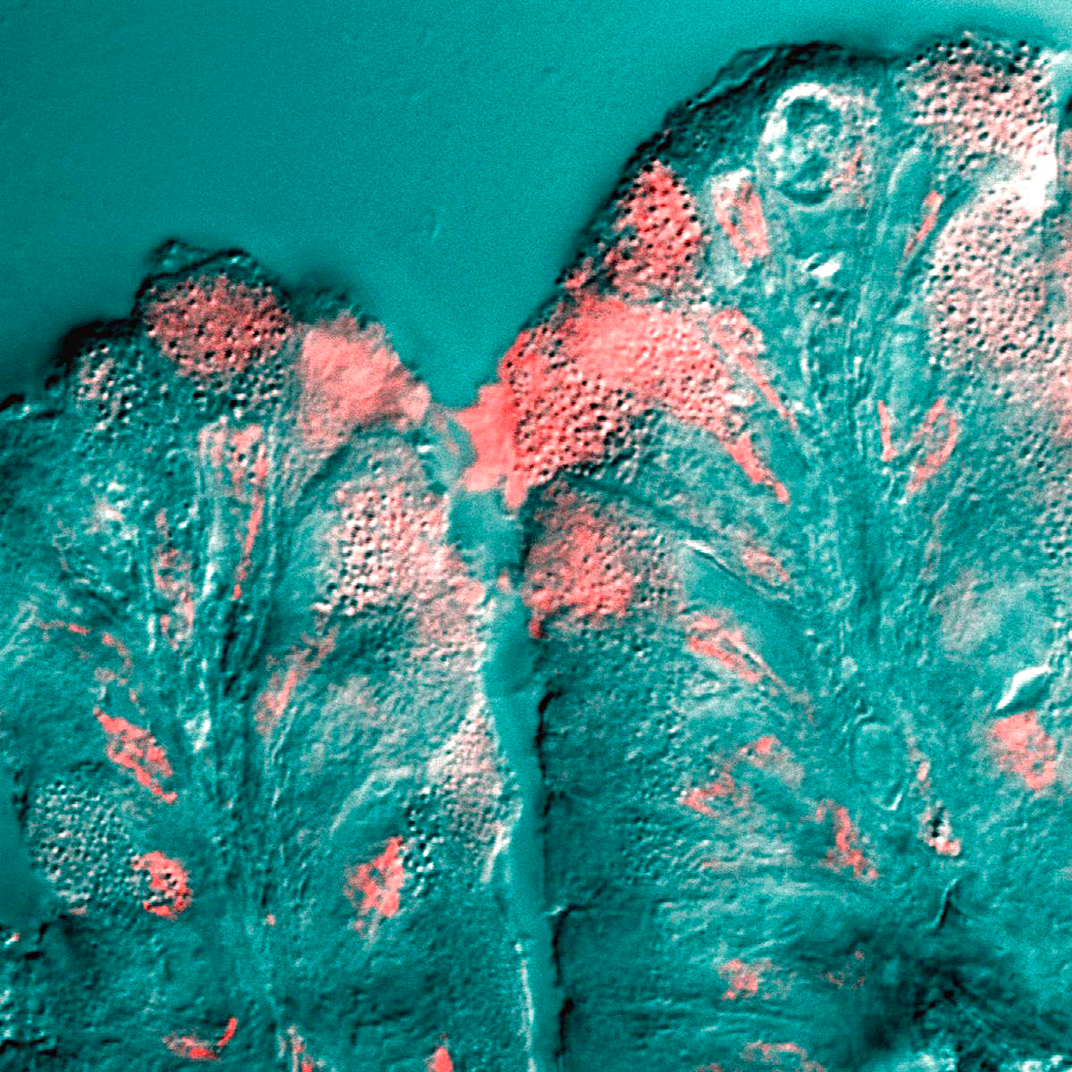
In the lancelet, Hatschek's pit, not to be confused with Hatschek's nephridium, is a deep ciliated fossa on the dorsal midline of the buccal cavity (the region of the gut behind the mouth). Among other things, it secretes mucus which entraps food particles from the water. It is named after Berthold Hatschek Berthold Hatschek (3 April 1854 in Skrbeň – 18 January 1941 in Vienna) was an Austrian zoologist remembered for embryological and morphological studies of invertebrates. Life He studied zoology in Vienna under Carl Claus (1835-1899), and in ..., an Austrian zoologist who worked on the lancelet. References Chordate anatomy {{chordate-stub ... [...More Info...] [...Related Items...] OR: [Wikipedia] [Google] [Baidu] |
Lancelet
The lancelets ( or ), also known as amphioxi (singular: amphioxus ), consist of some 30 to 35 species of "fish-like" benthic filter feeding chordates in the order Amphioxiformes. They are the modern representatives of the subphylum Cephalochordata. Lancelets closely resemble 530-million-year-old ''Pikaia'', fossils of which are known from the Burgess Shale. Zoologists are interested in them because they provide evolutionary insight into the origins of vertebrates. Lancelets contain many organs and organ systems that are closely related to those of modern fish, but in more primitive form. Therefore, they provide a number of examples of possible evolutionary exaptation. For example, the gill-slits of lancelets are used for feeding only, and not for respiration. The circulatory system carries food throughout their body, but does not have red blood cells or hemoglobin for transporting oxygen. Lancelet genomes hold clues about the early evolution of vertebrates: by comparing genes from ... [...More Info...] [...Related Items...] OR: [Wikipedia] [Google] [Baidu] |
Ciliated
The cilium, plural cilia (), is a membrane-bound organelle found on most types of eukaryotic cell, and certain microorganisms known as ciliates. Cilia are absent in bacteria and archaea. The cilium has the shape of a slender threadlike projection that extends from the surface of the much larger cell body. Eukaryotic flagella found on sperm cells and many protozoans have a similar structure to motile cilia that enables swimming through liquids; they are longer than cilia and have a different undulating motion. There are two major classes of cilia: ''motile'' and ''non-motile'' cilia, each with a subtype, giving four types in all. A cell will typically have one primary cilium or many motile cilia. The structure of the cilium core called the axoneme determines the cilium class. Most motile cilia have a central pair of single microtubules surrounded by nine pairs of double microtubules called a 9+2 axoneme. Most non-motile cilia have a 9+0 axoneme that lacks the central pair of mi ... [...More Info...] [...Related Items...] OR: [Wikipedia] [Google] [Baidu] |
Fossa (anatomy)
In anatomy, a fossa (; plural ''fossae'' ( or ); from Latin ''fossa'', "ditch" or "trench") is a depression or hollow, usually in a bone, such as the hypophyseal fossa (the depression in the sphenoid bone).Venieratos D, Anagnostopoulou S, Garidou A., A new morphometric method for the sella turcica and the hypophyseal fossa and its clinical relevance.;Folia Morphol (Warsz). 2005 Nov;64(4):240-7. Some examples include: In the Skull: * Cranial fossa ** Anterior cranial fossa ** Middle cranial fossa *** Interpeduncular fossa ** Posterior cranial fossa * Hypophyseal fossa * Temporal bone fossa ** Mandibular fossa ** Jugular fossa * Infratemporal fossa * Pterygopalatine fossa * Pterygoid fossa * Lacrimal fossa ** Fossa for lacrimal gland ** Fossa for lacrimal sac * Mandibular fossa * Scaphoid fossa * Jugular fossa * Condyloid fossa * Rhomboid fossa In the Mandible: * Retromolar fossa In the Torso: * Fossa ovalis (heart) * Infraclavicular fossa *Pyriform fossa * Substernal fossa * ... [...More Info...] [...Related Items...] OR: [Wikipedia] [Google] [Baidu] |
Dorsum (biology)
Standard anatomical terms of location are used to unambiguously describe the anatomy of animals, including humans. The terms, typically derived from Latin or Greek language, Greek roots, describe something in its standard anatomical position. This position provides a definition of what is at the front ("anterior"), behind ("posterior") and so on. As part of defining and describing terms, the body is described through the use of anatomical planes and anatomical axis, anatomical axes. The meaning of terms that are used can change depending on whether an organism is bipedal or quadrupedal. Additionally, for some animals such as invertebrates, some terms may not have any meaning at all; for example, an animal that is radially symmetrical will have no anterior surface, but can still have a description that a part is close to the middle ("proximal") or further from the middle ("distal"). International organisations have determined vocabularies that are often used as standard vocabular ... [...More Info...] [...Related Items...] OR: [Wikipedia] [Google] [Baidu] |
Buccal Cavity
The buccal space (also termed the buccinator space) is a fascial spaces of the head and neck, fascial space of the head and neck (sometimes also termed fascial tissue spaces or tissue spaces). It is a potential space in the cheek, and is paired on each side. The buccal space is superficial to the buccinator muscle and deep to the platysma muscle and the skin. The buccal space is part of the Subcutaneous tissue, subcutaneous space, which is continuous from head to toe. Structure Boundaries The boundaries of each buccal space are: * the angle of the mouth anteriorly, * the masseter muscle posteriorly, * the zygomatic process of the maxilla and the zygomaticus muscles superiorly, * the depressor anguli oris muscle and the attachment of the deep fascia to the mandible inferiorly, * the buccinator muscle medially (the buccal space is superficial to the buccinator), * the platysma muscle, subcutaneous tissue and skin laterally (the space is deep to platysma). Communications * to the pte ... [...More Info...] [...Related Items...] OR: [Wikipedia] [Google] [Baidu] |
Mucus
Mucus ( ) is a slippery aqueous secretion produced by, and covering, mucous membranes. It is typically produced from cells found in mucous glands, although it may also originate from mixed glands, which contain both serous and mucous cells. It is a viscous colloid containing inorganic salts, antimicrobial enzymes (such as lysozymes), immunoglobulins (especially IgA), and glycoproteins such as lactoferrin and mucins, which are produced by goblet cells in the mucous membranes and submucosal glands. Mucus serves to protect epithelial cells in the linings of the respiratory, digestive, and urogenital systems, and structures in the visual and auditory systems from pathogenic fungi, bacteria and viruses. Most of the mucus in the body is produced in the gastrointestinal tract. Amphibians, fish, snails, slugs, and some other invertebrates also produce external mucus from their epidermis as protection against pathogens, and to help in movement and is also produced in fish to line the ... [...More Info...] [...Related Items...] OR: [Wikipedia] [Google] [Baidu] |
Berthold Hatschek
Berthold Hatschek (3 April 1854 in Skrbeň – 18 January 1941 in Vienna) was an Austrian zoologist remembered for embryology, embryological and morphology (biology), morphological studies of invertebrates. Life He studied zoology in Vienna under Carl Friedrich Wilhelm Claus, Carl Claus (1835-1899), and in Leipzig with Rudolf Leuckart (1822-1898). He gained his doctorate at the University of Leipzig with a dissertation titled ''Beiträge zur Entwicklungsgeschichte der Lepidopteren''. Hatschek was deeply influenced by the works of Ernst Haeckel (1834-1919). In 1885 he was appointed professor of zoology at Charles University in Prague, and from 1896 was a professor and director of the second zoological institute at the University of Vienna. Hatschek suffered from severe depression, which greatly affected his work in the latter stages of his life. Hatschek is remembered for the so-called "trochophore theory", in which he explains the trochophore to be the larval form of a hypothetic ... [...More Info...] [...Related Items...] OR: [Wikipedia] [Google] [Baidu] |


