|
Gustav Albert Schwalbe
Gustav Albert Schwalbe, M.D. (1 August 1844 – 23 April 1916) was a German anatomist and anthropologist from Quedlinburg. He was educated at the universities of Berlin, Zurich, and Bonn (M.D. 1866), he became in 1870 privat-docent at the University of Halle, in 1871 privatdozent and prosector at the University of Freiburg in Baden, in 1872 assistant professor at the University of Leipzig, and then professor of anatomy successively at the universities of Jena (1873), Königsberg (1881), and Strassburg (1883) — at that time a German university, Alsace having been annexed to Germany. There he died. Known for his anthropological research of primitive man, Schwalbe considered the Neanderthal to be a direct ancestor of modern humans. Much to the dismay of the Dutch paleontologist Eugène Dubois (1858–1940) who had discovered Java Man, Schwalbe published in 1899 the influential treatise ''Studien über Pithecantropus Erectus'' (Study of Pithecantropus Erectus). In 1869 Schw ... [...More Info...] [...Related Items...] OR: [Wikipedia] [Google] [Baidu] |
Gustav Schwalbe (1792–1850), German writer, pastor and publisher
{{human name disambiguation ...
Gustav Schwalbe may refer to: *Gustav Christian Schwabe (1813–1897), German-born merchant and financier *Gustav Albert Schwalbe (1844–1916), German anatomist and anthropologist *Gustav Schwab Gustav Benjamin Schwab (19 June 1792 – 4 November 1850) was a German writer, pastor and publisher. Life Gustav Schwab was born in Stuttgart, the son of the philosopher Johann Christoph Schwab: he was introduced to the humanities early in li ... [...More Info...] [...Related Items...] OR: [Wikipedia] [Google] [Baidu] |
University Of Strasbourg
The University of Strasbourg (french: Université de Strasbourg, Unistra) is a public research university located in Strasbourg, Alsace, France, with over 52,000 students and 3,300 researchers. The French university traces its history to the earlier German-language ''Universität Straßburg'', which was founded in 1538, and was divided in the 1970s into three separate institutions: Louis Pasteur University, Marc Bloch University, and Robert Schuman University. On 1 January 2009, the fusion of these three universities reconstituted a united University of Strasbourg. With as many as 19 Nobel laureates, and two Fields Medal winners, the university is ranked among the best in the League of European Research Universities. History The university emerged from a Lutheran humanist German Gymnasium, founded in 1538 by Johannes Sturm in the Free Imperial City of Strassburg. It was transformed to a university in 1621 (german: Universität Straßburg) and elevated to the ranks of a royal u ... [...More Info...] [...Related Items...] OR: [Wikipedia] [Google] [Baidu] |
Collagen
Collagen () is the main structural protein in the extracellular matrix found in the body's various connective tissues. As the main component of connective tissue, it is the most abundant protein in mammals, making up from 25% to 35% of the whole-body protein content. Collagen consists of amino acids bound together to form a triple helix of elongated fibril known as a collagen helix. It is mostly found in connective tissue such as cartilage, bones, tendons, ligaments, and skin. Depending upon the degree of mineralization, collagen tissues may be rigid (bone) or compliant (tendon) or have a gradient from rigid to compliant (cartilage). Collagen is also abundant in corneas, blood vessels, the gut, intervertebral discs, and the dentin in teeth. In muscle tissue, it serves as a major component of the endomysium. Collagen constitutes one to two percent of muscle tissue and accounts for 6% of the weight of the skeletal muscle tissue. The fibroblast is the most common cell that crea ... [...More Info...] [...Related Items...] OR: [Wikipedia] [Google] [Baidu] |
Vestibular Nucleus
The vestibular nuclei (VN) are the cranial nuclei for the vestibular nerve located in the brainstem. In Terminologia Anatomica they are grouped in both the pons and the medulla in the brainstem. Structure Path The fibers of the vestibular nerve enter the medulla oblongata on the medial side of those of the cochlear, and pass between the inferior peduncle and the spinal tract of the trigeminal nerve. They then divide into ascending and descending fibers. The latter end by arborizing around the cells of the medial nucleus, which is situated in the area acustica of the rhomboid fossa. The ascending fibers either end in the same manner or in the lateral nucleus, which is situated lateral to the area acustica and farther from the ventricular floor. Some of the axons of the cells of the lateral nucleus, and possibly also of the medial nucleus, are continued upward through the inferior peduncle to the roof nuclei of the opposite side of the cerebellum, to which also other fibers of th ... [...More Info...] [...Related Items...] OR: [Wikipedia] [Google] [Baidu] |
Optic Nerve
In neuroanatomy, the optic nerve, also known as the second cranial nerve, cranial nerve II, or simply CN II, is a paired cranial nerve that transmits visual system, visual information from the retina to the brain. In humans, the optic nerve is derived from optic stalks during the seventh week of development and is composed of retinal ganglion cell axons and glial cells; it extends from the optic disc to the optic chiasma and continues as the optic tract to the lateral geniculate nucleus, Pretectal area, pretectal nuclei, and superior colliculus. Structure The optic nerve has been classified as the second of twelve paired cranial nerves, but it is technically part of the central nervous system, rather than the peripheral nervous system because it is derived from an out-pouching of the diencephalon (optic stalks) during embryonic development. As a consequence, the fibers of the optic nerve are covered with myelin produced by oligodendrocytes, rather than Schwann cells of the per ... [...More Info...] [...Related Items...] OR: [Wikipedia] [Google] [Baidu] |
Subdural Space
The subdural space (or subdural cavity) is a potential space that can be opened by the separation of the arachnoid mater from the dura mater as the result of trauma, pathologic process, or the absence of cerebrospinal fluid as seen in a cadaver. In the cadaver, due to the absence of cerebrospinal fluid in the subarachnoid space, the arachnoid mater falls away from the dura mater. It may also be the site of trauma, such as a subdural hematoma, causing abnormal separation of dura and arachnoid mater. Hence, the subdural space is referred to as " potential" or "artificial" space. See also * Epidural space * Subarachnoid space * Meninges * Subdural hematoma A subdural hematoma (SDH) is a type of bleeding in which a collection of blood—usually but not always associated with a traumatic brain injury—gathers between the inner layer of the dura mater and the arachnoid mater of the meninges surround ... References External links * * Meninges {{Neuroanatomy-stub ... [...More Info...] [...Related Items...] OR: [Wikipedia] [Google] [Baidu] |
Lymphatic System
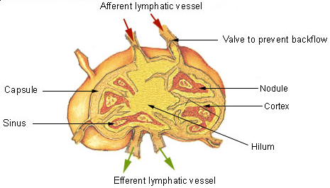
The lymphatic system, or lymphoid system, is an organ system in vertebrates that is part of the immune system, and complementary to the circulatory system. It consists of a large network of lymphatic vessels, lymph nodes, lymphatic or lymphoid organs, and lymphoid tissues. The vessels carry a clear fluid called lymph (the Latin word ''lympha'' refers to the deity of fresh water, "Lympha") back towards the heart, for re-circulation. Unlike the circulatory system that is a closed system, the lymphatic system is open. The human circulatory system processes an average of 20 litres of blood per day through capillary filtration, which removes plasma from the blood. Roughly 17 litres of the filtered blood is reabsorbed directly into the blood vessels, while the remaining three litres are left in the interstitial fluid. One of the main functions of the lymphatic system is to provide an accessory return route to the blood for the surplus three litres. The other main function is that of ... [...More Info...] [...Related Items...] OR: [Wikipedia] [Google] [Baidu] |
Cerebrospinal Fluid
Cerebrospinal fluid (CSF) is a clear, colorless body fluid found within the tissue that surrounds the brain and spinal cord of all vertebrates. CSF is produced by specialised ependymal cells in the choroid plexus of the ventricles of the brain, and absorbed in the arachnoid granulations. There is about 125 mL of CSF at any one time, and about 500 mL is generated every day. CSF acts as a shock absorber, cushion or buffer, providing basic mechanical and immunological protection to the brain inside the skull. CSF also serves a vital function in the cerebral autoregulation of cerebral blood flow. CSF occupies the subarachnoid space (between the arachnoid mater and the pia mater) and the ventricular system around and inside the brain and spinal cord. It fills the ventricles of the brain, cisterns, and sulci, as well as the central canal of the spinal cord. There is also a connection from the subarachnoid space to the bony labyrinth of the inner ear via the perilymphat ... [...More Info...] [...Related Items...] OR: [Wikipedia] [Google] [Baidu] |
Subarachnoid Space
In anatomy, the meninges (, ''singular:'' meninx ( or ), ) are the three membranes that envelop the brain and spinal cord. In mammals, the meninges are the dura mater, the arachnoid mater, and the pia mater. Cerebrospinal fluid is located in the subarachnoid space between the arachnoid mater and the pia mater. The primary function of the meninges is to protect the central nervous system. Structure Dura mater The dura mater ( la, tough mother) (also rarely called ''meninx fibrosa'' or ''pachymeninx'') is a thick, durable membrane, closest to the skull and vertebrae. The dura mater, the outermost part, is a loosely arranged, fibroelastic layer of cells, characterized by multiple interdigitating cell processes, no extracellular collagen, and significant extracellular spaces. The middle region is a mostly fibrous portion. It consists of two layers: the endosteal layer, which lies closest to the skull, and the inner meningeal layer, which lies closer to the brain. It contains large ... [...More Info...] [...Related Items...] OR: [Wikipedia] [Google] [Baidu] |
Berlin Blue
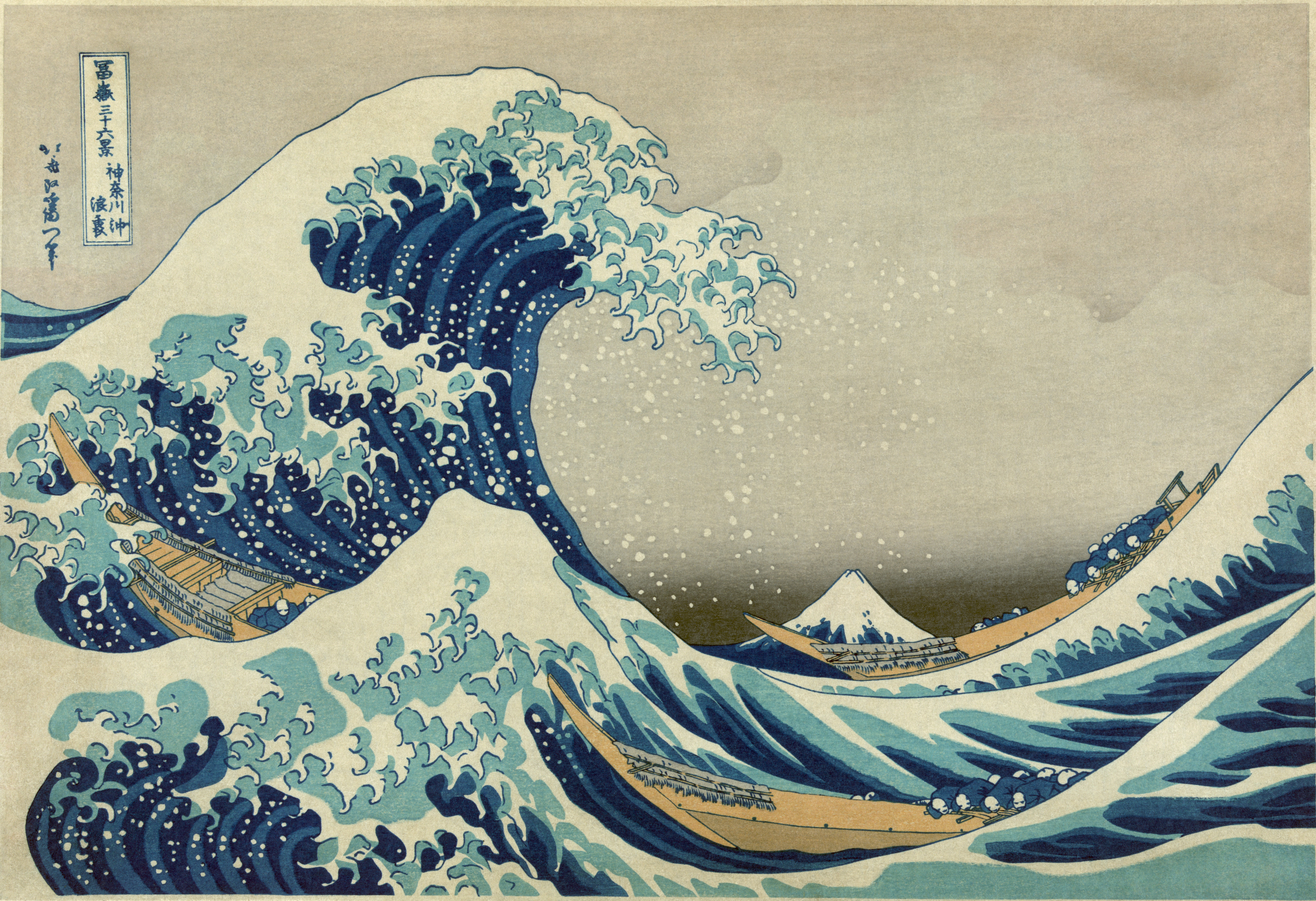
Prussian blue (also known as Berlin blue, Brandenburg blue or, in painting, Parisian or Paris blue) is a dark blue pigment produced by oxidation of ferrous ferrocyanide salts. It has the chemical formula Fe Cyanide.html" ;"title="e(Cyanide">CN) Turnbull's blue is chemically identical, but is made from different reagents, and its slightly different color stems from different impurities and particle sizes. Prussian blue was the first modern synthetic pigment. It is prepared as a very fine colloidal dispersion, because the compound is not soluble in water. It contains variable amounts of other ions and its appearance depends sensitively on the size of the colloidal particles. The pigment is used in paints, and it is the traditional "blue" in blueprints, and became prominent in 19th-century () Japanese woodblock prints. In medicine, orally administered Prussian blue is used as an antidote for certain kinds of heavy metal poisoning, e.g., by thallium(I) and radioactive isotopes o ... [...More Info...] [...Related Items...] OR: [Wikipedia] [Google] [Baidu] |
Pithecantropus Erectus
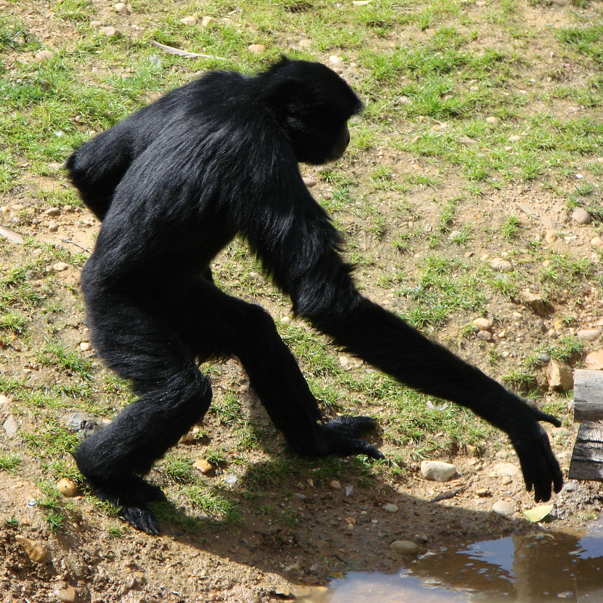
''Homo erectus'' (; meaning "upright man") is an extinct species of archaic human from the Pleistocene, with its earliest occurrence about 2 million years ago. Several human species, such as '' H. heidelbergensis'' and ''H. antecessor'' — with the former generally considered to have been the ancestor to Neanderthals, Denisovans, and modern humans — appear to have evolved from ''H. erectus''. Its specimens are among the first recognizable members of the genus ''Homo''. ''H. erectus'' was the first human ancestor to spread throughout Eurasia, with a continental range extending from the Iberian Peninsula to Java. Asian populations of ''H. erectus'' may be ancestral to '' H. floresiensis'' and possibly to '' H. luzonensis''. The last known population of ''H. erectus'' is '' H. e. soloensis'' from Java, around 117,000–108,000 years ago. ''H. erectus'' had a more modern gait and body proportions, and was the first human species to h ... [...More Info...] [...Related Items...] OR: [Wikipedia] [Google] [Baidu] |
Java Man
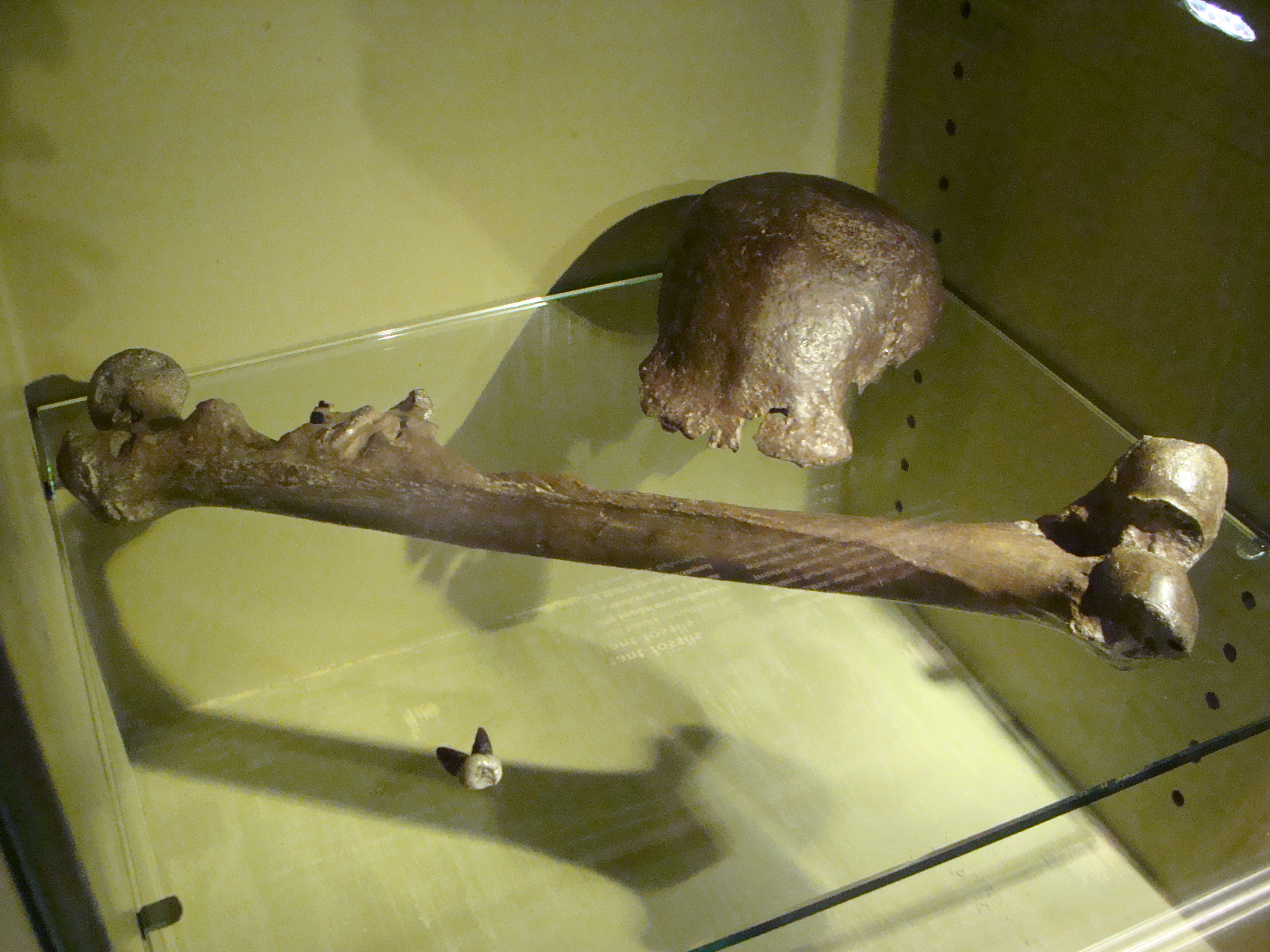
Java Man (''Homo erectus erectus'', formerly also ''Anthropopithecus erectus'', ''Pithecanthropus erectus'') is an early human fossil discovered in 1891 and 1892 on the island of Java (Dutch East Indies, now part of Indonesia). Estimated to be between 700,000 and 2,000,000 years old, it was, at the time of its discovery, the oldest hominid fossils ever found, and it remains the type specimen for ''Homo erectus''. Led by Eugène Dubois, the excavation team uncovered a tooth, a skullcap, and a thighbone at Trinil on the banks of the Solo River in East Java. Arguing that the fossils represented the " missing link" between apes and humans, Dubois gave the species the scientific name ''Anthropopithecus erectus'', then later renamed it ''Pithecanthropus erectus''. The fossil aroused much controversy. Less than ten years after 1891, almost eighty books or articles had been published on Dubois's finds. Despite Dubois's argument, few accepted that Java Man was a transitional form betwee ... [...More Info...] [...Related Items...] OR: [Wikipedia] [Google] [Baidu] |

.jpg)


