|
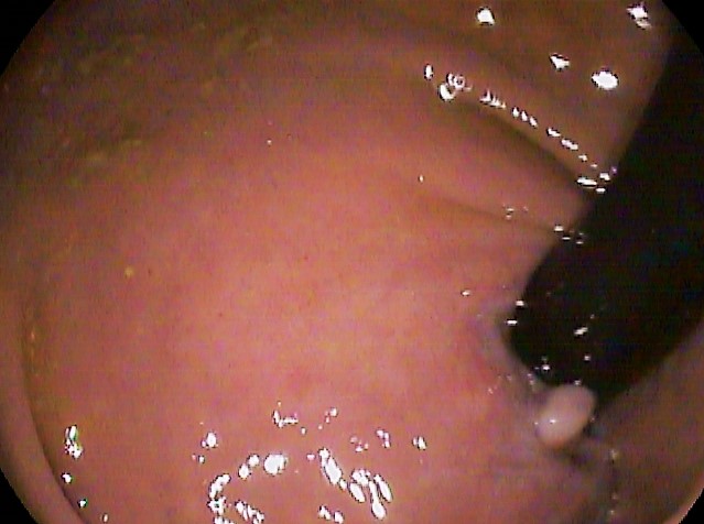
Griffith's Point
In the anatomy of the human digestive tract, there are two colic flexures, or curvatures in the transverse colon. The right colic flexure is also known as the hepatic flexure, and the left colic flexure is also known as the splenic flexure. Note that "right" refers to the patient's anatomical right, which may be depicted on the left of a diagram. Structure Right colic flexure The right colic flexure or hepatic flexure (as it is next to the liver) is the sharp bend between the ascending colon and the transverse colon. The hepatic flexure lies in the right upper quadrant of the human abdomen. It receives blood supply from the superior mesenteric artery. Left colic flexure The left colic flexure or splenic flexure (as it is close to the spleen) is the sharp bend between the transverse colon and the descending colon. The splenic flexure receives dual blood supply from the terminal branches of the superior mesenteric artery and the inferior mesenteric artery. Clinical significance ... [...More Info...] [...Related Items...] OR: [Wikipedia] [Google] [Baidu] |
Transverse Colon
In human anatomy, the transverse colon is the longest and most movable part of the colon. Anatomical position It crosses the abdomen from the ascending colon at the right colic flexure (hepatic flexure) with a downward convexity to the descending colon where it curves sharply on itself beneath the lower end of the spleen forming the left colic flexure (splenic flexure). In its course, it describes an arch, the concavity of which is directed backward and a little upward. Toward its splenic end there is often an abrupt U-shaped curve which may descend lower than the main curve. It is almost completely invested by the peritoneum, and is connected to the inferior border of the pancreas by a large and wide duplicature of that membrane, the transverse mesocolon. It is in relation, by its upper surface, with the liver and gall-bladder, the greater curvature of the stomach, and the lower end of the spleen; by its under surface, with the small intestine; by its anterior surface, with t ... [...More Info...] [...Related Items...] OR: [Wikipedia] [Google] [Baidu] |
Superior Mesenteric Artery
In human anatomy, the superior mesenteric artery (SMA) is an artery which arises from the anterior surface of the abdominal aorta, just inferior to the origin of the celiac trunk, and supplies blood to the intestine from the lower part of the duodenum through two-thirds of the transverse colon, as well as the pancreas. Structure It arises anterior to lower border of vertebra L1 in an adult. It is usually 1 cm lower than the celiac trunk. It initially travels in an anterior/inferior direction, passing behind/under the neck of the pancreas and the splenic vein. Located under this portion of the superior mesenteric artery, between it and the aorta, are the following: * left renal vein - travels between the left kidney and the inferior vena cava (can be compressed between the SMA and the abdominal aorta at this location, leading to nutcracker syndrome). * the third part of the duodenum, a segment of the small intestines (can be compressed by the SMA at this location, lea ... [...More Info...] [...Related Items...] OR: [Wikipedia] [Google] [Baidu] |
Sudeck's Point
The rectum is the final straight portion of the large intestine in humans and some other mammals, and the gut in others. The adult human rectum is about long, and begins at the rectosigmoid junction (the end of the sigmoid colon) at the level of the third sacral vertebra or the sacral promontory depending upon what definition is used. Its diameter is similar to that of the sigmoid colon at its commencement, but it is dilated near its termination, forming the rectal ampulla. It terminates at the level of the anorectal ring (the level of the puborectalis sling) or the dentate line, again depending upon which definition is used. In humans, the rectum is followed by the anal canal which is about long, before the gastrointestinal tract terminates at the anal verge. The word rectum comes from the Latin '' rectum intestinum'', meaning ''straight intestine''. Structure The rectum is a part of the lower gastrointestinal tract. The rectum is a continuation of the sigmoid co ... [...More Info...] [...Related Items...] OR: [Wikipedia] [Google] [Baidu] |
Rectum
The rectum is the final straight portion of the large intestine in humans and some other mammals, and the Gastrointestinal tract, gut in others. The adult human rectum is about long, and begins at the rectosigmoid junction (the end of the sigmoid colon) at the level of the third sacral vertebra or the sacral promontory depending upon what definition is used. Its diameter is similar to that of the sigmoid colon at its commencement, but it is dilated near its termination, forming the rectal ampulla. It terminates at the level of the anorectal ring (the level of the puborectalis sling) or the dentate line, again depending upon which definition is used. In humans, the rectum is followed by the anal canal which is about long, before the gastrointestinal tract terminates at the anal verge. The word rectum comes from the Latin ''Wikt:rectum, rectum Wikt:intestinum, intestinum'', meaning ''straight intestine''. Structure The rectum is a part of the lower gastrointestinal tract ... [...More Info...] [...Related Items...] OR: [Wikipedia] [Google] [Baidu] |
Ischemic Colitis
Ischemic colitis (also spelled ischaemic colitis) is a medical condition in which inflammation and injury of the large intestine result from inadequate blood supply. Although uncommon in the general population, ischemic colitis occurs with greater frequency in the elderly, and is the most common form of bowel ischemia. http://www.guideline.gov/summary/summary.aspx?ss=15&doc_id=3069&nbr=2295 Causes of the reduced blood flow can include changes in the systemic circulation (e.g. low blood pressure) or local factors such as constriction of blood vessels or a blood clot. In most cases, no specific cause can be identified. Ischemic colitis is usually suspected on the basis of the clinical setting, physical examination, and laboratory test results; the diagnosis can be confirmed by endoscopy or by using sigmoid or endoscopic placement of a visible light spectroscopic catheter (see Diagnosis). Ischemic colitis can span a wide spectrum of severity; most patients are treated supportively ... [...More Info...] [...Related Items...] OR: [Wikipedia] [Google] [Baidu] |
Bowel Ischemia
Intestinal ischemia is a medical condition in which injury to the large or small intestine occurs due to not enough blood supply. It can come on suddenly, known as acute intestinal ischemia, or gradually, known as chronic intestinal ischemia. The acute form of the disease often presents with sudden severe abdominal pain and is associated with a high risk of death. The chronic form typically presents more gradually with abdominal pain after eating, unintentional weight loss, vomiting, and fear of eating. Risk factors for acute intestinal ischemia include atrial fibrillation, heart failure, chronic kidney failure, being prone to forming blood clots, and previous myocardial infarction. There are four mechanisms by which poor blood flow occurs: a blood clot from elsewhere getting lodged in an artery, a new blood clot forming in an artery, a blood clot forming in the superior mesenteric vein, and insufficient blood flow due to low blood pressure or spasms of arteries. Chronic di ... [...More Info...] [...Related Items...] OR: [Wikipedia] [Google] [Baidu] |
Hypotension
Hypotension is low blood pressure. Blood pressure is the force of blood pushing against the walls of the arteries as the heart pumps out blood. Blood pressure is indicated by two numbers, the systolic blood pressure (the top number) and the diastolic blood pressure (the bottom number), which are the maximum and minimum blood pressures, respectively. A systolic blood pressure of less than 90 millimeters of mercury (mmHg) or diastolic of less than 60 mmHg is generally considered to be hypotension. Different numbers apply to children. However, in practice, blood pressure is considered too low only if noticeable symptoms are present. Symptoms include dizziness or lightheadedness, confusion, feeling tired, weakness, headache, blurred vision, nausea, neck or back pain, an irregular heartbeat or feeling that the heart is skipping beats or fluttering, or fainting. Hypotension is the opposite of hypertension, which is high blood pressure. It is best understood as a physiological st ... [...More Info...] [...Related Items...] OR: [Wikipedia] [Google] [Baidu] |
Watershed Area (medical)
Watershed area is the medical term referring to regions of the body, that receive dual blood supply from the most distal branches of two large arteries, such as the splenic flexure of the large intestine. The term refers metaphorically to a geological watershed, or drainage divide, which separates adjacent drainage basins. During times of blockage of one of the arteries that supply the watershed area, such as in atherosclerosis, these regions are spared from ischemia by virtue of their dual supply. However, during times of systemic hypoperfusion, such as in disseminated intravascular coagulation or heart failure, these regions are particularly vulnerable to ischemia because they are supplied by the most distal branches of their arteries, and thus the least likely to receive sufficient blood. Watershed areas are found in the brain, where areas are perfused by both the anterior and middle cerebral arteries, and in the intestines, where areas are perfused by both the superior and i ... [...More Info...] [...Related Items...] OR: [Wikipedia] [Google] [Baidu] |
Irritable Bowel Syndrome
Irritable bowel syndrome (IBS) is a "disorder of gut-brain interaction" characterized by a group of symptoms that commonly include abdominal pain and or abdominal bloating and changes in the consistency of bowel movements. These symptoms may occur over a long time, sometimes for years. IBS can negatively affect quality of life and may result in missed school or work (absenteeism) or reduced productivity at work (presenteeism). Disorders such as anxiety, major depression, and chronic fatigue syndrome are common among people with IBS.The cited review is based on sources ranging from 1988 to 2001 and is probably biased relative to a more recent research. The causes of IBS may well be multi-factorial. Theories include combinations of "gut–brain axis" problems, alterations in gut motility, visceral hypersensitivity, infections including small intestinal bacterial overgrowth, neurotransmitters, genetic factors, and food sensitivity. Onset may be triggered by an intestinal infec ... [...More Info...] [...Related Items...] OR: [Wikipedia] [Google] [Baidu] |
Inferior Mesenteric Artery
In human anatomy, the inferior mesenteric artery, often abbreviated as IMA, is the third main branch of the abdominal aorta and arises at the level of L3, supplying the large intestine from the distal transverse colon to the upper part of the anal canal. The regions supplied by the IMA are the descending colon, the sigmoid colon, and part of the rectum. Structure Proximally, its territory of distribution overlaps (forms a watershed) with the middle colic artery, and therefore the superior mesenteric artery. The SMA and IMA anastomose via the marginal artery of the colon (artery of Drummond) and via Riolan's arcade (also called the "meandering artery", an arterial connection between the left colic artery and the middle colic artery). The territory of distribution of the IMA is more or less equivalent to the embryonic hindgut. Branches The IMA branches off the anterior surface of the abdominal aorta below the renal artery branch points, 3-4 cm above the aortic bifurcation (into t ... [...More Info...] [...Related Items...] OR: [Wikipedia] [Google] [Baidu] |
Descending Colon
In the anatomy of humans and homologous primates, the descending colon is the part of the colon extending from the left colic flexure to the level of the iliac crest (whereupon it transitions into the sigmoid colon). The function of the descending colon in the digestive system is to store the remains of digested food that will be emptied into the rectum. The descending colon is on the left side of the body (barring any malformations). The term left colon is hypernymous to ''descending colon'' in precise use; many casual mentions of the left colon chiefly concern the descending colon. Structure The descending colon extends from the left colic flexure at the upper left part of the abdomen inferior-ward through the left hypochondrium and lumbar regions, along the outer border of the left kidney, ending at the level of the iliac crest at the lower left part of the abdomen, being contunued thenceforth as the sigmoid colon. It usually retroperitoneal (being lined by peritoneum on ... [...More Info...] [...Related Items...] OR: [Wikipedia] [Google] [Baidu] |
Spleen
The spleen is an organ found in almost all vertebrates. Similar in structure to a large lymph node, it acts primarily as a blood filter. The word spleen comes .σπλήν Henry George Liddell, Robert Scott, ''A Greek-English Lexicon'', on Perseus Digital Library The spleen plays very important roles in regard to s (erythrocytes) and the . It removes old red blood cells and holds a reserve of blood, which can be valuable in case of |


.png)
