|
Glomerulosclerosis, Focal
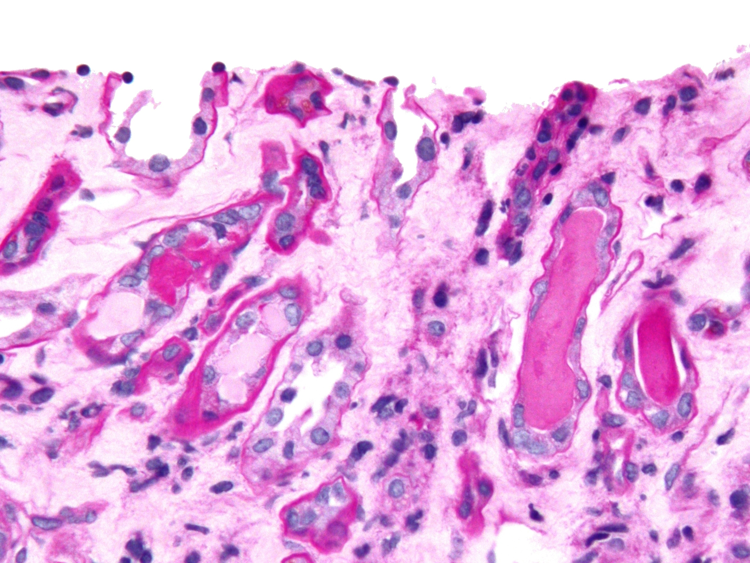
Focal segmental glomerulosclerosis (FSGS) is a histopathologic finding of scarring (sclerosis) of glomeruli and damage to renal podocytes.Rosenberg, Avi Z.; Kopp, Jeffrey B. (2017-03-07). "Focal Segmental Glomerulosclerosis". ''Clinical Journal of the American Society of Nephrology''. 12 (3): 502–517. doi:10.2215/CJN.05960616. ISSN 1555-9041. PMC 5338705. PMID 28242845.D'Agati V. The many masks of focal segmental glomerulosclerosis. Kidney Int. 1994 Oct;46(4):1223-41. doi: 10.1038/ki.1994.388. . This process damages the filtration function of the kidney, resulting in protein loss in the urine. FSGS is a leading cause of excess protein loss—nephrotic syndrome—in children and adults.Kitiyakara C, Eggers P, Kopp JB. Twenty-one-year trend in ESRD due to focal segmental glomerulosclerosis in the United States. Am J Kidney Dis. 2004 Nov;44(5):815-25. . Signs and symptoms include proteinuria, water retention, and edema.Rydel JJ, Korbet SM, Borok RZ, Schwartz MM. Focal segment ... [...More Info...] [...Related Items...] OR: [Wikipedia] [Google] [Baidu] |
Micrograph
A micrograph or photomicrograph is a photograph or digital image taken through a microscope or similar device to show a magnified image of an object. This is opposed to a macrograph or photomacrograph, an image which is also taken on a microscope but is only slightly magnified, usually less than 10 times. Micrography is the practice or art of using microscopes to make photographs. A micrograph contains extensive details of microstructure. A wealth of information can be obtained from a simple micrograph like behavior of the material under different conditions, the phases found in the system, failure analysis, grain size estimation, elemental analysis and so on. Micrographs are widely used in all fields of microscopy. Types Photomicrograph A light micrograph or photomicrograph is a micrograph prepared using an optical microscope, a process referred to as ''photomicroscopy''. At a basic level, photomicroscopy may be performed simply by connecting a camera to a microscope, th ... [...More Info...] [...Related Items...] OR: [Wikipedia] [Google] [Baidu] |
Antibody
An antibody (Ab), also known as an immunoglobulin (Ig), is a large, Y-shaped protein used by the immune system to identify and neutralize foreign objects such as pathogenic bacteria and viruses. The antibody recognizes a unique molecule of the pathogen, called an antigen. Each tip of the "Y" of an antibody contains a paratope (analogous to a lock) that is specific for one particular epitope (analogous to a key) on an antigen, allowing these two structures to bind together with precision. Using this binding mechanism, an antibody can ''tag'' a microbe or an infected cell for attack by other parts of the immune system, or can neutralize it directly (for example, by blocking a part of a virus that is essential for its invasion). To allow the immune system to recognize millions of different antigens, the antigen-binding sites at both tips of the antibody come in an equally wide variety. In contrast, the remainder of the antibody is relatively constant. It only occurs in a few varia ... [...More Info...] [...Related Items...] OR: [Wikipedia] [Google] [Baidu] |
Glomerulosclerosis
Glomerulosclerosis is the hardening of the glomeruli in the kidney. It is a general term to describe scarring of the kidneys' tiny blood vessels, the glomeruli, the functional units in the kidney that filter urea from the blood. Proteinuria (large amounts of protein in the urine) is one of the signs of glomerulosclerosis. Scarring disturbs the filtering process of the kidneys and allows protein to leak from the blood into the urine. However, glomerulosclerosis is one of many causes of proteinuria. A kidney biopsy (the removal of a tiny part of the kidney with a needle) may be necessary to determine whether a patient has glomerulosclerosis or another kidney problem. About 15 percent of people with proteinuria turn out to have glomerulosclerosis. Both children and adults can develop glomerulosclerosis, which can result in different types of kidney conditions. One frequently encountered type of glomerulosclerosis is caused by diabetes. Drug use or infections may cause focal segmen ... [...More Info...] [...Related Items...] OR: [Wikipedia] [Google] [Baidu] |
Agnes Fogo
Agnes B. Fogo is a professor of renal pathology at the Vanderbilt University Medical Center. Biography Fogo graduated from the University of Oslo, Norway, and the University of Tennessee, USA. She completed her M.D. from Vanderbilt University School of Medicine before going on to do residency and a fellowship in renal pathology. Appointments Fogo works at the Vanderbilt University Medical Center and is the John L. Shapiro Professor of Pathology, Microbiology and Immunology, Professor of Medicine and Pediatrics, and director of the Renal/Electron Microscopy Laboratory. In 2021 she also became the International Society of Nephrology president for a 2 year term. Awards *2011 Robert G. Narins award from the American Society of Nephrology for "substantial accomplishments in the development and leadership of educational courses and resources" *2019 Roscoe R. Robinson award from the International Society of Nephrology The International Society of Nephrology (ISN) is an organization ... [...More Info...] [...Related Items...] OR: [Wikipedia] [Google] [Baidu] |
Nanometre
330px, Different lengths as in respect to the molecular scale. The nanometre (international spelling as used by the International Bureau of Weights and Measures; SI symbol: nm) or nanometer (American and British English spelling differences#-re, -er, American spelling) is a units of measurement, unit of length in the International System of Units (SI), equal to one billionth (short scale) of a metre () and to 1000 picometres. One nanometre can be expressed in scientific notation as , and as metres. History The nanometre was formerly known as the millimicrometre – or, more commonly, the millimicron for short – since it is of a micron (micrometre), and was often denoted by the symbol mμ or (more rarely and confusingly, since it logically should refer to a ''millionth'' of a micron) as μμ. Etymology The name combines the SI prefix ''nano-'' (from the Ancient Greek , ', "dwarf") with the parent unit name ''metre'' (from Greek , ', "unit of measurement"). ... [...More Info...] [...Related Items...] OR: [Wikipedia] [Google] [Baidu] |
Bowman's Capsule
Bowman's capsule (or the Bowman capsule, capsula glomeruli, or glomerular capsule) is a cup-like sac at the beginning of the tubular component of a nephron in the mammalian kidney that performs the first step in the filtration of blood to form urine. A glomerulus is enclosed in the sac. Fluids from blood in the glomerulus are collected in the Bowman's capsule. Structure Outside the capsule, there are two "poles": * The vascular pole is the side with the afferent arteriole and efferent arteriole. * The urinary pole is the side with the proximal convoluted tubule. Inside the capsule, the layers are as follows, from outside to inside: *''Parietal layer''—A single layer of simple squamous epithelium. Does not function in filtration. *''Bowman's space (or "urinary space", or "capsular space")''—Between the visceral and parietal layers, into which the filtrate enters after passing through the filtration slits. *''Visceral layer''—Lies just above the thickened glomerular baseme ... [...More Info...] [...Related Items...] OR: [Wikipedia] [Google] [Baidu] |
Podocyte
Podocytes are cells in Bowman's capsule in the kidneys that wrap around capillaries of the glomerulus. Podocytes make up the epithelial lining of Bowman's capsule, the third layer through which filtration of blood takes place. Bowman's capsule filters the blood, retaining large molecules such as proteins while smaller molecules such as water, salts, and sugars are filtered as the first step in the formation of urine. Although various viscera have epithelial layers, the name visceral epithelial cells usually refers specifically to podocytes, which are specialized epithelial cells that reside in the visceral layer of the capsule. One type of specialized epithelial cell is podocalyxin. The podocytes have long foot processes called ''pedicels'', for which the cells are named (''podo-'' + '' -cyte''). The pedicels wrap around the capillaries and leave slits between them. Blood is filtered through these slits, each known as a filtration slit, slit diaphragm, or slit pore. Several pr ... [...More Info...] [...Related Items...] OR: [Wikipedia] [Google] [Baidu] |
Glomerulus (kidney)
The glomerulus (plural glomeruli) is a network of small blood vessels (capillaries) known as a ''tuft'', located at the beginning of a nephron in the kidney. Each of the two kidneys contains about one million nephrons. The tuft is structurally supported by the mesangium (the space between the blood vessels), composed of intraglomerular mesangial cells. The blood is filtered across the capillary walls of this tuft through the glomerular filtration barrier, which yields its filtrate of water and soluble substances to a cup-like sac known as Bowman's capsule. The filtrate then enters the renal tubule of the nephron. The glomerulus receives its blood supply from an afferent arteriole of the renal arterial circulation. Unlike most capillary beds, the glomerular capillaries exit into efferent arterioles rather than venules. The resistance of the efferent arterioles causes sufficient hydrostatic pressure within the glomerulus to provide the force for ultrafiltration. The glomerulus and ... [...More Info...] [...Related Items...] OR: [Wikipedia] [Google] [Baidu] |
Urinary Cast
Urinary casts are microscopic cylindrical structures produced by the kidney and present in the urine in certain disease states. They form in the distal convoluted tubule and collecting ducts of nephrons, then dislodge and pass into the urine, where they can be detected by microscopy. They form via precipitation of Tamm–Horsfall mucoprotein, which is secreted by renal tubule cells, and sometimes also by albumin in conditions of proteinuria. Cast formation is pronounced in environments favoring protein denaturation and precipitation (low flow, concentrated salts, low pH). Tamm–Horsfall protein is particularly susceptible to precipitation in these conditions. Casts were first described by Henry Bence Jones (1813–1873). As reflected in their cylindrical form, casts are generated in the small distal convoluted tubules and collecting ducts of the kidney, and generally maintain their shape and composition as they pass through the urinary system. Although the most common forms ar ... [...More Info...] [...Related Items...] OR: [Wikipedia] [Google] [Baidu] |
Oncotic Pressure
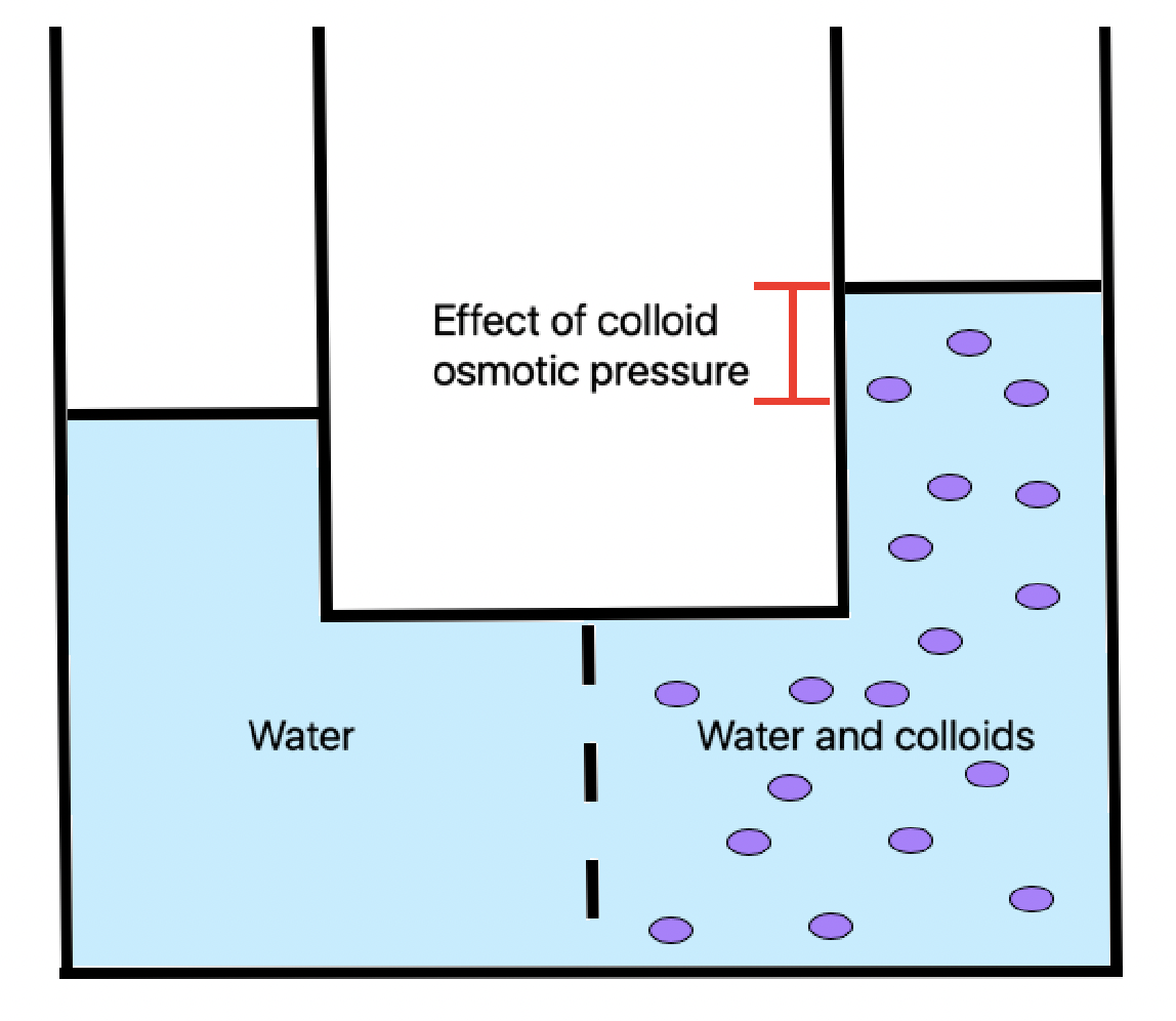
Oncotic pressure, or colloid osmotic-pressure, is a form of osmotic pressure induced by the proteins, notably albumin, in a blood vessel's plasma (blood/liquid) that causes a pull on fluid back into the capillary. Participating colloids displace water molecules, thus creating a relative water molecule deficit with water molecules moving back into the circulatory system within the lower venous pressure end of capillaries. It has the opposing effect of both hydrostatic blood pressure pushing water and small molecules out of the blood into the interstitial spaces within the arterial end of capillaries and interstitial colloidal osmotic pressure. These interacting factors determine the partition balancing of extracellular water between the blood plasma and outside the blood stream. Oncotic pressure strongly affects the physiological function of the circulatory system. It is suspected to have a major effect on the pressure across the glomerular filter. However, this concept has been ... [...More Info...] [...Related Items...] OR: [Wikipedia] [Google] [Baidu] |
Hypercholesterolemia
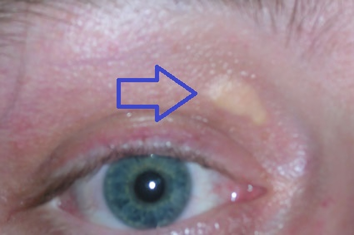
Hypercholesterolemia, also called high cholesterol, is the presence of high levels of cholesterol in the blood. It is a form of hyperlipidemia (high levels of lipids in the blood), hyperlipoproteinemia (high levels of lipoproteins in the blood), and dyslipidemia (any abnormalities of lipid and lipoprotein levels in the blood). Elevated levels of non-HDL cholesterol and LDL in the blood may be a consequence of diet, obesity, inherited (genetic) diseases (such as LDL receptor mutations in familial hypercholesterolemia), or the presence of other diseases such as type 2 diabetes and an underactive thyroid. Cholesterol is one of three major classes of lipids produced and used by all animal cells to form membranes. Plant cells manufacture phytosterols (similar to cholesterol), but in rather small quantities. Cholesterol is the precursor of the steroid hormones and bile acids. Since cholesterol is insoluble in water, it is transported in the blood plasma within protein particles ... [...More Info...] [...Related Items...] OR: [Wikipedia] [Google] [Baidu] |


