|
Giantin
Giantin or Golgin subfamily B member 1 is a protein that in humans is encoded by the ''GOLGB1'' gene. Giantin is located at the cis-medial rims of the Golgi apparatus and is part of the Golgi matrix that is responsible for membrane trafficking in secretory pathway of proteins. This function is key for proper localisation of proteins at the plasma membrane and outside the cell (extracellular region) which is important for cell function that is dependent on for example receptors and the extracellular matrix function. Recent animal model knockout studies of ''GOLGB1'' in mice, rat, and zebrafish have shown that phenotypes are different between species ranging from mild to severe craniofacial defects in the rodent models to just minor size defects in zebrafish. However, in adult zebrafish a tumoral calcinosis-like phenotype was observed, and in humans such phenotype has been linked to defective glycosyltransferase function (e.g. GALNT3 protein). Function and Interactions Giantin is ... [...More Info...] [...Related Items...] OR: [Wikipedia] [Google] [Baidu] |
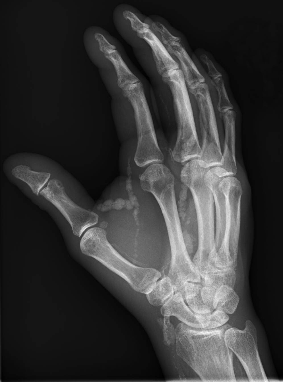
Tumoral Calcinosis
Tumoral calcinosis is a rare condition in which there is calcium deposition in the soft tissue in periarticular location, around joints, outside the joint capsule. They are frequently (0.5–3%) seen in patients undergoing renal dialysis. Clinically also known as hyperphosphatemic familial tumoral calcinosis (HFTC), is often caused by genetic mutations in genes that regulate phosphate physiology in the body (leading to too much phosphate (hyperphosphatemia)). Best described genes that harbour mutations in humans are FGF-23, Klotho (KL), or GALNT3. A zebrafish animal model with reduced GALNT3 expression also showed HFTC-like phenotype, indicating an evolutionary conserved mechanism that is involved in developing tumoral calcinosis. Clinical features The name indicates calcinosis (calcium deposition) which resembles tumor (like a new growth). They are not true neoplasms – they don't have dividing cells. They are just deposition of inorganic calcium with serum exudate. Childr ... [...More Info...] [...Related Items...] OR: [Wikipedia] [Google] [Baidu] |
Golgi Matrix
The Golgi matrix is a collection of proteins involved in the structure and function of the Golgi apparatus. The matrix was first isolated in 1994 as an amorphous collection of 12 proteins that remained associated together in the presence of Triton X-100, detergent (which removed Golgi membranes) and 150 Milli-, mMolar concentration, M NaCl (which removed weakly associated proteins). Treatment with a Proteinase K, protease enzyme removed the matrix, which confirmed the importance of proteins for the matrix structure. Modern Electron microscope#Sample preparation, freeze etch electron microscopy (EM) clearly shows a mesh connecting Golgi cisternae and associated Vesicle (biology and chemistry), vesicles. Further support for the existence of a matrix comes from EM images showing that ribosomes are excluded from regions between and near Golgi cisternae.Fig. 14 in Structure and function The first individual protein component of the matrix was identified in 1995 as Golgin A2 (then cal ... [...More Info...] [...Related Items...] OR: [Wikipedia] [Google] [Baidu] |

