|
Germ Cell Neoplasia In Situ
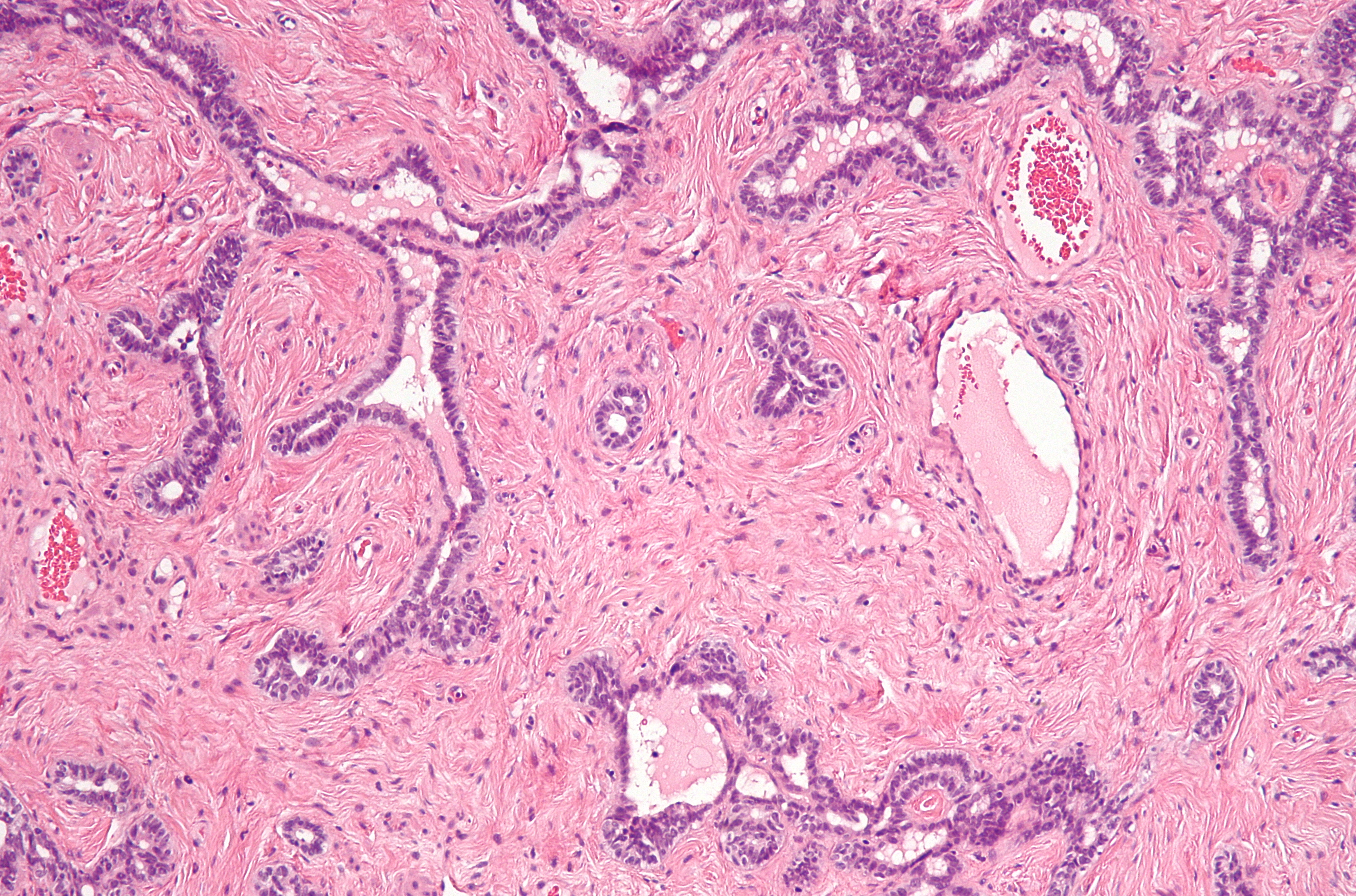
Germ cell neoplasia in situ (GCNIS) represents the precursor lesion for many types of testicular germ cell tumors. The term GCNIS was introduced with the 2016 edition of the WHO classification of urological tumours. GCNIS more accurate describes the lesion as it arises between the basement membrane and Sertoli cells (the cells that 'nurse' the developing germ cell). The common, unspecified variant of the entity was once considered to be a carcinoma in situ, although the term "carcinoma ''in situ''" is now largely historical as it is not an accurate description of the process.Mills, S (ed.) 2009.Sternberg's Diagnostic Pathology. 5th Edition. Classification The World Health Organization classification of testicular tumoursEble J.N., Sauter G., Epstein J.I., Sesterhenn I.A. (Eds.): World Health Organization Classification of Tumours. Pathology and Genetics of Tumours of the Urinary System and Male Genital Organs. IARC Press: Lyon 2004. subdivides ITGCN into (1) a more common, uns ... [...More Info...] [...Related Items...] OR: [Wikipedia] [Google] [Baidu] |
H&E Stain
Hematoxylin and eosin stain ( or haematoxylin and eosin stain or hematoxylin-eosin stain; often abbreviated as H&E stain or HE stain) is one of the principal tissue stains used in histology. It is the most widely used stain in medical diagnosis and is often the gold standard. For example, when a pathologist looks at a biopsy of a suspected cancer, the histological section is likely to be stained with H&E. H&E is the combination of two histological stains: hematoxylin and eosin. The hematoxylin stains cell nuclei a purplish blue, and eosin stains the extracellular matrix and cytoplasm pink, with other structures taking on different shades, hues, and combinations of these colors. Hence a pathologist can easily differentiate between the nuclear and cytoplasmic parts of a cell, and additionally, the overall patterns of coloration from the stain show the general layout and distribution of cells and provides a general overview of a tissue sample's structure. Thus, pattern recogniti ... [...More Info...] [...Related Items...] OR: [Wikipedia] [Google] [Baidu] |
Nucleolus
The nucleolus (, plural: nucleoli ) is the largest structure in the nucleus of eukaryotic cells. It is best known as the site of ribosome biogenesis, which is the synthesis of ribosomes. The nucleolus also participates in the formation of signal recognition particles and plays a role in the cell's response to stress. Nucleoli are made of proteins, DNA and RNA, and form around specific chromosomal regions called nucleolar organizing regions. Malfunction of nucleoli can be the cause of several human conditions called "nucleolopathies" and the nucleolus is being investigated as a target for cancer chemotherapy. History The nucleolus was identified by bright-field microscopy during the 1830s. Little was known about the function of the nucleolus until 1964, when a study of nucleoli by John Gurdon and Donald Brown in the African clawed frog ''Xenopus laevis'' generated increasing interest in the function and detailed structure of the nucleolus. They found that 25% of the frog e ... [...More Info...] [...Related Items...] OR: [Wikipedia] [Google] [Baidu] |
Carcinoma In Situ
Carcinoma ''in situ'' (CIS) is a group of abnormal cells. While they are a form of neoplasm, there is disagreement over whether CIS should be classified as cancer. This controversy also depends on the exact CIS in question (i.e. cervical, skin, breast). Some authors do not classify them as cancer, however, recognizing that they can potentially become cancer. Others classify certain types as a non-invasive form of cancer. The term "pre-cancer" has also been used. These abnormal cells grow in their normal place, thus "''in situ''" (from Latin for "in its place"). For example, carcinoma ''in situ'' of the skin, also called Bowen's disease, is the accumulation of dysplastic epidermal cells within the epidermis only, that has failed to penetrate into the deeper dermis. For this reason, CIS will usually not form a tumor. Rather, the lesion is flat (in the skin, cervix, etc.) or follows the existing architecture of the organ (in the breast, lung, etc.). Exceptions include CIS of the colon ... [...More Info...] [...Related Items...] OR: [Wikipedia] [Google] [Baidu] |
Metastatic
Metastasis is a pathogenic agent's spread from an initial or primary site to a different or secondary site within the host's body; the term is typically used when referring to metastasis by a cancerous tumor. The newly pathological sites, then, are metastases (mets). It is generally distinguished from cancer invasion, which is the direct extension and penetration by cancer cells into neighboring tissues. Cancer occurs after cells are genetically altered to proliferate rapidly and indefinitely. This uncontrolled proliferation by mitosis produces a primary heterogeneic tumour. The cells which constitute the tumor eventually undergo metaplasia, followed by dysplasia then anaplasia, resulting in a malignant phenotype. This malignancy allows for invasion into the circulation, followed by invasion to a second site for tumorigenesis. Some cancer cells known as circulating tumor cells acquire the ability to penetrate the walls of lymphatic or blood vessels, after which they are able ... [...More Info...] [...Related Items...] OR: [Wikipedia] [Google] [Baidu] |
Chemotherapy
Chemotherapy (often abbreviated to chemo and sometimes CTX or CTx) is a type of cancer treatment that uses one or more anti-cancer drugs (chemotherapeutic agents or alkylating agents) as part of a standardized chemotherapy regimen. Chemotherapy may be given with a curative intent (which almost always involves combinations of drugs) or it may aim to prolong life or to reduce symptoms ( palliative chemotherapy). Chemotherapy is one of the major categories of the medical discipline specifically devoted to pharmacotherapy for cancer, which is called ''medical oncology''. The term ''chemotherapy'' has come to connote non-specific usage of intracellular poisons to inhibit mitosis (cell division) or induce DNA damage, which is why inhibition of DNA repair can augment chemotherapy. The connotation of the word chemotherapy excludes more selective agents that block extracellular signals (signal transduction). The development of therapies with specific molecular or genetic targets, wh ... [...More Info...] [...Related Items...] OR: [Wikipedia] [Google] [Baidu] |
Orchiectomy
Orchiectomy (also named orchidectomy, and sometimes shortened as orchi or orchie) is a surgical procedure in which one or both testicles are removed. The surgery is performed as treatment for testicular cancer, as part of surgery for transgender women, as management for advanced prostate cancer, and to remove damaged testes after testicular torsion. Less frequently, orchiectomy may be performed following a trauma, or due to wasting away of the testis or testes. Procedure Simple orchiectomy A simple orchiectomy is commonly performed as part of gender reassignment surgery for transgender women, or as palliative treatment for advanced cases of prostate cancer. A simple orchiectomy may also be required in the event of testicular torsion. For the procedure, the person lies flat on an operating table with the penis taped against the abdomen. The nurse shaves a small area for the incision. After anesthetic has been administered, the surgeon makes an incision in the midpoint of the ... [...More Info...] [...Related Items...] OR: [Wikipedia] [Google] [Baidu] |
Sensitivity And Specificity
''Sensitivity'' and ''specificity'' mathematically describe the accuracy of a test which reports the presence or absence of a condition. Individuals for which the condition is satisfied are considered "positive" and those for which it is not are considered "negative". *Sensitivity (true positive rate) refers to the probability of a positive test, conditioned on truly being positive. *Specificity (true negative rate) refers to the probability of a negative test, conditioned on truly being negative. If the true condition can not be known, a " gold standard test" is assumed to be correct. In a diagnostic test, sensitivity is a measure of how well a test can identify true positives and specificity is a measure of how well a test can identify true negatives. For all testing, both diagnostic and screening, there is usually a trade-off between sensitivity and specificity, such that higher sensitivities will mean lower specificities and vice versa. If the goal is to return the ratio at w ... [...More Info...] [...Related Items...] OR: [Wikipedia] [Google] [Baidu] |
Immunostaining
In biochemistry, immunostaining is any use of an antibody-based method to detect a specific protein in a sample. The term "immunostaining" was originally used to refer to the immunohistochemical staining of tissue sections, as first described by Albert Coons in 1941. However, immunostaining now encompasses a broad range of techniques used in histology, cell biology, and molecular biology that use antibody-based staining methods. Techniques Immunohistochemistry Immunohistochemistry or IHC staining of tissue sections (or immunocytochemistry, which is the staining of cells), is perhaps the most commonly applied immunostaining technique. While the first cases of IHC staining used fluorescent dyes (see '' immunofluorescence''), other non-fluorescent methods using enzymes such as peroxidase (see '' immunoperoxidase staining'') and alkaline phosphatase are now used. These enzymes are capable of catalysing reactions that give a coloured product that is easily detectable by light m ... [...More Info...] [...Related Items...] OR: [Wikipedia] [Google] [Baidu] |
Rete Testis
The rete testis ( ) is an anastomosing network of delicate tubules located in the hilum of the testicle (mediastinum testis) that carries sperm from the seminiferous tubules to the efferent ducts. It is the counterpart of the rete ovarii in females. Its function is to provide a site for fluid reabsorption. Structure The rete testis is the network of interconnecting tubules where the straight seminiferous tubules (the terminal part of the seminiferous tubules) empty. It is located within a highly vascular connective tissue in the mediastinum testis. The epithelial cells form a single layer that lines the inner surface of the tubules. These cells are cuboidal, with microvilli and a single cilium on their surface. Development In the development of the urinary and reproductive organs, the testis is developed in much the same way as the ovary, originating from mesothelium as well as mesonephros. Like the ovary, in its earliest stages it consists of a central mass covered by a surfac ... [...More Info...] [...Related Items...] OR: [Wikipedia] [Google] [Baidu] |
Pagetoid
Pagetoid is a term used in dermatology to refer to "upward spreading" of abnormal cells in the epidermis (ie from bottom to top). It is uncommon and a possible indication of a precancerous or cancerous condition. Cells display pagetoid growth when they invade the upper epidermis from below. Squamous cell carcinoma, melanoma in situ, Pagetoid Bowen's disease, ocular sebaceous carcinoma, and other carcinomas can all display pagetoid growth. The term ''pagetoid'' (i.e., 'Paget-like') is derived from the extramammary Paget's disease, wherein the large tumour cells are arranged singly or in small clusters within the epidermis and its appendages. These cells are distinguished by a clear halo from the surrounding epithelial cells and a finely granular cytoplasm. This proliferation of cells in the epidermis is responsible for the "buckshot scatter" pattern.{{cite web, url=http://www.dermnetnz.org/pathology/pathology-glossary.html, title=Dermatopathological terminology - DermNet New Zealand ... [...More Info...] [...Related Items...] OR: [Wikipedia] [Google] [Baidu] |
Spermatogenesis
Spermatogenesis is the process by which haploid spermatozoa develop from germ cells in the seminiferous tubules of the testis. This process starts with the mitotic division of the stem cells located close to the basement membrane of the tubules. These cells are called spermatogonial stem cells. The mitotic division of these produces two types of cells. Type A cells replenish the stem cells, and type B cells differentiate into primary spermatocytes. The primary spermatocyte divides meiotically (Meiosis I) into two secondary spermatocytes; each secondary spermatocyte divides into two equal haploid spermatids by Meiosis II. The spermatids are transformed into spermatozoa (sperm) by the process of spermiogenesis. These develop into mature spermatozoa, also known as sperm cells. Thus, the primary spermatocyte gives rise to two cells, the secondary spermatocytes, and the two secondary spermatocytes by their subdivision produce four spermatozoa and four haploid cells. Spermatozoa are ... [...More Info...] [...Related Items...] OR: [Wikipedia] [Google] [Baidu] |
Sertoli Cells
Sertoli cells are a type of sustentacular "nurse" cell found in human testes which contribute to the process of spermatogenesis (the production of sperm) as a structural component of the seminiferous tubules. They are activated by follicle-stimulating hormone (FSH) secreted by the adenohypophysis and express FSH receptor on their membranes. History Sertoli cells are named after Enrico Sertoli, an Italian physiologist who discovered them while studying medicine at the University of Pavia, Italy. He published a description of his eponymous cell in 1865. The cell was discovered by Sertoli with a Belthle microscope which had been purchased in 1862. In the 1865 publication, his first description used the terms "tree-like cell" or "stringy cell"; most importantly, he referred to these as "mother cells". Other scientists later used Enrico's family name to label these cells in publications, beginning in 1888. As of 2006, two textbooks that are devoted specifically to the Sertoli cell ha ... [...More Info...] [...Related Items...] OR: [Wikipedia] [Google] [Baidu] |


_(14779654884).jpg)


