|
GPR132
G protein coupled receptor 132, also termed G2A, is classified as a member of the proton sensing G protein coupled receptor (GPR) subfamily. Like other members of this subfamily, i.e. GPR4, GPR68 (OGR1), and GPR65 (TDAG8), G2A is a G protein coupled receptor that resides in the cell surface membrane, senses changes in extracellular pH, and can alter cellular function as a consequence of these changes. Subsequently, G2A was suggested to be a receptor for lysophosphatidylcholine (LPC). However, the roles of G2A as a pH-sensor or LPC receptor are disputed. Rather, current studies suggest that it is a receptor for certain metabolites of the polyunsaturated fatty acid, linoleic acid. The G2A gene G2A in humans is encoded by the ''GPR132'' gene. The G2A gene is located on chromosome 14q32.3 codes for two alternative splice variants, the original one, G2A-a, and G2A-b, that consist of 380 and 371 amino acids, respectively; the two receptor variants, when expressed in Chinese ha ... [...More Info...] [...Related Items...] OR: [Wikipedia] [Google] [Baidu] |
GPR4
G-protein coupled receptor 4 is a protein that in humans is encoded by the ''GPR4'' gene. See also *Proton-sensing G protein-coupled receptors Proton-sensing G protein-coupled receptors are transmembrane receptors which sense acidic pH and include GPR132 (G2A), GPR4, GPR68 (OGR1) and GPR65 (TDAG8). These G protein-coupled receptors are activated when extracellular pH falls into the rang ... References Further reading * * * * * * * * * * * * * G protein-coupled receptors {{Transmembranereceptor-stub ... [...More Info...] [...Related Items...] OR: [Wikipedia] [Google] [Baidu] |
HL-60
The HL-60 cell line is a human leukemia cell line that has been used for laboratory research on blood cell formation and physiology. HL-60 proliferates continuously in suspension culture in nutrient and antibiotic chemicals. The doubling time is about 36–48 hours. The cell line was derived from a 36-year-old woman who was originally reported to have acute promyelocytic leukemia at the MD Anderson Cancer Center. HL-60 cells predominantly show neutrophilic promyelocytic morphology. Subsequent evaluation, including the karyotype that showed absence of the defining t(15;17) translocation, concluded that HL-60 cells are from a case of AML FAB-M2 (now referred to as AML with maturation (WHO)). Proliferation of HL-60 cells occurs through the transferrin and insulin receptors, which are expressed on cell surface. The requirement for insulin and transferrin is absolute, as HL-60 proliferation immediately ceases if either of these compounds is removed from the serum-free culture medi ... [...More Info...] [...Related Items...] OR: [Wikipedia] [Google] [Baidu] |
Ischemia
Ischemia or ischaemia is a restriction in blood supply to any tissue, muscle group, or organ of the body, causing a shortage of oxygen that is needed for cellular metabolism (to keep tissue alive). Ischemia is generally caused by problems with blood vessels, with resultant damage to or dysfunction of tissue i.e. hypoxia and microvascular dysfunction. It also implies local hypoxia in a part of a body resulting from constriction (such as vasoconstriction, thrombosis, or embolism). Ischemia causes not only insufficiency of oxygen, but also reduced availability of nutrients and inadequate removal of metabolic wastes. Ischemia can be partial (poor perfusion) or total blockage. The inadequate delivery of oxygenated blood to the organs must be resolved either by treating the cause of the inadequate delivery or reducing the oxygen demand of the system that needs it. For example, patients with myocardial ischemia have a decreased blood flow to the heart and are prescribed wit ... [...More Info...] [...Related Items...] OR: [Wikipedia] [Google] [Baidu] |
Gene Knockout
A gene knockout (abbreviation: KO) is a genetic technique in which one of an organism's genes is made inoperative ("knocked out" of the organism). However, KO can also refer to the gene that is knocked out or the organism that carries the gene knockout. Knockout organisms or simply knockouts are used to study gene function, usually by investigating the effect of gene loss. Researchers draw inferences from the difference between the knockout organism and normal individuals. The KO technique is essentially the opposite of a gene knock-in. Knocking out two genes simultaneously in an organism is known as a double knockout (DKO). Similarly the terms triple knockout (TKO) and quadruple knockouts (QKO) are used to describe three or four knocked out genes, respectively. However, one needs to distinguish between heterozygous and homozygous KOs. In the former, only one of two gene copies ( alleles) is knocked out, in the latter both are knocked out. Methods Knockouts are accomplished thr ... [...More Info...] [...Related Items...] OR: [Wikipedia] [Google] [Baidu] |
G2-M DNA Damage Checkpoint
The G2-M DNA damage checkpoint is an important cell cycle checkpoint in eukaryotic organisms that ensures that cells don't initiate mitosis until damaged or incompletely replicated DNA is sufficiently repaired. Cells with a defective G2-M checkpoint will undergo apoptosis or death after cell division if they enter the M phase before repairing their DNA. The defining biochemical feature of this checkpoint is the activation of M-phase cyclin-CDK complexes, which phosphorylate proteins that promote spindle assembly and bring the cell to metaphase. Cyclin B-CDK 1 activity The cell cycle is driven by proteins called cyclin dependent kinases that associate with cyclin regulatory proteins at different checkpoints of the cell cycle. Different phases of the cell cycle experience activation and/or deactivation of specific cyclin-CDK complexes. CyclinB-CDK1 activity is specific to the G2/M checkpoint. Accumulation of cyclin B increases the activity of the cyclin dependent kinase Cdk1 hu ... [...More Info...] [...Related Items...] OR: [Wikipedia] [Google] [Baidu] |
Cell Cycle
The cell cycle, or cell-division cycle, is the series of events that take place in a cell that cause it to divide into two daughter cells. These events include the duplication of its DNA ( DNA replication) and some of its organelles, and subsequently the partitioning of its cytoplasm, chromosomes and other components into two daughter cells in a process called cell division. In cells with nuclei (eukaryotes, i.e., animal, plant, fungal, and protist cells), the cell cycle is divided into two main stages: interphase and the mitotic (M) phase (including mitosis and cytokinesis). During interphase, the cell grows, accumulating nutrients needed for mitosis, and replicates its DNA and some of its organelles. During the mitotic phase, the replicated chromosomes, organelles, and cytoplasm separate into two new daughter cells. To ensure the proper replication of cellular components and division, there are control mechanisms known as cell cycle checkpoints after each of the ke ... [...More Info...] [...Related Items...] OR: [Wikipedia] [Google] [Baidu] |
Immunoglobulin Heavy Chain
The immunoglobulin heavy chain (IgH) is the large polypeptide subunit of an antibody (immunoglobulin). In human genome, the IgH gene loci are on chromosome 14. A typical antibody is composed of two immunoglobulin (Ig) heavy chains and two Ig light chains. Several different types of heavy chain exist that define the class or isotype of an antibody. These heavy chain types vary between different animals. All heavy chains contain a series of immunoglobulin domains, usually with one variable domain (VH) that is important for binding antigen and several constant domains (CH1, CH2, etc.). Production of a viable heavy chain is a key step in B cell maturation. If the heavy chain is able to bind to a surrogate light chain and move to the plasma membrane, then the developing B cell can begin producing its light chain. The heavy chain doesn't always have to bind to a light chain. Pre-B lymphocytes can synthesize heavy chain in the absence of light chain, which then can allow the heavy c ... [...More Info...] [...Related Items...] OR: [Wikipedia] [Google] [Baidu] |
Acute Myelocytic Leukemia
Acute myeloid leukemia (AML) is a cancer of the myeloid line of blood cells, characterized by the rapid growth of abnormal cells that build up in the bone marrow and blood and interfere with normal blood cell production. Symptoms may include feeling tired, shortness of breath, easy bruising and bleeding, and increased risk of infection. Occasionally, spread may occur to the brain, skin, or gums. As an acute leukemia, AML progresses rapidly, and is typically fatal within weeks or months if left untreated. Risk factors include smoking, previous chemotherapy or radiation therapy, myelodysplastic syndrome, and exposure to the chemical benzene. The underlying mechanism involves replacement of normal bone marrow with leukemia cells, which results in a drop in red blood cells, platelets, and normal white blood cells. Diagnosis is generally based on bone marrow aspiration and specific blood tests. AML has several subtypes for which treatments and outcomes may vary. The first-li ... [...More Info...] [...Related Items...] OR: [Wikipedia] [Google] [Baidu] |
Acute Lymphocytic Leukemia
Acute lymphoblastic leukemia (ALL) is a cancer of the lymphoid line of blood cells characterized by the development of large numbers of immature lymphocytes. Symptoms may include feeling tired, pale skin color, fever, easy bleeding or bruising, enlarged lymph nodes, or bone pain. As an acute leukemia, ALL progresses rapidly and is typically fatal within weeks or months if left untreated. In most cases, the cause is unknown. Genetic risk factors may include Down syndrome, Li-Fraumeni syndrome, or neurofibromatosis type 1. Environmental risk factors may include significant radiation exposure or prior chemotherapy. Evidence regarding electromagnetic fields or pesticides is unclear. Some hypothesize that an abnormal immune response to a common infection may be a trigger. The underlying mechanism involves multiple genetic mutations that results in rapid cell division. The excessive immature lymphocytes in the bone marrow interfere with the production of new red blood cells, ... [...More Info...] [...Related Items...] OR: [Wikipedia] [Google] [Baidu] |
Chronic Myelogenous Leukemia
Chronic myelogenous leukemia (CML), also known as chronic myeloid leukemia, is a cancer of the white blood cells. It is a form of leukemia characterized by the increased and unregulated growth of myeloid cells in the bone marrow and the accumulation of these cells in the blood. CML is a clonal bone marrow stem cell disorder in which a proliferation of mature granulocytes ( neutrophils, eosinophils and basophils) and their precursors is found. It is a type of myeloproliferative neoplasm associated with a characteristic chromosomal translocation called the Philadelphia chromosome. CML is largely treated with targeted drugs called tyrosine-kinase inhibitors (TKIs) which have led to dramatically improved long-term survival rates since 2001. These drugs have revolutionized treatment of this disease and allow most patients to have a good quality of life when compared to the former chemotherapy drugs. In Western countries, CML accounts for 15–25% of all adult leukemias and 14% o ... [...More Info...] [...Related Items...] OR: [Wikipedia] [Google] [Baidu] |
Philadelphia Chromosome
The Philadelphia chromosome or Philadelphia translocation (Ph) is a specific genetic abnormality in Chromosome 22 (human), chromosome 22 of Leukemia, leukemia cancer cells (particularly chronic myeloid leukemia (CML) cells). This chromosome is defective and unusually short because of Chromosomal translocation, reciprocal translocation, t(9;22)(q34;q11), of genetic material between chromosome 9 (human), chromosome 9 and chromosome 22, and contains a fusion gene called ''BCR-ABL1''. This gene is the ''ABL (gene), ABL1'' gene of chromosome 9 juxtaposed onto the breakpoint cluster region ''BCR (gene), BCR'' gene of chromosome 22, Gene expression, coding for a hybrid protein: a tyrosine kinase Cell signaling, signaling protein that is "always on", causing the cell to Cell division, divide uncontrollably by interrupting the stability of the genome and impairing various signaling pathways governing the cell cycle. The presence of this translocation is required for diagnosis of CML; in othe ... [...More Info...] [...Related Items...] OR: [Wikipedia] [Google] [Baidu] |
Oncogene
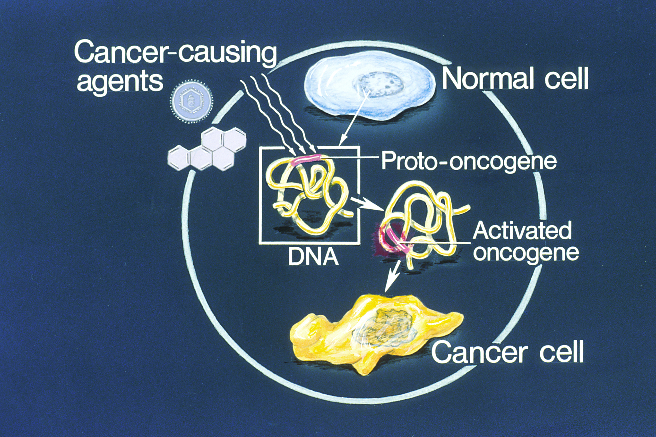
An oncogene is a gene that has the potential to cause cancer. In tumor cells, these genes are often mutated, or expressed at high levels.Kimball's Biology Pages. "Oncogenes" Free full text Most normal cells will undergo a programmed form of rapid cell death ( apoptosis) when critical functions are altered and malfunctioning. Activated oncogenes can cause those cells designated for apoptosis to survive and proliferate instead. Most oncogenes began as proto-oncogenes: normal genes involved in cell growth and proliferation or inhibition of apoptosis. If, through mutation, normal genes promoting cellular growth are up-regulated (gain-of-function mutation), they will predispose the cell to cancer; thus, t ... [...More Info...] [...Related Items...] OR: [Wikipedia] [Google] [Baidu] |


