|
GABAB Receptor Agonists
GABAB receptors (GABABR) are G-protein coupled receptors for gamma-aminobutyric acid (GABA), therefore making them metabotropic receptors, that are linked via G-proteins to potassium channels. The changing potassium concentrations hyperpolarize the cell at the end of an action potential. The reversal potential of the GABAB-mediated IPSP (inhibitory postsynaptic potential) is –100 mV, which is much more hyperpolarized than the GABAA IPSP. GABAB receptors are found in the central nervous system and the autonomic division of the peripheral nervous system. The receptors were first named in 1981 when their distribution in the CNS was determined, which was determined by Norman Bowery and his team using radioactively labelled baclofen. Functions GABABRs stimulate the opening of K+ channels, specifically GIRKs, which brings the neuron closer to the equilibrium potential of K+. This reduces the frequency of action potentials which reduces neurotransmitter release. Thus GABAB ... [...More Info...] [...Related Items...] OR: [Wikipedia] [Google] [Baidu] |
GABBR1
Gamma-aminobutyric acid B receptor, 1 (GABAB1), is a G-protein coupled receptor subunit encoded by the ''GABBR1'' gene. Function GABAB1 is a receptor for Gamma-aminobutyric acid. Upon binding, GABAB1 will produce a slow and prolonged inhibitory effect. GABAB1 is one part of a heterodimer, which is the GABAB receptor, consisting of it and the related GABAB2 protein. The GABA(B) receptor 1 gene is mapped to chromosome 6p21.3 within the HLA class I region close to the HLA-F gene. Susceptibility loci for multiple sclerosis, epilepsy, and schizophrenia have also been mapped in this region. Alternative splicing of this gene generates 4 transcript variants. Interactions GABBR1 has been shown to interact with ATF4 and GABBR2. See also * GABAB receptor GABAB receptors (GABABR) are G-protein coupled receptors for gamma-aminobutyric acid (GABA), therefore making them metabotropic receptors, that are linked via G-proteins to potassium channels. The changing potassium concen ... [...More Info...] [...Related Items...] OR: [Wikipedia] [Google] [Baidu] |
Action Potential
An action potential occurs when the membrane potential of a specific cell location rapidly rises and falls. This depolarization then causes adjacent locations to similarly depolarize. Action potentials occur in several types of animal cells, called excitable cells, which include neurons, muscle cells, and in some plant cells. Certain endocrine cells such as pancreatic beta cells, and certain cells of the anterior pituitary gland are also excitable cells. In neurons, action potentials play a central role in cell-cell communication by providing for—or with regard to saltatory conduction, assisting—the propagation of signals along the neuron's axon toward synaptic boutons situated at the ends of an axon; these signals can then connect with other neurons at synapses, or to motor cells or glands. In other types of cells, their main function is to activate intracellular processes. In muscle cells, for example, an action potential is the first step in the chain of events l ... [...More Info...] [...Related Items...] OR: [Wikipedia] [Google] [Baidu] |
Cryo-electron Microscopy
Cryogenic electron microscopy (cryo-EM) is a cryomicroscopy technique applied on samples cooled to cryogenic temperatures. For biological specimens, the structure is preserved by embedding in an environment of vitreous ice. An aqueous sample solution is applied to a grid-mesh and plunge-frozen in liquid ethane or a mixture of liquid ethane and propane. While development of the technique began in the 1970s, recent advances in detector technology and software algorithms have allowed for the determination of biomolecular structures at near-atomic resolution. This has attracted wide attention to the approach as an alternative to X-ray crystallography or NMR spectroscopy for macromolecular structure determination without the need for crystallization. In 2017, the Nobel Prize in Chemistry was awarded to Jacques Dubochet, Joachim Frank, and Richard Henderson "for developing cryo-electron microscopy for the high-resolution structure determination of biomolecules in solution." ''Nature ... [...More Info...] [...Related Items...] OR: [Wikipedia] [Google] [Baidu] |
Protein Isoform
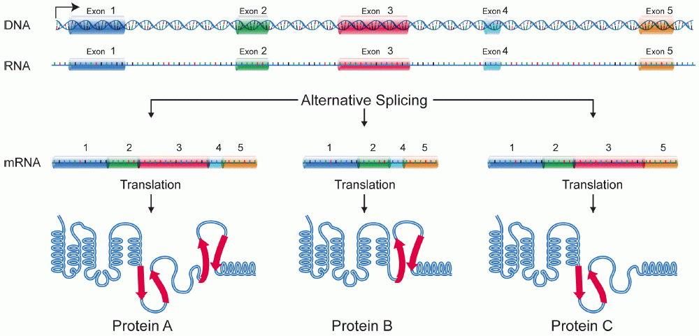
A protein isoform, or "protein variant", is a member of a set of highly similar proteins that originate from a single gene or gene family and are the result of genetic differences. While many perform the same or similar biological roles, some isoforms have unique functions. A set of protein isoforms may be formed from alternative splicings, variable promoter usage, or other post-transcriptional modifications of a single gene; post-translational modifications are generally not considered. (For that, see Proteoforms.) Through RNA splicing mechanisms, mRNA has the ability to select different protein-coding segments (exons) of a gene, or even different parts of exons from RNA to form different mRNA sequences. Each unique sequence produces a specific form of a protein. The discovery of isoforms could explain the discrepancy between the small number of protein coding regions genes revealed by the human genome project and the large diversity of proteins seen in an organism: different p ... [...More Info...] [...Related Items...] OR: [Wikipedia] [Google] [Baidu] |
C Terminus
The C-terminus (also known as the carboxyl-terminus, carboxy-terminus, C-terminal tail, C-terminal end, or COOH-terminus) is the end of an amino acid chain (protein or polypeptide), terminated by a free carboxyl group (-COOH). When the protein is translated from messenger RNA, it is created from N-terminus to C-terminus. The convention for writing peptide sequences is to put the C-terminal end on the right and write the sequence from N- to C-terminus. Chemistry Each amino acid has a carboxyl group and an amine group. Amino acids link to one another to form a chain by a dehydration reaction which joins the amine group of one amino acid to the carboxyl group of the next. Thus polypeptide chains have an end with an unbound carboxyl group, the C-terminus, and an end with an unbound amine group, the N-terminus. Proteins are naturally synthesized starting from the N-terminus and ending at the C-terminus. Function C-terminal retention signals While the N-terminus of a protein often conta ... [...More Info...] [...Related Items...] OR: [Wikipedia] [Google] [Baidu] |
Heterodimer
In biochemistry, a protein dimer is a macromolecular complex formed by two protein monomers, or single proteins, which are usually non-covalently bound. Many macromolecules, such as proteins or nucleic acids, form dimers. The word ''dimer'' has roots meaning "two parts", '' di-'' + '' -mer''. A protein dimer is a type of protein quaternary structure. A protein homodimer is formed by two identical proteins. A protein heterodimer is formed by two different proteins. Most protein dimers in biochemistry are not connected by covalent bonds. An example of a non-covalent heterodimer is the enzyme reverse transcriptase, which is composed of two different amino acid chains. An exception is dimers that are linked by disulfide bridges such as the homodimeric protein NEMO. Some proteins contain specialized domains to ensure dimerization (dimerization domains) and specificity. The G protein-coupled cannabinoid receptors have the ability to form both homo- and heterodimers with severa ... [...More Info...] [...Related Items...] OR: [Wikipedia] [Google] [Baidu] |
Metabotropic Glutamate Receptor
The metabotropic glutamate receptors, or mGluRs, are a type of glutamate receptor that are active through an indirect metabotropic process. They are members of the group C family of G-protein-coupled receptors, or GPCRs. Like all glutamate receptors, mGluRs bind with glutamate, an amino acid that functions as an excitatory neurotransmitter. Function and structure The mGluRs perform a variety of functions in the central and peripheral nervous systems: For example, they are involved in learning, memory, anxiety, and the perception of pain. They are found in pre- and postsynaptic neurons in synapses of the hippocampus, cerebellum, and the cerebral cortex, as well as other parts of the brain and in peripheral tissues. Like other metabotropic receptors, mGluRs have seven transmembrane domains that span the cell membrane. Unlike ionotropic receptors, metabotropic glutamate receptors are not ion channels. Instead, they activate biochemical cascades, leading to the modification ... [...More Info...] [...Related Items...] OR: [Wikipedia] [Google] [Baidu] |
Gamma-Hydroxybutyric Acid
''gamma''-Hydroxybutyric acid (or γ-hydroxybutyric acid (GHB), also known as 4-hydroxybutanoic acid) is a naturally occurring neurotransmitter and a depressant drug. It is a precursor to GABA, glutamate, and glycine in certain brain areas. It acts on the GHB receptor and is a weak agonist at the GABAB receptor. GHB has been used in the medical setting as a general anesthetic and as treatment for cataplexy, narcolepsy, and alcoholism. It is also used illegally as an intoxicant, as an athletic-performance enhancer, as a date-rape drug, and as a recreational drug. It is commonly used in the form of a salt, such as sodium γ-hydroxybutyrate (NaGHB, sodium oxybate, or Xyrem) or potassium γ-hydroxybutyrate (KGHB, potassium oxybate). GHB is also produced as a result of fermentation, and is found in small quantities in some beers and wines, beef, and small citrus fruits. Succinic semialdehyde dehydrogenase deficiency is a disease that causes GHB to accumulate in the blood ... [...More Info...] [...Related Items...] OR: [Wikipedia] [Google] [Baidu] |
Ethanol
Ethanol (abbr. EtOH; also called ethyl alcohol, grain alcohol, drinking alcohol, or simply alcohol) is an organic compound. It is an alcohol with the chemical formula . Its formula can be also written as or (an ethyl group linked to a hydroxyl group). Ethanol is a volatile, flammable, colorless liquid with a characteristic wine-like odor and pungent taste. It is a psychoactive recreational drug, the active ingredient in alcoholic drinks. Ethanol is naturally produced by the fermentation process of sugars by yeasts or via petrochemical processes such as ethylene hydration. It has medical applications as an antiseptic and disinfectant. It is used as a chemical solvent and in the synthesis of organic compounds, and as a fuel source. Ethanol also can be dehydrated to make ethylene, an important chemical feedstock. As of 2006, world production of ethanol was , coming mostly from Brazil and the U.S. Etymology ''Ethanol'' is the systematic name defined by the ... [...More Info...] [...Related Items...] OR: [Wikipedia] [Google] [Baidu] |
GiG0 Alpha Subunits
Gi protein alpha subunit is a family of heterotrimeric G protein alpha subunits. This family is also commonly called the Gi/o (Gi /Go ) family or Gi/o/z/t family to include closely related family members. G alpha subunits may be referred to as Gi alpha, Gαi, or Giα. Family members There are four distinct subtypes of alpha subunits in the Gi/o/z/t alpha subunit family that define four families of heterotrimeric G proteins: * Gi proteins: Gi1α, Gi2α, and Gi3α * Go protein: Goα (in mouse there is alternative splicing to generate Go1α and Go2α) * Gz protein: Gzα * Transducins (Gt proteins): Gt1α, Gt2α, Gt3α Giα proteins Gi1α Gi1α is encoded by the gene GNAI1. Gi2α Gi2α is encoded by the gene GNAI2. Gi3α Gi3α is encoded by the gene GNAI3. Goα protein Go1α is encoded by the gene GNAO1. Gzα protein Gzα is encoded by the gene GNAZ. Transducin proteins Gt1α Transducin/Gt1α is encoded by the gene GNAT1. Gt2α Transducin 2/Gt2 ... [...More Info...] [...Related Items...] OR: [Wikipedia] [Google] [Baidu] |


