|
Gymnopilus Luteofolius
''Gymnopilus luteofolius'', known as the yellow-gilled gymnopilus, is a large and widely distributed mushroom that grows in dense clusters on dead hardwoods and conifers. It grows in late July to November in the east and in the winter on the west coast of North America. It has a rusty orange spore print and a bitter taste. Systematics ''Gymnopilus luteofolius'' was first described as ''Agaricus luteofolius'' by Charles Horton Peck in 1875. It was renamed ''Pholiota luteofolius'' by Pier Andrea Saccardo in 1887, and was given its current name by mycologist Rolf Singer in 1951. Description The fruit bodies of ''Gymnopilus luteofolius'' have reddish to purplish to yellow caps in diameter, which often develop green stains. This cap surface is covered with fasciculate scales that start out purplish, soon fade to brick red, and finally fades to yellow as the mushroom matures. The context is reddish to light lavender, fading to yellowish as the mushroom matures. The gills have a ... [...More Info...] [...Related Items...] OR: [Wikipedia] [Google] [Baidu] |
Fungi
A fungus ( : fungi or funguses) is any member of the group of eukaryotic organisms that includes microorganisms such as yeasts and molds, as well as the more familiar mushrooms. These organisms are classified as a kingdom, separately from the other eukaryotic kingdoms, which by one traditional classification include Plantae, Animalia, Protozoa, and Chromista. A characteristic that places fungi in a different kingdom from plants, bacteria, and some protists is chitin in their cell walls. Fungi, like animals, are heterotrophs; they acquire their food by absorbing dissolved molecules, typically by secreting digestive enzymes into their environment. Fungi do not photosynthesize. Growth is their means of mobility, except for spores (a few of which are flagellated), which may travel through the air or water. Fungi are the principal decomposers in ecological systems. These and other differences place fungi in a single group of related organisms, named the ''Eumycota'' (''t ... [...More Info...] [...Related Items...] OR: [Wikipedia] [Google] [Baidu] |
Stipe (mycology)
In mycology, a stipe () is the stem or stalk-like feature supporting the cap of a mushroom. Like all tissues of the mushroom other than the hymenium, the stipe is composed of sterile hyphal tissue. In many instances, however, the fertile hymenium extends down the stipe some distance. Fungi that have stipes are said to be stipitate. The evolutionary benefit of a stipe is generally considered to be in mediating spore dispersal. An elevated mushroom will more easily release its spores into wind currents or onto passing animals. Nevertheless, many mushrooms do not have stipes, including cup fungi, puffballs, earthstars, some polypores, jelly fungi, ergots, and smuts. It is often the case that features of the stipe are required to make a positive identification of a mushroom. Such distinguishing characters include: # the texture of the stipe (fibrous, brittle, chalky, leathery, firm, etc.) # whether it has remains of a partial veil (such as an annulus or cortina) or universal ve ... [...More Info...] [...Related Items...] OR: [Wikipedia] [Google] [Baidu] |
Cystidia
A cystidium (plural cystidia) is a relatively large cell found on the sporocarp of a basidiomycete (for example, on the surface of a mushroom gill), often between clusters of basidia. Since cystidia have highly varied and distinct shapes that are often unique to a particular species or genus, they are a useful micromorphological characteristic in the identification of basidiomycetes. In general, the adaptive significance of cystidia is not well understood. Classification of cystidia By position Cystidia may occur on the edge of a lamella (or analogous hymenophoral structure) (cheilocystidia), on the face of a lamella (pleurocystidia), on the surface of the cap (dermatocystidia or pileocystidia), on the margin of the cap (circumcystidia) or on the stipe (caulocystidia). Especially the pleurocystidia and cheilocystidia are important for identification within many genera. Sometimes the cheilocystidia give the gill edge a distinct colour which is visible to the naked eye or wit ... [...More Info...] [...Related Items...] OR: [Wikipedia] [Google] [Baidu] |
Ventricose
Ventricose is an adjective describing the condition of a mushroom, gastropod or plant that it is "swollen, distended, or inflated especially on one side". Mycology In mycology, ventricose is a condition in which the cystidia, lamella or stipe of a mushroom A mushroom or toadstool is the fleshy, spore-bearing fruiting body of a fungus, typically produced above ground, on soil, or on its food source. ''Toadstool'' generally denotes one poisonous to humans. The standard for the name "mushroom" is t ... is swollen in the middle. Gastropods In gastropods, if the shell of a snail is ventricose or subventricose, it means the whorl of the shell is swollen. References Mycology Fungal morphology and anatomy {{Basidiomycota-stub ... [...More Info...] [...Related Items...] OR: [Wikipedia] [Google] [Baidu] |
Cylindrical
A cylinder (from ) has traditionally been a three-dimensional solid, one of the most basic of curvilinear geometric shapes. In elementary geometry, it is considered a prism with a circle as its base. A cylinder may also be defined as an infinite curvilinear surface in various modern branches of geometry and topology. The shift in the basic meaning—solid versus surface (as in ball and sphere)—has created some ambiguity with terminology. The two concepts may be distinguished by referring to solid cylinders and cylindrical surfaces. In the literature the unadorned term cylinder could refer to either of these or to an even more specialized object, the ''right circular cylinder''. Types The definitions and results in this section are taken from the 1913 text ''Plane and Solid Geometry'' by George Wentworth and David Eugene Smith . A ' is a surface consisting of all the points on all the lines which are parallel to a given line and which pass through a fixed plane curve in a pla ... [...More Info...] [...Related Items...] OR: [Wikipedia] [Google] [Baidu] |
Cap Cuticle
The pileipellis is the uppermost layer of hyphae in the pileus of a fungal fruit body. It covers the trama, the fleshy tissue of the fruit body. The pileipellis is more or less synonymous with the cuticle, but the cuticle generally describes this layer as a macroscopic feature, while pileipellis refers to this structure as a microscopic layer. Pileipellis type is an important character in the identification of fungi. Pileipellis types include the cutis, trichoderm, epithelium, and hymeniderm types. Types Cutis A cutis is a type of pileipellis characterized by hyphae that are repent, that is, that run parallel to the pileus surface. In an ixocutis, the hyphae are gelatinous. Trichoderm In a trichoderm, the outermost hyphae emerge roughly parallel, like hairs, perpendicular to the cap surface. The prefix "tricho-" comes from a Greek word for "hair". In an ixotrichodermium, the outermost hyphae are gelatinous. Epithelium An epithelium is a pileipellis consisting of rounded ce ... [...More Info...] [...Related Items...] OR: [Wikipedia] [Google] [Baidu] |
Biological Pigment
Biological pigments, also known simply as pigments or biochromes, are substances produced by living organisms that have a color resulting from selective color absorption. Biological pigments include plant pigments and flower pigments. Many biological structures, such as skin, eyes, feathers, fur and hair contain pigments such as melanin in specialized cells called chromatophores. In some species, pigments accrue over very long periods during an individual's lifespan. Pigment color differs from structural color in that it is the same for all viewing angles, whereas structural color is the result of selective reflection or iridescence, usually because of multilayer structures. For example, butterfly wings typically contain structural color, although many butterflies have cells that contain pigment as well. Biological pigments See conjugated systems for electron bond chemistry that causes these molecules to have pigment. * Heme/porphyrin-based: chlorophyll, bilirubin, hemocy ... [...More Info...] [...Related Items...] OR: [Wikipedia] [Google] [Baidu] |
Septate
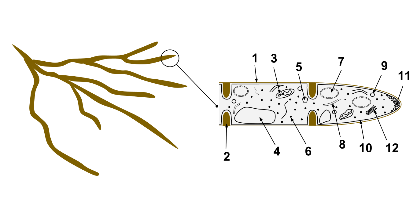
In biology, a septum (Latin for ''something that encloses''; plural septa) is a wall, dividing a cavity or structure into smaller ones. A cavity or structure divided in this way may be referred to as septate. Examples Human anatomy * Interatrial septum, the wall of tissue that is a sectional part of the left and right atria of the heart * Interventricular septum, the wall separating the left and right ventricles of the heart * Lingual septum, a vertical layer of fibrous tissue that separates the halves of the tongue. *Nasal septum: the cartilage wall separating the nostrils of the nose * Alveolar septum: the thin wall which separates the alveoli from each other in the lungs * Orbital septum, a palpebral ligament in the upper and lower eyelids * Septum pellucidum or septum lucidum, a thin structure separating two fluid pockets in the brain * Uterine septum, a malformation of the uterus * Vaginal septum, a lateral or transverse partition inside the vagina * Intermuscular sep ... [...More Info...] [...Related Items...] OR: [Wikipedia] [Google] [Baidu] |
Trama (mycology)
In mycology, the term trama is used in two ways. In the broad sense, it is the inner, fleshy portion of a mushroom's basidiocarp, or fruit body. It is distinct from the outer layer of tissue, known as the pileipellis or cuticle, and from the spore-bearing tissue layer known as the hymenium. In essence, the trama is the tissue that is commonly referred to as the "flesh" of mushrooms and similar fungi.Largent D, Johnson D, Watling R. 1977. ''How to Identify Mushrooms to Genus III: Microscopic Features''. Arcata, CA: Mad River Press. . pp. 60–70. The second use is more specific, and refers to the "hymenophoral trama" that supports the hymenium. It is similarly interior, connective tissue, but it is more specifically the central layer of hyphae running from the underside of the mushroom cap to the lamella or gill, upon which the hymenium rests. Various types have been classified by their structure, including trametoid, cantharelloid, boletoid, and agaricoid, with agaricoid the ... [...More Info...] [...Related Items...] OR: [Wikipedia] [Google] [Baidu] |
Capitate
The capitate bone is a bone in the human wrist found in the center of the carpal bone region, located at the distal end of the radius and ulna bones. It articulates with the third metacarpal bone (the middle finger) and forms the third carpometacarpal joint. The capitate bone is the largest of the carpal bones in the human hand. It presents, above, a rounded portion or head, which is received into the concavity formed by the scaphoid and lunate bones; a constricted portion or neck; and below this, the body.''Gray's Anatomy'' (1918). See infobox. The bone is also found in many other mammals, and is homologous with the "third distal carpal" of reptiles and amphibians. Structure The capitate is the largest carpal bone found within the hand. The capitate is found within the distal row of carpal bones. The capitate lies directly adjacent to the metacarpal of the ring finger on its distal surface, has the hamate on its ulnar surface and trapezoid on its radial surface, and abuts the ... [...More Info...] [...Related Items...] OR: [Wikipedia] [Google] [Baidu] |
Cystidia
A cystidium (plural cystidia) is a relatively large cell found on the sporocarp of a basidiomycete (for example, on the surface of a mushroom gill), often between clusters of basidia. Since cystidia have highly varied and distinct shapes that are often unique to a particular species or genus, they are a useful micromorphological characteristic in the identification of basidiomycetes. In general, the adaptive significance of cystidia is not well understood. Classification of cystidia By position Cystidia may occur on the edge of a lamella (or analogous hymenophoral structure) (cheilocystidia), on the face of a lamella (pleurocystidia), on the surface of the cap (dermatocystidia or pileocystidia), on the margin of the cap (circumcystidia) or on the stipe (caulocystidia). Especially the pleurocystidia and cheilocystidia are important for identification within many genera. Sometimes the cheilocystidia give the gill edge a distinct colour which is visible to the naked eye or wit ... [...More Info...] [...Related Items...] OR: [Wikipedia] [Google] [Baidu] |
Basidium
A basidium () is a microscopic sporangium (a spore-producing structure) found on the hymenophore of fruiting bodies of basidiomycete fungi which are also called tertiary mycelium, developed from secondary mycelium. Tertiary mycelium is highly-coiled secondary myceliuma dikaryon. The presence of basidia is one of the main characteristic features of the Basidiomycota. A basidium usually bears four sexual spores called basidiospores; occasionally the number may be two or even eight. In a typical basidium, each basidiospore is borne at the tip of a narrow prong or horn called a sterigma (), and is forcibly discharged upon maturity. The word ''basidium'' literally means "little pedestal", from the way in which the basidium supports the spores. However, some biologists suggest that the structure more closely resembles a club. An immature basidium is known as a basidiole. Structure Most basidiomycota have single celled basidia (holobasidia), but in some groups basidia can be multicel ... [...More Info...] [...Related Items...] OR: [Wikipedia] [Google] [Baidu] |


