|
Glandular Metaplasia
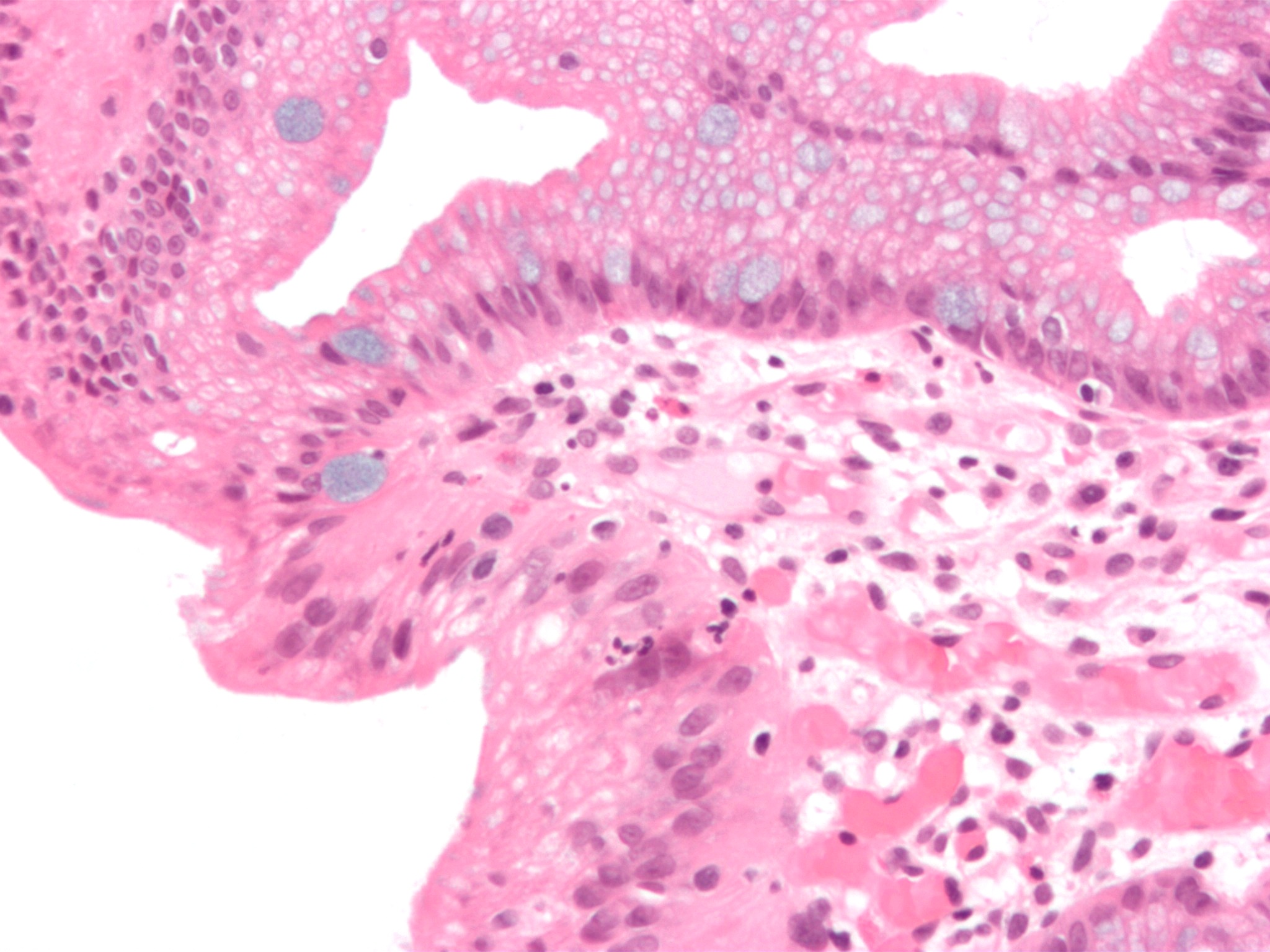
Glandular metaplasia is a type of metaplasia where irritated tissue converts to a glandular form. An example occurs in the esophagus, where tissue becomes more similar to the tissue of the stomach. Another example occurs in the urinary bladder. See also *Intestinal metaplasia *Squamous metaplasia Additional images Image:Barretts esophagus alcian blue high mag.jpg, Micrograph of Barrett's esophagus Barrett's esophagus is a condition in which there is an abnormal (metaplastic) change in the mucosal cells lining the lower portion of the esophagus, from stratified squamous epithelium to simple columnar epithelium with interspersed goblet cells ..., an example of glandular metaplasia. References {{Pathology Histopathology ... [...More Info...] [...Related Items...] OR: [Wikipedia] [Google] [Baidu] |
Barretts Alcian Blue
Barret, Barrett, or Barretts may refer to: People * Barrett (name), including a list of people with the surname * Barrett Brown (born 1981), American journalist and activist) * Barrett Foa, American actor Court cases * ''Barrett v. Rosenthal'', a 2006 California Supreme Court case concerning online defamation * ''Barrett v. United States'', an 1898 Supreme Court case regarding subdivision of South Carolina into judicial districts Fictional characters *Brenda Barrett, a character on the daytime soap opera ''General Hospital'' * Dana Barrett, a character in the films ''Ghostbusters'' and ''Ghostbusters II'', played by Sigourney Weaver *Elcid Barrett, captain of the ''Antelope'' in the folk song " Barrett's Privateers" *Betty Barrett, a character on the TV show '' Atomic Betty'' *Oliver Barrett, a character in the book ''Love Story'' and its film and musical adaptations *Barret Wallace, a character in the video game ''Final Fantasy VII'' Organizations * Barrett, The Honors College ... [...More Info...] [...Related Items...] OR: [Wikipedia] [Google] [Baidu] |
Metaplasia
Metaplasia ( gr, "change in form") is the transformation of one differentiated cell type to another differentiated cell type. The change from one type of cell to another may be part of a normal maturation process, or caused by some sort of abnormal stimulus. In simplistic terms, it is as if the original cells are not robust enough to withstand their environment, so they transform into another cell type better suited to their environment. If the stimulus causing metaplasia is removed or ceases, tissues return to their normal pattern of differentiation. Metaplasia is not synonymous with dysplasia, and is not considered to be an actual cancer. It is also contrasted with heteroplasia, which is the spontaneous abnormal growth of cytologic and histologic elements. Today, metaplastic changes are usually considered to be an early phase of carcinogenesis, specifically for those with a history of cancers or who are known to be susceptible to carcinogenic changes. Metaplastic change is t ... [...More Info...] [...Related Items...] OR: [Wikipedia] [Google] [Baidu] |
Esophagus
The esophagus (American English) or oesophagus (British English; both ), non-technically known also as the food pipe or gullet, is an organ in vertebrates through which food passes, aided by peristaltic contractions, from the pharynx to the stomach. The esophagus is a fibromuscular tube, about long in adults, that travels behind the trachea and heart, passes through the diaphragm, and empties into the uppermost region of the stomach. During swallowing, the epiglottis tilts backwards to prevent food from going down the larynx and lungs. The word ''oesophagus'' is from Ancient Greek οἰσοφάγος (oisophágos), from οἴσω (oísō), future form of φέρω (phérō, “I carry”) + ἔφαγον (éphagon, “I ate”). The wall of the esophagus from the lumen outwards consists of mucosa, submucosa (connective tissue), layers of muscle fibers between layers of fibrous tissue, and an outer layer of connective tissue. The mucosa is a stratified squamous epithel ... [...More Info...] [...Related Items...] OR: [Wikipedia] [Google] [Baidu] |
Stomach
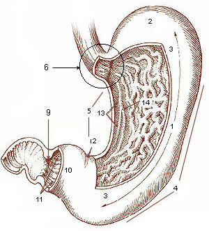
The stomach is a muscular, hollow organ in the gastrointestinal tract of humans and many other animals, including several invertebrates. The stomach has a dilated structure and functions as a vital organ in the digestive system. The stomach is involved in the gastric phase of digestion, following chewing. It performs a chemical breakdown by means of enzymes and hydrochloric acid. In humans and many other animals, the stomach is located between the oesophagus and the small intestine. The stomach secretes digestive enzymes and gastric acid to aid in food digestion. The pyloric sphincter controls the passage of partially digested food ( chyme) from the stomach into the duodenum, where peristalsis takes over to move this through the rest of intestines. Structure In the human digestive system, the stomach lies between the oesophagus and the duodenum (the first part of the small intestine). It is in the left upper quadrant of the abdominal cavity. The top of the stomach lies ag ... [...More Info...] [...Related Items...] OR: [Wikipedia] [Google] [Baidu] |
Urinary Bladder
The urinary bladder, or simply bladder, is a hollow organ in humans and other vertebrates that stores urine from the kidneys before disposal by urination. In humans the bladder is a distensible organ that sits on the pelvic floor. Urine enters the bladder via the ureters and exits via the urethra. The typical adult human bladder will hold between 300 and (10.14 and ) before the urge to empty occurs, but can hold considerably more. The Latin phrase for "urinary bladder" is ''vesica urinaria'', and the term ''vesical'' or prefix ''vesico -'' appear in connection with associated structures such as vesical veins. The modern Latin word for "bladder" – ''cystis'' – appears in associated terms such as cystitis (inflammation of the bladder). Structure In humans, the bladder is a hollow muscular organ situated at the base of the pelvis. In gross anatomy, the bladder can be divided into a broad , a body, an apex, and a neck. The apex (also called the vertex) is directed forward ... [...More Info...] [...Related Items...] OR: [Wikipedia] [Google] [Baidu] |
Intestinal Metaplasia
Intestinal metaplasia is the transformation (metaplasia) of epithelium (usually of the stomach or the esophagus) into a type of epithelium resembling that found in the intestine. In the esophagus, this is called Barrett's esophagus. Chronic inflammation caused by ''H. pylori'' infection in the stomach and GERD in the esophagus are seen as the primary instigators of metaplasia and subsequent adenocarcinoma formation. Initially, the transformed epithelium resembles the small intestine lining; in the later stages it resembles the lining of the colon. It is characterized by the appearance of goblet cells and expression of intestinal cell markers such as the transcription factor, CDX2. Risk factors Although it was originally reported that people of East Asian ethnicity with gastric intestinal metaplasia are at increased risk of stomach cancer Stomach cancer, also known as gastric cancer, is a cancer that develops from the lining of the stomach. Most cases of stomach cancers are gas ... [...More Info...] [...Related Items...] OR: [Wikipedia] [Google] [Baidu] |
Squamous Metaplasia
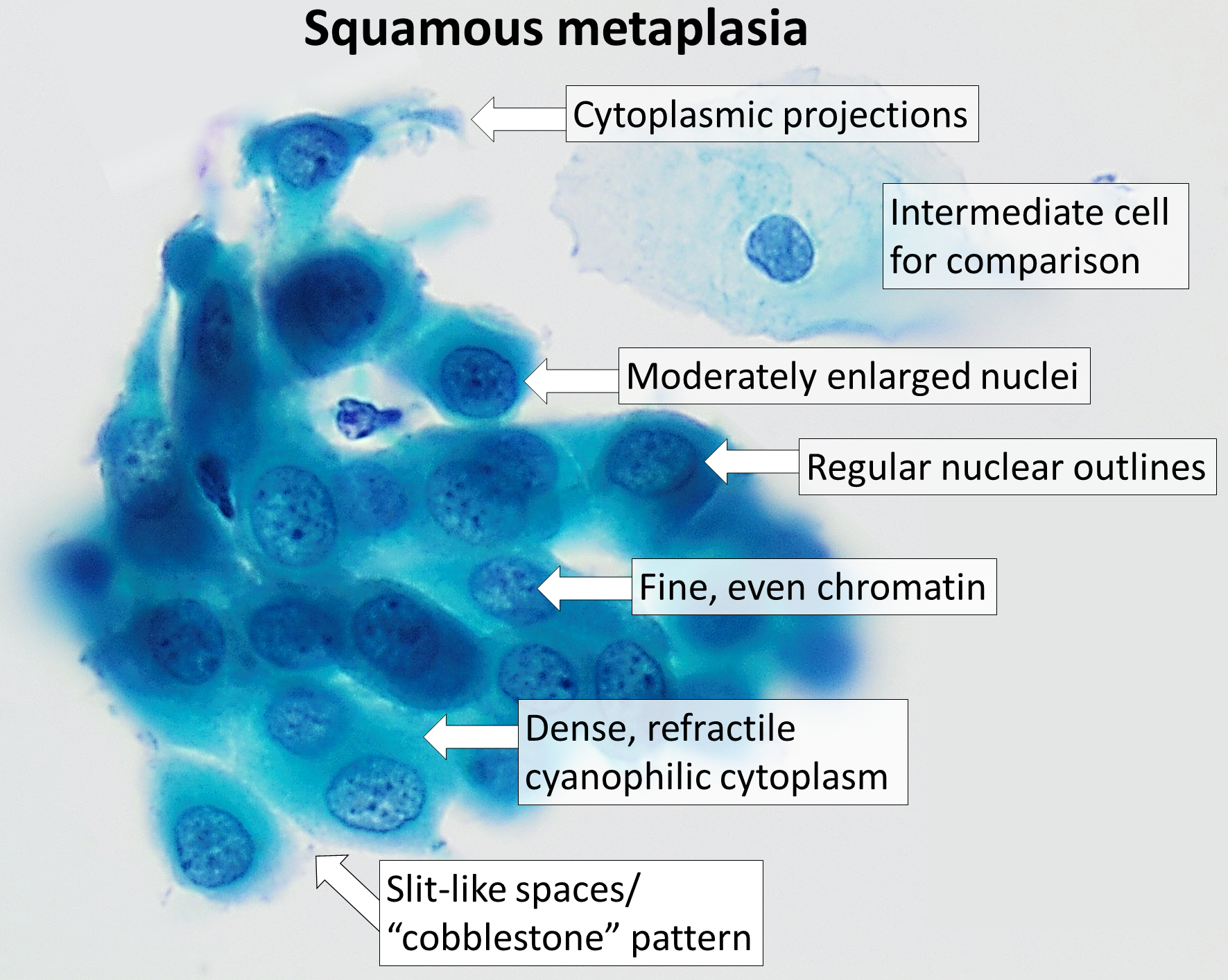
Squamous metaplasia is a benign non-cancerous change (metaplasia) of surfacing lining cells (epithelium) to a squamous morphology. Location Common sites for squamous metaplasia include the bladder and cervix. Smokers often exhibit squamous metaplasia in the linings of their airways. These changes don't signify a specific disease, but rather usually represent the body's response to stress or irritation. Vitamin A deficiency or overdose can also lead to squamous metaplasia. Uterine cervix In regard to the cervix, squamous metaplasia can sometimes be found in the endocervix, as it is composed of simple columnar epithelium, whereas the ectocervix is composed of stratified squamous non-keratinized epithelium.Kumar, Vinay; Abbas, Abul K.; Fausto, Nelson; & Mitchell, Richard N. (2007) ''Robbins Basic Pathology'' (8th ed.). Saunders Elsevier. pp. 716-720 Significance Squamous metaplasia may be seen in the context of benign lesions (e.g., atypical polypoid adenomyoma), chronic irri ... [...More Info...] [...Related Items...] OR: [Wikipedia] [Google] [Baidu] |
Micrograph
A micrograph or photomicrograph is a photograph or digital image taken through a microscope or similar device to show a magnified image of an object. This is opposed to a macrograph or photomacrograph, an image which is also taken on a microscope but is only slightly magnified, usually less than 10 times. Micrography is the practice or art of using microscopes to make photographs. A micrograph contains extensive details of microstructure. A wealth of information can be obtained from a simple micrograph like behavior of the material under different conditions, the phases found in the system, failure analysis, grain size estimation, elemental analysis and so on. Micrographs are widely used in all fields of microscopy. Types Photomicrograph A light micrograph or photomicrograph is a micrograph prepared using an optical microscope, a process referred to as ''photomicroscopy''. At a basic level, photomicroscopy may be performed simply by connecting a camera to a microscope, th ... [...More Info...] [...Related Items...] OR: [Wikipedia] [Google] [Baidu] |
Barrett's Esophagus
Barrett's esophagus is a condition in which there is an abnormal (metaplastic) change in the mucosal cells lining the lower portion of the esophagus, from stratified squamous epithelium to simple columnar epithelium with interspersed goblet cells that are normally present only in the small intestine and large intestine. This change is considered to be a premalignant condition because it is associated with a high incidence of further transition to esophageal adenocarcinoma, an often-deadly cancer. The main cause of Barrett's esophagus is thought to be an adaptation to chronic acid exposure from reflux esophagitis. Barrett's esophagus is diagnosed by endoscopy: observing the characteristic appearance of this condition by direct inspection of the lower esophagus; followed by microscopic examination of tissue from the affected area obtained from biopsy. The cells of Barrett's esophagus are classified into four categories: nondysplastic, low-grade dysplasia, high-grade dysplasia, and fr ... [...More Info...] [...Related Items...] OR: [Wikipedia] [Google] [Baidu] |


