|
Full Blood Count
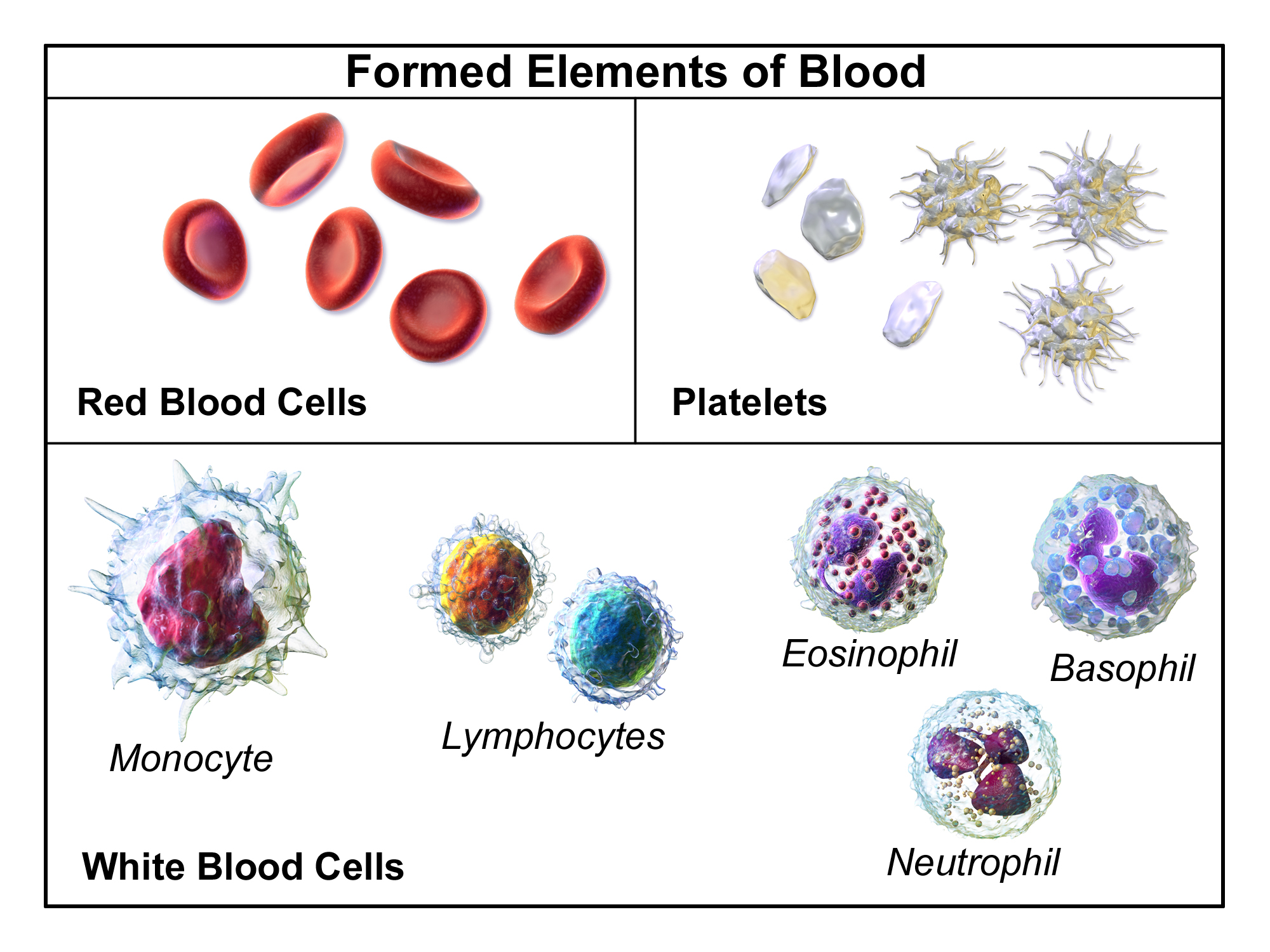
A complete blood count (CBC), also known as a full blood count (FBC), is a set of medical laboratory tests that provide information about the cells in a person's blood. The CBC indicates the counts of white blood cells, red blood cells and platelets, the concentration of hemoglobin, and the hematocrit (the volume percentage of red blood cells). The red blood cell indices, which indicate the average size and hemoglobin content of red blood cells, are also reported, and a white blood cell differential, which counts the different types of white blood cells, may be included. The CBC is often carried out as part of a medical assessment and can be used to monitor health or diagnose diseases. The results are interpreted by comparing them to reference ranges, which vary with sex and age. Conditions like anemia and thrombocytopenia are defined by abnormal complete blood count results. The red blood cell indices can provide information about the cause of a person's anemia such as iron d ... [...More Info...] [...Related Items...] OR: [Wikipedia] [Google] [Baidu] |
Cancer Research UK
Cancer Research UK (CRUK) is the world's largest independent cancer research organization. It is registered as a charity in the United Kingdom and Isle of Man, and was formed on 4 February 2002 by the merger of The Cancer Research Campaign and the Imperial Cancer Research Fund. Cancer Research UK conducts research using both its own staff and grant-funded researchers. It also provides information about cancer and runs campaigns aimed at raising awareness and influencing public policy. The organisation's work is almost entirely funded by the public. It raises money through donations, legacies, community fundraising, events, retail and corporate partnerships. Over 40,000 people are regular volunteers. History The Imperial Cancer Research Fund (ICRF) was founded in 1902 as the Cancer Research Fund, changing its name to the Imperial Cancer Research Fund in 1904. It grew over the next twenty years to become one of the world's leading cancer research charities. Its flagship laborato ... [...More Info...] [...Related Items...] OR: [Wikipedia] [Google] [Baidu] |
Bacterial Infection
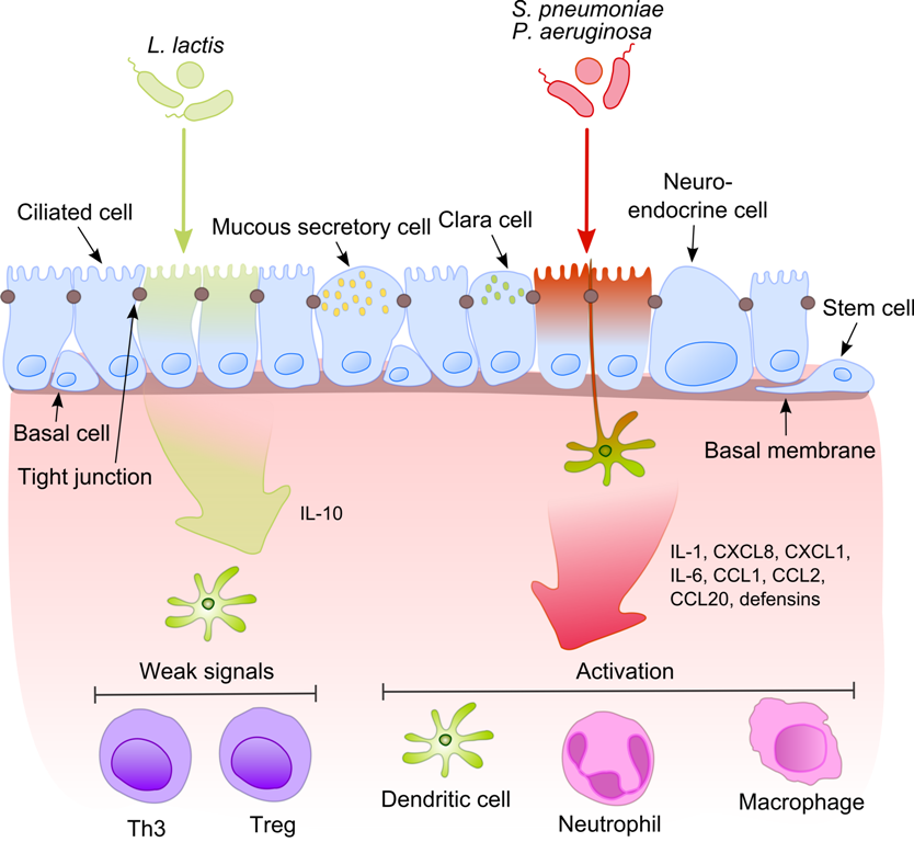
Pathogenic bacteria are bacteria that can cause disease. This article focuses on the bacteria that are pathogenic to humans. Most species of bacteria are harmless and are often beneficial but others can cause infectious diseases. The number of these pathogenic species in humans is estimated to be fewer than a hundred. By contrast, several thousand species are part of the gut flora present in the digestive tract. The body is continually exposed to many species of bacteria, including beneficial commensals, which grow on the skin and mucous membranes, and saprophytes, which grow mainly in the soil and in decaying matter. The blood and tissue fluids contain nutrients sufficient to sustain the growth of many bacteria. The body has defence mechanisms that enable it to resist microbial invasion of its tissues and give it a natural immunity or innate resistance against many microorganisms. Pathogenic bacteria are specially adapted and endowed with mechanisms for overcoming the ... [...More Info...] [...Related Items...] OR: [Wikipedia] [Google] [Baidu] |
Paul Ehrlich
Paul Ehrlich (; 14 March 1854 – 20 August 1915) was a Nobel Prize-winning German physician and scientist who worked in the fields of hematology, immunology, and antimicrobial chemotherapy. Among his foremost achievements were finding a cure for syphilis in 1909 and inventing the precursor technique to Gram staining bacteria. The methods he developed for staining tissue made it possible to distinguish between different types of blood cells, which led to the ability to diagnose numerous blood diseases. His laboratory discovered arsphenamine (Salvarsan), the first effective medicinal treatment for syphilis, thereby initiating and also naming the concept of chemotherapy. Ehrlich popularized the concept of a magic bullet. He also made a decisive contribution to the development of an antiserum to combat diphtheria and conceived a method for standardizing therapeutic serums. In 1908, he received the Nobel Prize in Physiology or Medicine for his contributions to immunology. ... [...More Info...] [...Related Items...] OR: [Wikipedia] [Google] [Baidu] |
Louis-Charles Malassez
Louis-Charles Malassez (21 September 1842 – 22 December 1909) was a French anatomist and histologist born in Nevers, department of Nièvre. He studied medicine in Paris, where he worked as an ''interne'' from 1867. He served with the 5th Ambulance Corps during the Franco-Prussian War, afterwards returning to Paris, where he worked with distinguished physicians that included Claude Bernard, Jean-Martin Charcot and Pierre Potain. In 1875, he attained the chair of anatomy at Collège de France, and in 1894 he became a member of the '' Académie de Médecine''. He conducted histological research of the blood, and is credited for design of the hemocytometer, a device used to quantitatively measure blood cells. In the field of dentistry, he described residual cells of the epithelial root sheath in the periodontal ligament. These remaining cells are referred to as epithelial cell rests of Malassez (ERM). A genus of fungi called '' Malassezia'' bears his name. The species in the ... [...More Info...] [...Related Items...] OR: [Wikipedia] [Google] [Baidu] |
Karl Vierordt
Karl von Vierordt (July 1, 1818 – November 22, 1884) was a German physiologist. Vierordt was born in Lahr, Baden. He studied at the universities of Berlin, Göttingen, Vienna, and Heidelberg, and began a practice in Karlsruhe in 1842. In 1849 he became a professor of theoretical medicine at the University of Tübingen, and in 1853 a professor of physiology. Vierordt developed techniques and tools for the monitoring of blood circulation. He is credited with the construction of an early "hemotachometer", an apparatus for monitoring the velocity of blood flow. In 1854, he created a device called a sphygmograph, a mechanism consisting of weights and levers used to estimate blood pressure, and considered to be a forerunner of the modern sphygmomanometer. One of his better known written works was a treatise on the arterial pulse, titled ''Die Lehre vom Arterienpuls in gesunden und kranken Zuständen''. Vierordt also made substantial contributions to the psychology of time per ... [...More Info...] [...Related Items...] OR: [Wikipedia] [Google] [Baidu] |
Hemocytometer
The hemocytometer (or haemocytometer) is a counting-chamber device originally designed and usually used for counting blood cells. The hemocytometer was invented by Louis-Charles Malassez and consists of a thick glass microscope slide with a rectangular indentation that creates a precision volume chamber. This chamber is engraved with a laser-etched grid of perpendicular lines. The device is carefully crafted so that the area bounded by the lines is known, and the depth of the chamber is also known. By observing a defined area of the grid, it is therefore possible to count the number of cells or particles in a specific volume of fluid, and thereby calculate the concentration of cells in the fluid overall. A well used type of hemocytometer is the ''Neubauer'' counting chamber. Other types of hemocytometers with different rulings are in use for different applications. Fuchs-Rosenthal rulings, commonly used for spinal fluid counting, Howard Mold rulings used for mold on food and ... [...More Info...] [...Related Items...] OR: [Wikipedia] [Google] [Baidu] |
Centrifuge
A centrifuge is a device that uses centrifugal force to separate various components of a fluid. This is achieved by spinning the fluid at high speed within a container, thereby separating fluids of different densities (e.g. cream from milk) or liquids from solids. It works by causing denser substances and particles to move outward in the radial direction. At the same time, objects that are less dense are displaced and moved to the centre. In a laboratory centrifuge that uses sample tubes, the radial acceleration causes denser particles to settle to the bottom of the tube, while low-density substances rise to the top. A centrifuge can be a very effective filter that separates contaminants from the main body of fluid. Industrial scale centrifuges are commonly used in manufacturing and waste processing to sediment suspended solids, or to separate immiscible liquids. An example is the cream separator found in dairies. Very high speed centrifuges and ultracentrifuges able to prov ... [...More Info...] [...Related Items...] OR: [Wikipedia] [Google] [Baidu] |
Microscope
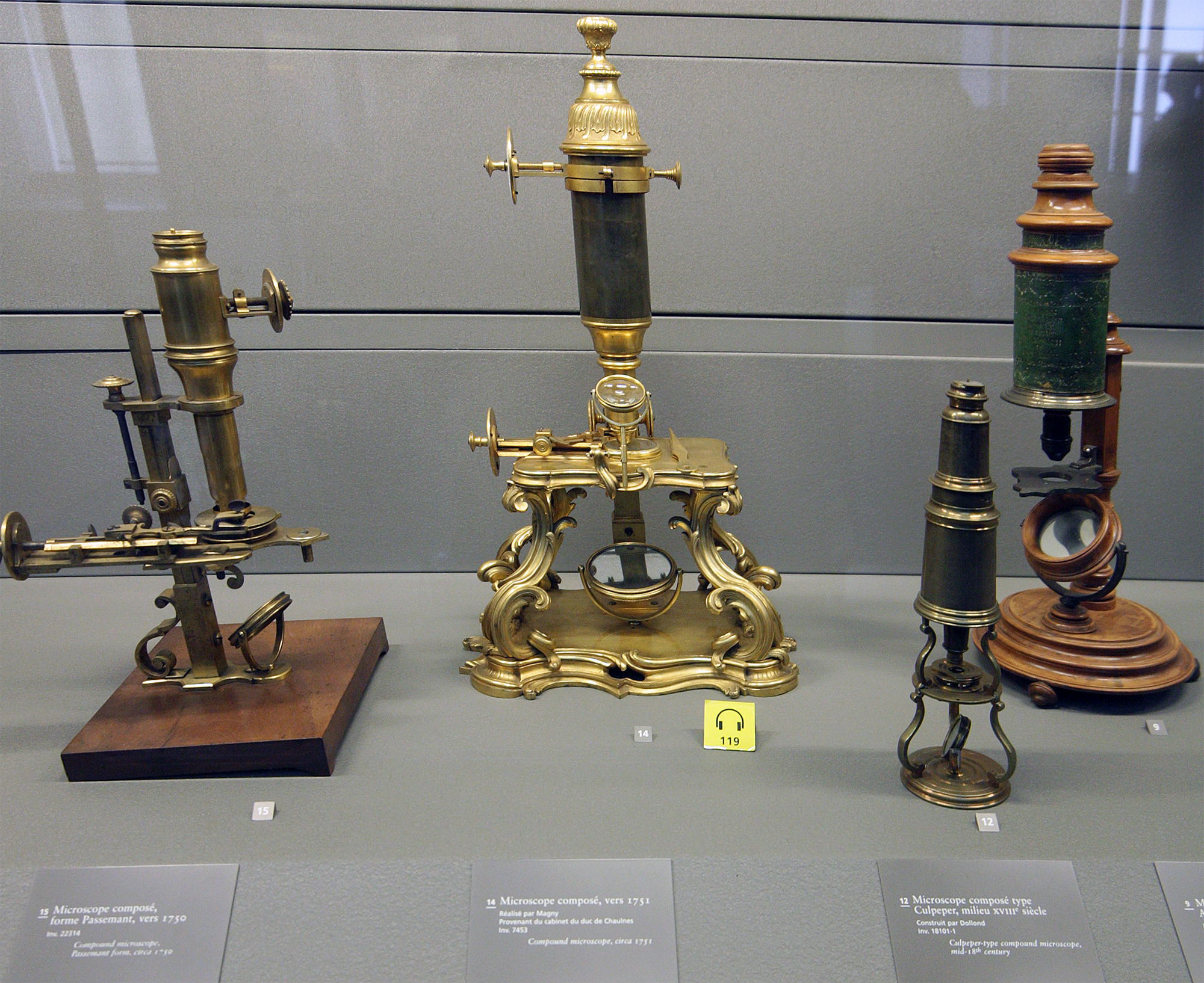
A microscope () is a laboratory instrument used to examine objects that are too small to be seen by the naked eye. Microscopy is the science of investigating small objects and structures using a microscope. Microscopic means being invisible to the eye unless aided by a microscope. There are many types of microscopes, and they may be grouped in different ways. One way is to describe the method an instrument uses to interact with a sample and produce images, either by sending a beam of light or electrons through a sample in its optical path, by detecting photon emissions from a sample, or by scanning across and a short distance from the surface of a sample using a probe. The most common microscope (and the first to be invented) is the optical microscope, which uses lenses to refract visible light that passed through a thinly sectioned sample to produce an observable image. Other major types of microscopes are the fluorescence microscope, electron microscope (both the ... [...More Info...] [...Related Items...] OR: [Wikipedia] [Google] [Baidu] |
Romanowsky Stain
Romanowsky staining, also known as Romanowsky–Giemsa staining, is a prototypical staining technique that was the forerunner of several distinct but similar stains widely used in hematology (the study of blood) and cytopathology (the study of diseased cells). Romanowsky-type stains are used to differentiate cells for microscopic examination in pathological specimens, especially blood and bone marrow films, and to detect parasites such as malaria within the blood. Stains that are related to or derived from the Romanowsky-type stains include Giemsa, Jenner, Wright, Field, May–Grünwald and Leishman stains. The staining technique is named after the Russian physician Dmitri Leonidovich Romanowsky (1861–1921), who was one of the first to recognize its potential for use as a blood stain. Mechanism The value of Romanowsky staining lies in its ability to produce a wide range of hues, allowing cellular components to be easily differentiated. This phenomenon is referred to as ... [...More Info...] [...Related Items...] OR: [Wikipedia] [Google] [Baidu] |
Blood Smear
A blood smear, peripheral blood smear or blood film is a thin layer of blood smeared on a glass microscope slide and then stained in such a way as to allow the various blood cells to be examined microscopically. Blood smears are examined in the investigation of hematological (blood) disorders and are routinely employed to look for blood parasites, such as those of malaria and filariasis. Preparation A blood smear is made by placing a drop of blood on one end of a slide, and using a ''spreader slide'' to disperse the blood over the slide's length. The aim is to get a region, called a monolayer, where the cells are spaced far enough apart to be counted and differentiated. The monolayer is found in the "feathered edge" created by the spreader slide as it draws the blood forward. The slide is left to air dry, after which the blood is fixed to the slide by immersing it briefly in methanol. The fixative is essential for good staining and presentation of cellular detail. After fix ... [...More Info...] [...Related Items...] OR: [Wikipedia] [Google] [Baidu] |
Cell Counting
Cell counting is any of various methods for the counting or similar quantification of cells in the life sciences, including medical diagnosis and treatment. It is an important subset of cytometry, with applications in research and clinical practice. For example, the complete blood count can help a physician to determine why a patient feels unwell and what to do to help. Cell counts within liquid media (such as blood, plasma, lymph, or laboratory rinsate) are usually expressed as a number of cells per unit of volume, thus expressing a concentration (for example, 5,000 cells per milliliter). Uses Numerous procedures in biology and medicine require the counting of cells. By the counting of cells in a known small volume, the concentration can be mediated. Examples of the need for cell counting include: * In medicine, the concentration of various blood cells, such as red blood cells and white blood cells, can give crucial information regarding the health situation of a person ... [...More Info...] [...Related Items...] OR: [Wikipedia] [Google] [Baidu] |
Hematology Analyzer
Hematology analyzers ( also spelled haematology analysers in British English) are used to count and identify blood cells at high speed with accuracy. During the 1950s, laboratory technicians counted each individual blood cell underneath a microscope. Tedious and inconsistent, this was replaced with the first, very basic hematology analyzer, engineered by Wallace H. Coulter. The early hematology analyzers relied on Coulter's Principle (see Coulter counter). However, they have evolved to encompass numerous techniques. Uses Hematology analyzers are used to conduct a complete blood count (CBC), which is usually the first test requested by physicians to determine a patients general health status. A complete blood count includes red blood cell (RBC), white blood cell (WBC), hemoglobin, and platelet counts, as well as hematocrit levels. Other analyses include: * RBC distribution width * Mean corpuscular volume * Mean corpuscular hemoglobin * Mean corpuscular hemoglobin concentrations ... [...More Info...] [...Related Items...] OR: [Wikipedia] [Google] [Baidu] |


.jpg)


