|
Foramen Spinosum
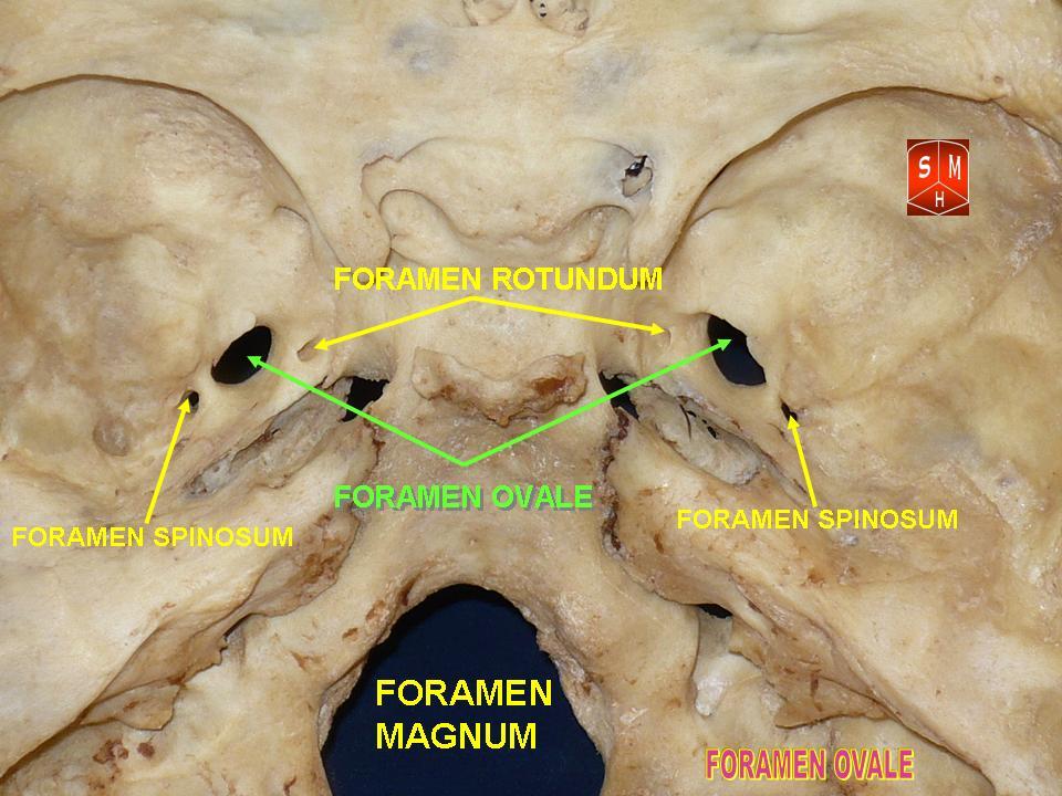
The foramen spinosum is a hole located in the greater wing of the sphenoid. It is located posterolateral to the foramen ovale and anterior to the sphenoidal spine. It allows the passage of the middle meningeal artery, middle meningeal vein and usually the meningeal branch of the mandibular nerve (sometimes it passes through the foramen ovale). The foramen spinosum is often used as a landmark in neurosurgery, due to its close relations with other cranial foramina. It was first described by Jakob Benignus Winslow in the 18th century. Structure The foramen spinosum is a foramen in the sphenoid bone of the skull. It connects the middle cranial fossa to the infratemporal fossa. It is located posterolateral to the foramen ovale, and anterior to the sphenoidal spine. Variation The foramen spinosum varies in size and location. The foramen is rarely absent, usually unilaterally, in which case the middle meningeal artery enters the cranial cavity through the foramen ovale. It ma ... [...More Info...] [...Related Items...] OR: [Wikipedia] [Google] [Baidu] |
Sphenoid Bone
The sphenoid bone is an unpaired bone of the neurocranium. It is situated in the middle of the skull towards the front, in front of the basilar part of occipital bone, basilar part of the occipital bone. The sphenoid bone is one of the seven bones that articulate to form the orbit (anatomy), orbit. Its shape somewhat resembles that of a butterfly or bat with its wings extended. Structure It is divided into the following parts: * a median portion, known as the body of sphenoid bone, containing the sella turcica, which houses the pituitary gland as well as the paired paranasal sinuses, the sphenoidal sinuses * two Greater wing of sphenoid bone, greater wings on the lateral side of the body and two Lesser wing of sphenoid bone, lesser wings from the anterior side. * Pterygoid processes of the sphenoides, directed downwards from the junction of the body and the greater wings. Two sphenoidal conchae are situated at the anterior and inferior part of the body. Intrinsic ligaments of ... [...More Info...] [...Related Items...] OR: [Wikipedia] [Google] [Baidu] |
Foramen Ovale (skull)
The foramen ovale (Latin: oval window) is a hole in the posterior part of the sphenoid bone, posterolateral to the foramen rotundum. It is one of the larger of the several holes (the foramina) in the skull. It transmits the mandibular nerve, a branch of the trigeminal nerve. Structure The foramen ovale is an opening in the greater wing of the sphenoid bone. The foramen ovale is one of two cranial foramina in the greater wing, the other being the foramen spinosum. The foramen ovale is posterolateral to the foramen rotundum and anteromedial to the foramen spinosum. Posterior and medial to the foramen is the opening for the carotid canal. Variation Similar to other foramina, the foramen ovale differs in shape and size throughout the natural life. The earliest perfect ring-shaped formation of the foramen ovale was observed in the 7th fetal month and the latest in 3 years after birth, in a study using over 350 skulls.In a study conducted on 100 skulls, the foramen ovale was d ... [...More Info...] [...Related Items...] OR: [Wikipedia] [Google] [Baidu] |
Saunders (imprint)
Saunders is an American academic publisher based in the United States. It is currently an imprint of Elsevier. Formerly independent, the W. B. Saunders company was acquired by CBS in 1968, who added it to their publishing division Holt, Rinehart & Winston. When CBS left the publishing field in 1986, it sold the academic publishing units to Harcourt Brace Jovanovich. Harcourt was acquired by Reed Elsevier in 2001. . . Retrieved May 2, 2015. W. B. Saunders published the Kinsey Reports and |
Ligature (medicine)
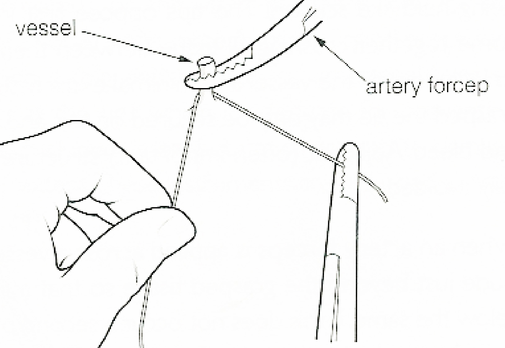
In surgery or medical procedure, a ligature consists of a piece of thread ( suture) tied around an anatomical structure, usually a blood vessel or another hollow structure (e.g. urethra) to shut it off. History The principle of ligation is attributed to Hippocrates and Galen. In ancient Rome, ligatures were used to treat hemorrhoids. The concept of a ligature was reintroduced some 1,500 years later by Ambroise Paré, and finally it found its modern use in 1870–80, made popular by Jules-Émile Péan. Procedure With a blood vessel the surgeon will clamp the vessel perpendicular to the axis of the artery or vein with a hemostat, then secure it by ligating it; i.e. using a piece of suture around it before dividing the structure and releasing the hemostat. It is different from a tourniquet in that the tourniquet will not be secured by knots and it can therefore be released/tightened at will. Ligature is one of the remedies to treat skin tag, or acrochorda. It is done by tying str ... [...More Info...] [...Related Items...] OR: [Wikipedia] [Google] [Baidu] |
Haemostasis
In biology, hemostasis American and British English spelling differences#ae and oe, or haemostasis is a process to prevent and stop bleeding, meaning to keep blood within a damaged blood vessel (the opposite of hemostasis is Bleeding, hemorrhage). It is the first stage of wound healing. This involves coagulation, which changes blood from a liquid to a gel. Intact blood vessels are central to moderating blood's tendency to form thrombus, clots. The endothelium, endothelial cells of intact vessels prevent blood clotting with a heparin-like molecule and thrombomodulin, and prevent platelet aggregation with nitric oxide and prostacyclin. When endothelium of a blood vessel is damaged, the endothelial cells stop secretion of coagulation and aggregation inhibitors and instead secrete von Willebrand factor, which initiate the maintenance of hemostasis after injury. Hemostasis involves three major steps: * vasoconstriction * temporary blockage of a hole in a damaged blood vessel by a platelet ... [...More Info...] [...Related Items...] OR: [Wikipedia] [Google] [Baidu] |
Trigeminal Ganglion
A trigeminal ganglion (or Gasserian ganglion, or semilunar ganglion, or Gasser's ganglion) is the sensory ganglion at the base of each of the two trigeminal nerves (CN V), occupying a cavity (Meckel's cave) in the dura mater, covering the trigeminal impression near the apex of the petrous part of the temporal bone. Structure It is somewhat crescent-shaped, with its convexity directed forward: Medially, it is in relation with the internal carotid artery and the posterior part of the cavernous sinus. The motor root runs in front of and medial to the sensory root, and passes beneath the ganglion; it leaves the skull through the foramen ovale, and, immediately below this foramen, joins the mandibular nerve. The greater superficial petrosal nerve lies also underneath the ganglion. The ganglion receives, on its medial side, filaments from the carotid plexus of the sympathetic. It gives off minute branches to the tentorium cerebelli, and to the dura mater in the middle fossa of th ... [...More Info...] [...Related Items...] OR: [Wikipedia] [Google] [Baidu] |
Mandibular Nerve
In neuroanatomy, the mandibular nerve (V) is the largest of the three divisions of the trigeminal nerve, the fifth cranial nerve (CN V). Unlike the other divisions of the trigeminal nerve (ophthalmic nerve, maxillary nerve) which contain only afferent fibers, the mandibular nerve contains both afferent and efferent fibers. These nerve fibers innervate structures of the lower jaw and face, such as the tongue, lower lip, and chin. The mandibular nerve also innervates the muscles of mastication. Structure The large sensory root emerges from the lateral part of the trigeminal ganglion and exits the cranial cavity through the foramen ovale. Portio minor, the small motor root of the trigeminal nerve, passes under the trigeminal ganglion and through the foramen ovale to unite with the sensory root just outside the skull. The mandibular nerve immediately passes between tensor veli palatini, which is medial, and lateral pterygoid, which is lateral, and gives off a meningeal branch (n ... [...More Info...] [...Related Items...] OR: [Wikipedia] [Google] [Baidu] |
Anatomical Landmark
Anatomical terminology is a form of scientific terminology used by anatomists, zoologists, and health professionals such as doctors. Anatomical terminology uses many unique terms, suffixes, and prefixes deriving from Ancient Greek and Latin. These terms can be confusing to those unfamiliar with them, but can be more precise, reducing ambiguity and errors. Also, since these anatomical terms are not used in everyday conversation, their meanings are less likely to change, and less likely to be misinterpreted. To illustrate how inexact day-to-day language can be: a scar "above the wrist" could be located on the forearm two or three inches away from the hand or at the base of the hand; and could be on the palm-side or back-side of the arm. By using precise anatomical terminology such ambiguity is eliminated. An international standard for anatomical terminology, ''Terminologia Anatomica'' has been created. Word formation Anatomical terminology has quite regular morphology: the same ... [...More Info...] [...Related Items...] OR: [Wikipedia] [Google] [Baidu] |
Middle Meningeal Vein
The pterygoid plexus (; in Merriam-Webster Online Dictionary '. from ''pteryx'', "wing" and ''eidos'', "shape") is a of considerable size, and is situated between the and |
Sphenosquamosal Suture
The sphenosquamosal suture is a cranial suture between the sphenoid bone and the squama of the temporal bone The temporal bones are situated at the sides and base of the skull, and lateral to the temporal lobes of the cerebral cortex. The temporal bones are overlaid by the sides of the head known as the temples, and house the structures of the ears. Th .... Additional images References External links * * Bones of the head and neck Cranial sutures Human head and neck Joints Joints of the head and neck Skeletal system Skull {{Portal bar, Anatomy ... [...More Info...] [...Related Items...] OR: [Wikipedia] [Google] [Baidu] |
Squama Temporalis
The squamous part of temporal bone, or temporal squama, forms the front and upper part of the temporal bone, and is scale-like, thin, and translucent. Surfaces Its outer surface is smooth and convex; it affords attachment to the temporal muscle, and forms part of the temporal fossa; on its hinder part is a vertical groove for the middle temporal artery. A curved line, the ''temporal line'', or ''supramastoid crest'', runs backward and upward across its posterior part; it serves for the attachment of the temporal fascia, and limits the origin of the temporalis muscle. The boundary between the squamous part and the mastoid portion of the bone, as indicated by traces of the original suture, lies about 1 cm. below this line. Projecting from the lower part of the squamous part is a long, arched process, the ''zygomatic process''. This process is at first directed lateralward, its two surfaces looking upward and downward; it then appears as if twisted inward upon itself, and runs ... [...More Info...] [...Related Items...] OR: [Wikipedia] [Google] [Baidu] |
Temporal Bone
The temporal bones are situated at the sides and base of the skull, and lateral to the temporal lobes of the cerebral cortex. The temporal bones are overlaid by the sides of the head known as the temples, and house the structures of the ears. The lower seven cranial nerves and the major vessels to and from the brain traverse the temporal bone. Structure The temporal bone consists of four parts— the squamous, mastoid, petrous and tympanic parts. The squamous part is the largest and most superiorly positioned relative to the rest of the bone. The zygomatic process is a long, arched process projecting from the lower region of the squamous part and it articulates with the zygomatic bone. Posteroinferior to the squamous is the mastoid part. Fused with the squamous and mastoid parts and between the sphenoid and occipital bones lies the petrous part, which is shaped like a pyramid. The tympanic part is relatively small and lies inferior to the squamous part, anterior to the mast ... [...More Info...] [...Related Items...] OR: [Wikipedia] [Google] [Baidu] |


