|
Foot Drop
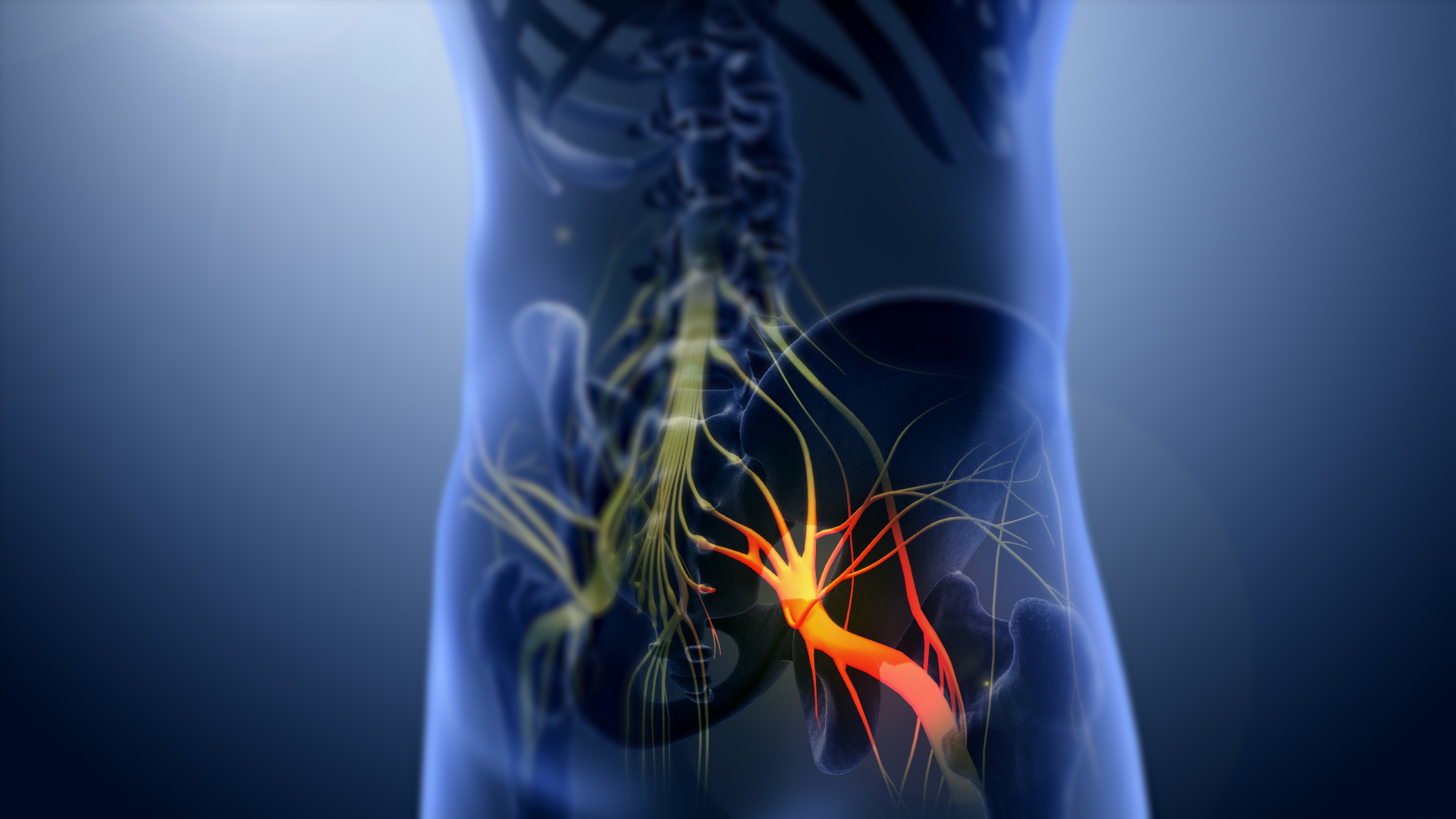
Foot drop is a gait abnormality in which the dropping of the forefoot happens due to weakness, irritation or damage to the deep fibular nerve (deep peroneal), including the sciatic nerve, or paralysis of the muscles in the anterior portion of the lower leg. It is usually a symptom of a greater problem, not a disease in itself. Foot drop is characterized by inability or impaired ability to raise the toes or raise the foot from the ankle (dorsiflexion). Foot drop may be temporary or permanent, depending on the extent of muscle weakness or paralysis and it can occur in one or both feet. In walking, the raised leg is slightly bent at the knee to prevent the foot from dragging along the ground. Foot drop can be caused by nerve damage alone or by muscle or spinal cord trauma, abnormal anatomy, atoxins, or disease. Toxins include organophosphate compounds which have been used as pesticides and as chemical agents in warfare. The poison can lead to further damage to the body such as a n ... [...More Info...] [...Related Items...] OR: [Wikipedia] [Google] [Baidu] |
Peroneal Nerve
The common fibular nerve (also known as the common peroneal nerve, external popliteal nerve, or lateral popliteal nerve) is a nerve in the lower leg that provides sensation over the posterolateral part of the leg and the knee joint. It divides at the knee into two terminal branches: the superficial fibular nerve and deep fibular nerve, which innervate the muscles of the lateral and anterior compartments of the leg respectively. When the common fibular nerve is damaged or compressed, foot drop can ensue. Structure The common fibular nerve is the smaller terminal branch of the sciatic nerve. The common fibular nerve has root values of L4, L5, S1, and S2. It arises from the superior angle of the popliteal fossa and extends to the lateral angle of the popliteal fossa, along the medial border of the biceps femoris. It then winds around the neck of the fibula to pierce the fibularis longus and divides into terminal branches of the superficial fibular nerve and the deep fibular nerve. Bef ... [...More Info...] [...Related Items...] OR: [Wikipedia] [Google] [Baidu] |
Lumbosacral Plexus
The anterior divisions of the lumbar nerves, sacral nerves, and coccygeal nerve form the lumbosacral plexus, the first lumbar nerve being frequently joined by a branch from the twelfth thoracic. For descriptive purposes this plexus is usually divided into three parts: * lumbar plexus * sacral plexus * pudendal plexus Injuries to the lumbosacral plexus are predominantly witnessed as bone injuries. Lumbosacral trunk and sacral plexus In human anatomy, the sacral plexus is a nerve plexus which provides motor and sensory nerves for the posterior thigh, most of the lower leg and foot, and part of the pelvis. It is part of the lumbosacral plexus and emerges from the lumbar verte ... palsies are common injury patterns. References External links * - "Lumbosacral Plexus" Additional Images File:Slide2Anat.JPG, Lumbosacral plexus Deep dissection. File:Slide4Anat.JPG, Lumbosacral plexus Deep dissection. Nerve plexus Nerves of the lower limb and lower torso {{Neuroana ... [...More Info...] [...Related Items...] OR: [Wikipedia] [Google] [Baidu] |
Sciatic Nerve
The sciatic nerve, also called the ischiadic nerve, is a large nerve in humans and other vertebrate animals which is the largest branch of the sacral plexus and runs alongside the hip joint and down the lower limb. It is the longest and widest single nerve in the human body, going from the top of the leg to the foot on the posterior aspect. The sciatic nerve has no cutaneous branches for the thigh. This nerve provides the connection to the nervous system for the skin of the lateral leg and the whole foot, the muscles of the back of the thigh, and those of the leg and foot. It is derived from spinal nerves L4 to S3. It contains fibers from both the anterior and posterior divisions of the lumbosacral plexus. Structure In humans, the sciatic nerve is formed from the L4 to S3 segments of the sacral plexus, a collection of nerve fibres that emerge from the sacral part of the spinal cord. The lumbosacral trunk from the L4 and L5 roots descends between the sacral promontory and ala and ... [...More Info...] [...Related Items...] OR: [Wikipedia] [Google] [Baidu] |
Neuromuscular Disease
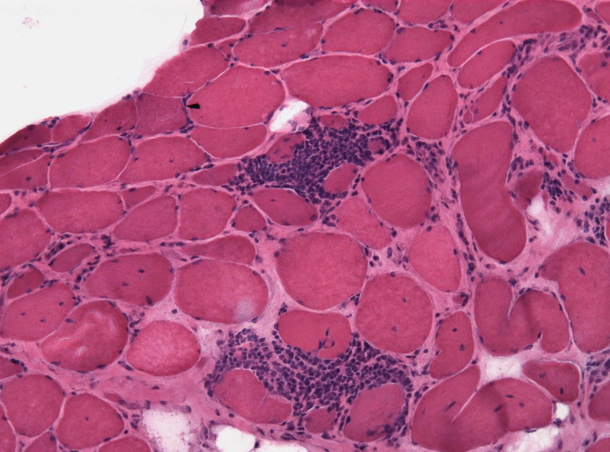
A neuromuscular disease is any disease affecting the peripheral nervous system (PNS), the neuromuscular junction, or skeletal muscle, all of which are components of the motor unit. Damage to any of these structures can cause muscle atrophy and weakness. Issues with sensation can also occur. Neuromuscular diseases can be acquired or genetic. Mutations of more than 500 genes have shown to be causes of neuromuscular diseases. Other causes include nerve or muscle degeneration, autoimmunity, toxins, medications, malnutrition, metabolic derangements, hormone imbalances, infection, nerve compression/entrapment, comprised blood supply, and trauma. Signs and symptoms Symptoms of neuromuscular disease may include numbness, paresthesia, muscle weakness, muscle atrophy, myalgia (muscle pain), and fasciculations (muscle twitches). Causes Neuromuscular disease can be caused by autoimmune disorders, genetic/hereditary disorders and some forms of the collagen disorder Ehlers–Dan ... [...More Info...] [...Related Items...] OR: [Wikipedia] [Google] [Baidu] |
Lumbar Plexus
The lumbar plexus is a web of nerves (a nervous plexus) in the lumbar region of the body which forms part of the larger lumbosacral plexus. It is formed by the Ventral ramus of spinal nerve, divisions of the first four lumbar nerves (L1-L4) and from contributions of the subcostal nerve (T12), which is the last Thoracic nerves, thoracic nerve. Additionally, the ventral rami of the fourth lumbar nerve pass communicating branches, the lumbosacral trunk, to the sacral plexus. The nerves of the lumbar plexus pass in front of the hip joint and mainly support the anterior part of the thigh.''Thieme Atlas of anatomy'' (2006), pp 470-471 The plexus is formed lateral to the intervertebral foramina and passes through Psoas major muscle, psoas major. Its smaller motor branches are distributed directly to psoas major, while the larger branches leave the muscle at various sites to run obliquely down through the pelvis to leave under the inguinal ligament with the exception of the obturator n ... [...More Info...] [...Related Items...] OR: [Wikipedia] [Google] [Baidu] |
Extensor Hallucis Longus
The extensor hallucis longus muscle is a thin skeletal muscle, situated between the tibialis anterior and the extensor digitorum longus. It extends the big toe and dorsiflects the foot. It also assists with foot eversion and inversion. Structure The extensor hallucis longus muscle arises from the anterior surface of the fibula for about the middle two-fourths of its extent, medial to the origin of the extensor digitorum longus muscle. It also arises from the interosseous membrane of the leg to a similar extent. The anterior tibial vessels and deep fibular nerve lie between it and the tibialis anterior. The fibers pass downward, and end in a tendon, which occupies the anterior border of the muscle, passes through a distinct compartment in the cruciate crural ligament, crosses from the lateral to the medial side of the anterior tibial vessels near the bend of the ankle, and is inserted into the base of the distal phalanx of the great toe. Opposite the metatarsophalangeal art ... [...More Info...] [...Related Items...] OR: [Wikipedia] [Google] [Baidu] |
Extensor Digitorum Longus
The extensor digitorum longus is a pennate muscle, situated at the lateral part of the front of the leg. Origin and insertion It arises from the lateral condyle of the tibia; from the upper three-quarters of the anterior surface of the body of the fibula; from the upper part of the interosseous membrane; from the deep surface of the fascia; and from the intermuscular septa between it and the tibialis anterior on the medial, and the peroneal muscles on the lateral side. Between it and the tibialis anterior are the upper portions of the anterior tibial vessels and deep peroneal nerve. The muscle passes under the superior and inferior extensor retinaculum of foot in company with the fibularis tertius, and divides into four slips, which run forward on the dorsum of the foot, and are inserted into the second and third phalanges of the four lesser toes. The tendons to the second, third, and fourth toes are each joined, opposite the metatarsophalangeal articulations, on the lateral si ... [...More Info...] [...Related Items...] OR: [Wikipedia] [Google] [Baidu] |
Fibularis Tertius
In human anatomy, the fibularis tertius (also known as the peroneus tertius) is a muscle in the anterior compartment of the leg. It acts to tilt the sole of the foot away from the midline of the body ( eversion) and to pull the foot upward toward the body (dorsiflexion). Structure The fibularis tertius arises from the lower third of the front surface of the fibula, the lower part of the interosseous membrane, and septum, or connective tissue, between it and the fibularis brevis. The septum is sometimes called the intermuscular septum of Otto. The muscle passes downward and ends in a tendon that passes under the superior extensor retinaculum and the inferior extensor retinaculum of the foot in the same canal as the extensor digitorum longus muscle. It may be mistaken as a fifth tendon of the extensor digitorum longus. The tendon inserts into the medial part of the posterior surface of the shaft of the fifth metatarsal bone. The fibularis tertius is supplied by the deep fibula ... [...More Info...] [...Related Items...] OR: [Wikipedia] [Google] [Baidu] |
Anterior Tibialis
The tibialis anterior muscle is a muscle in humans that originates along the upper two-thirds of the lateral (outside) surface of the tibia and inserts into the medial cuneiform and first metatarsal bones of the foot. It acts to dorsiflex and invert the foot. This muscle is mostly located near the shin. It is situated on the lateral side of the tibia; it is thick and fleshy above, tendinous below. The tibialis anterior overlaps the anterior tibial vessels and deep peroneal nerve in the upper part of the leg. Structure The tibialis anterior muscle arises from: * the lateral condyle of the tibia. * the upper 2/3 of the lateral surface of the tibia. * the adjoining part of the interosseous membrane. * the deep surface of the fascia. * the intermuscular septum between it and the extensor digitorum longus. The fibers of this circumpennate muscle are relatively parallel to the plane of insertion, ending in a tendon, apparent on the anteriomedial dorsal aspect of the foot close to t ... [...More Info...] [...Related Items...] OR: [Wikipedia] [Google] [Baidu] |


