|
Follicular Dendritic Cell
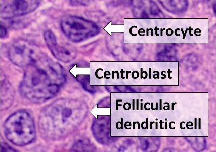
Follicular dendritic cells (FDC) are cells of the immune system found in primary and secondary lymph follicles (lymph nodes) of the B cell areas of the lymphoid tissue. Unlike dendritic cells (DC), FDCs are not derived from the bone-marrow hematopoietic stem cell, but are of mesenchymal origin. Possible functions of FDC include: organizing lymphoid tissue's cells and microarchitecture, capturing antigen to support B cell, promoting debris removal from germinal centers, and protecting against autoimmunity. Disease processes that FDC may contribute include primary FDC-tumor, chronic inflammatory conditions, HIV-1 infection development, and neuroinvasive scrapie. Location and molecular markers Follicular DCs are a non-migratory population found in primary and secondary follicles of the B cell areas of lymph nodes, spleen, and mucosa-associated lymphoid tissue (MALT). They form a stable network due to intercellular connections between FDCs processes and intimate interaction with f ... [...More Info...] [...Related Items...] OR: [Wikipedia] [Google] [Baidu] |
Mucosa-associated Lymphoid Tissue
The mucosa-associated lymphoid tissue (MALT), also called mucosa-associated lymphatic tissue, is a diffuse system of small concentrations of lymphoid tissue found in various submucosal membrane sites of the body, such as the gastrointestinal tract, nasopharynx, thyroid, breast, lung, salivary glands, eye, and skin. MALT is populated by lymphocytes such as T cells and B cells, as well as plasma cells and macrophages, each of which is well situated to encounter antigens passing through the mucosal epithelium. In the case of intestinal MALT, M cells are also present, which sample antigen from the lumen and deliver it to the lymphoid tissue. MALT constitute about 50% of the lymphoid tissue in human body. Immune responses that occur at mucous membranes are studied by mucosal immunology. Categorization The components of MALT are sometimes subdivided into the following: * GALT (gut-associated lymphoid tissue. Peyer's patches are a component of GALT found in the lining of the small in ... [...More Info...] [...Related Items...] OR: [Wikipedia] [Google] [Baidu] |
Synovial Joint
A synovial joint, also known as diarthrosis, joins bones or cartilage with a fibrous joint capsule that is continuous with the periosteum of the joined bones, constitutes the outer boundary of a synovial cavity, and surrounds the bones' articulating surfaces. This joint unites long bones and permits free bone movement and greater mobility. The synovial cavity/joint is filled with synovial fluid. The joint capsule is made up of an outer layer of fibrous membrane, which keeps the bones together structurally, and an inner layer, the synovial membrane, which seals in the synovial fluid. They are the most common and most movable type of joint in the body of a mammal. As with most other joints, synovial joints achieve movement at the point of contact of the articulating bones. Structure Synovial joints contain the following structures: * Synovial cavity: all diarthroses have the characteristic space between the bones that is filled with synovial fluid * Joint capsule: the fibrous ... [...More Info...] [...Related Items...] OR: [Wikipedia] [Google] [Baidu] |
Trends (journals)
''Trends'' is a series of 16 review journals in a range of areas of biology and chemistry published under its Cell Press imprint by Elsevier. The publisher in lieu is Danielle Loughlin. The ''Trends'' series was established in 1976 with ''Trends in Biochemical Sciences'', rapidly followed by ''Trends in Neurosciences'', ''Trends in Pharmacological Sciences'', and ''Immunology Today''. ''Immunology Today'', ''Parasitology Today'', and ''Molecular Medicine Today'' changed their names to ''Trends in...'' in 2001. ''Drug Discovery Today ''Drug Discovery Today'' is a monthly peer-reviewed scientific journal that is published by Elsevier. It was established in 1996 and publishes reviews on all aspects of preclinical drug discovery from target identification and validation through h ...'' was spun off as an independent brand. Titles The current set of ''Trends'' journals are all published monthly: References External links * {{Reed Elsevier, state=collapsed Academic journal ... [...More Info...] [...Related Items...] OR: [Wikipedia] [Google] [Baidu] |
VLA-4
Integrin α4β1 (very late antigen-4) is an integrin dimer. It is composed of CD49d (alpha 4) and CD29 (beta 1). The alpha 4 subunit is 155 kDa, and the beta 1 subunit is 150 kDa. Function The integrin VLA-4 is expressed on the cell surfaces of stem cells, progenitor cells, T and B cells, monocytes, natural killer cells, eosinophils, but not neutrophils. It functions to promote an inflammatory response by the immune system by assisting in the movement of leukocytes to tissue that requires inflammation. It is a key player in cell adhesion. However, VLA-4 does not adhere to its appropriate ligands until the leukocytes are activated by chemotactic agents or other stimuli (often produced by the endothelium or other cells at the site of injury). VLA-4's primary ligands include VCAM-1 and fibronectin. One activating chemokine is SDF-1. Following SDF-1 binding, the integrin undergoes a conformational change of the alpha and beta domains that is necessary to confer high binding af ... [...More Info...] [...Related Items...] OR: [Wikipedia] [Google] [Baidu] |
Lymphocyte Function-associated Antigen 1
Lymphocyte function-associated antigen 1 (LFA-1) is an integrin found on lymphocytes and other leukocytes. LFA-1 plays a key role in emigration, which is the process by which leukocytes leave the bloodstream to enter the tissues. LFA-1 also mediates firm arrest of leukocytes. Additionally, LFA-1 is involved in the process of cytotoxic T cell mediated killing as well as antibody mediated killing by granulocytes and monocytes. As of 2007, LFA-1 has 6 known ligands: ICAM-1, ICAM-2, ICAM-3, ICAM-4, ICAM-5, and JAM-A. LFA-1/ICAM-1 interactions have recently been shown to stimulate signaling pathways that influence T cell differentiation. LFA-1 belongs to the integrin superfamily of adhesion molecules. Structure LFA-1 is a heterodimeric glycoprotein with non-covalently linked subunits. LFA-1 has two subunits designated as the alpha subunit and beta subunit. The alpha subunit was named aL in 1983. The alpha subunit is designated CD11a; and the beta subunit, unique to leukocytes, is beta ... [...More Info...] [...Related Items...] OR: [Wikipedia] [Google] [Baidu] |
Apoptosis
Apoptosis (from grc, ἀπόπτωσις, apóptōsis, 'falling off') is a form of programmed cell death that occurs in multicellular organisms. Biochemical events lead to characteristic cell changes (morphology) and death. These changes include blebbing, cell shrinkage, nuclear fragmentation, chromatin condensation, DNA fragmentation, and mRNA decay. The average adult human loses between 50 and 70 billion cells each day due to apoptosis. For an average human child between eight and fourteen years old, approximately twenty to thirty billion cells die per day. In contrast to necrosis, which is a form of traumatic cell death that results from acute cellular injury, apoptosis is a highly regulated and controlled process that confers advantages during an organism's life cycle. For example, the separation of fingers and toes in a developing human embryo occurs because cells between the digits undergo apoptosis. Unlike necrosis, apoptosis produces cell fragments called apoptotic ... [...More Info...] [...Related Items...] OR: [Wikipedia] [Google] [Baidu] |
CXCL13
Chemokine (C-X-C motif) ligand 13 (CXCL13), also known as B lymphocyte chemoattractant (BLC) or B cell-attracting chemokine 1 (BCA-1), is a protein ligand that in humans is encoded by the ''CXCL13'' gene. Function CXCL13 is a small chemokine belonging to the CXC chemokine family. As its other names suggest, this chemokine is selectively chemotactic for B cells belonging to both the B-1 and B-2 subsets, and elicits its effects by interacting with chemokine receptor CXCR5. CXCL13 and its receptor CXCR5 control the organization of B cells within follicles of lymphoid tissues and is expressed highly in the liver, spleen, lymph nodes, and gut of humans. The gene for CXCL13 is located on human chromosome 4 in a cluster of other CXC chemokines. In T lymphocytes, CXCL13 expression is thought to reflect a germinal center origin of the T cell, particularly a subset of T cells called follicular B helper T cells Follicular helper T cells (also known as follicular B helper T cel ... [...More Info...] [...Related Items...] OR: [Wikipedia] [Google] [Baidu] |
Lymphotoxin Beta Receptor
Lymphotoxin beta receptor (LTBR), also known as tumor necrosis factor receptor superfamily member 3 (TNFRSF3), is a cell surface receptor for lymphotoxin involved in apoptosis and cytokine release. It is a member of the tumor necrosis factor receptor superfamily. Function The protein encoded by this gene is a member of the tumor necrosis factor (TNF) family of receptors. It is expressed on the surface of most cell types, including cells of epithelial and myeloid lineages, but not on T and B lymphocytes. The protein specifically binds the lymphotoxin membrane form (a complex of lymphotoxin-alpha and lymphotoxin-beta). The encoded protein and its ligand play a role in the development and organization of lymphoid tissue and transformed cells. Activation of the encoded protein can trigger apoptosis. Not only does the LTBR help trigger apoptosis, it can lead to the release of the cytokine interleukin 8. Overexpression of LTBR in HEK293 cells increases IL-8 promoter activity and le ... [...More Info...] [...Related Items...] OR: [Wikipedia] [Google] [Baidu] |
TNFRSF1A
Tumor necrosis factor receptor 1 (TNFR1), also known as tumor necrosis factor receptor superfamily member 1A (TNFRSF1A) and CD120a, is a ubiquitous membrane receptor that binds tumor necrosis factor-alpha (TNFα). Function The protein encoded by this gene is a member of the tumor necrosis factor receptor superfamily, which also contains TNFRSF1B. This protein is one of the major receptors for the tumor necrosis factor-alpha. This receptor can activate the transcription factor NF-κB, mediate apoptosis, and function as a regulator of inflammation. Antiapoptotic protein BCL2-associated athanogene 4 (BAG4/SODD) and adaptor proteins TRADD and TRAF2 have been shown to interact with this receptor, and thus play regulatory roles in the signal transduction mediated by the receptor. Clinical significance Germline mutations of the extracellular domains of this receptor were found to be associated with the human genetic disorder called tumor necrosis factor associated periodic syndrome ... [...More Info...] [...Related Items...] OR: [Wikipedia] [Google] [Baidu] |
Lymphotoxin
Lymphotoxin is a member of the tumor necrosis factor (TNF) superfamily of cytokines, whose members are responsible for regulating the growth and function of lymphocytes and are expressed by a wide variety of cells in the body. Lymphotoxin plays a critical role in developing and preserving the framework of lymphoid organs and of gastrointestinal immune responses, as well as in the activation signaling of both the innate and adaptive immune responses. Lymphotoxin alpha (LT-α, previously known as TNF-beta) and lymphotoxin beta (LT-β), the two forms of lymphotoxin, each have distinctive structural characteristics and perform specific functions. Structure and function Each LT-α/LT-β subunit is a trimer and assembles into homotrimers or heterotrimers. LT-α binds with LT-β to form membrane-bound heterotrimers LT-α1-β2 and LT-α2-β1, which are commonly referred to as lymphotoxin beta. LT-α1-β2 is the most prevalent form of lymphotoxin beta. LT-α also forms a homotrimer, LT ... [...More Info...] [...Related Items...] OR: [Wikipedia] [Google] [Baidu] |
Tumor Necrosis Factor-alpha
Tumor necrosis factor (TNF, cachexin, or cachectin; formerly known as tumor necrosis factor alpha or TNF-α) is an adipokine and a cytokine. TNF is a member of the TNF superfamily, which consists of various transmembrane proteins with a homologous TNF domain. As an adipokine, TNF promotes insulin resistance, and is associated with obesity-induced type 2 diabetes. As a cytokine, TNF is used by the immune system for cell signaling. If macrophages (certain white blood cells) detect an infection, they release TNF to alert other immune system cells as part of an inflammatory response. TNF signaling occurs through two receptors: TNFR1 and TNFR2. TNFR1 is constituitively expressed on most cell types, whereas TNFR2 is restricted primarily to endothelial, epithelial, and subsets of immune cells. TNFR1 signaling tends to be pro-inflammatory and apoptotic, whereas TNFR2 signaling is anti-inflammatory and promotes cell proliferation. Suppression of TNFR1 signaling has been important fo ... [...More Info...] [...Related Items...] OR: [Wikipedia] [Google] [Baidu] |


