|
Foam Cell
Foam cells, also called lipid-laden macrophages, are a type of cell that contain cholesterol. These can form a plaque that can lead to atherosclerosis and trigger heart attacks and stroke. Foam cells are fat-laden cells with a M2 macrophage-like phenotype. They contain low density lipoproteins (LDL) and can only be truly detected by examining a fatty plaque under a microscope after it is removed from the body. They are named because the lipoproteins give the cell a foamy appearance. Despite the connection with cardiovascular diseases they might not be inherently dangerous. Some foam cells are derived from smooth muscle cells and present a limited macrophage-like phenotype. Formation Foam cell formation is triggered by a number of factors including the uncontrolled uptake of modified low density lipoproteins (LDL), the upregulation of cholesterol esterification and the impairment of mechanisms associated with cholesterol release. Foam cells are formed when circulating monocyte ... [...More Info...] [...Related Items...] OR: [Wikipedia] [Google] [Baidu] |
Cholesterolosis
In surgical pathology, strawberry gallbladder, more formally cholesterolosis of the gallbladder and gallbladder cholesterolosis, is a change in the gallbladder wall due to excess cholesterol. - cancerweb.ncl.ac.uk. The name ''strawberry gallbladder'' comes from the typically stippled appearance of the mucosal surface on gross examination, which resembles a strawberry. Cholesterolosis results from abnormal deposits of cholesterol esters in macrophages within the lamina propria (foam cells) and in mucosal epithelium. The gallbladder may be affected in a patchy localized form or in a diffuse form. The diffuse form macroscopically appears as a bright red mucosa with yellow mottling (due to lipid), hence the term ''strawberry'' gallbladder. It is not tied to cholelithiasis (gallstones) or cholecystitis (infla ... [...More Info...] [...Related Items...] OR: [Wikipedia] [Google] [Baidu] |
Monocyte
Monocytes are a type of leukocyte or white blood cell. They are the largest type of leukocyte in blood and can differentiate into macrophages and conventional dendritic cells. As a part of the vertebrate innate immune system monocytes also influence adaptive immune responses and exert tissue repair functions. There are at least three subclasses of monocytes in human blood based on their phenotypic receptors. Structure Monocytes are amoeboid in appearance, and have nongranulated cytoplasm. Thus they are classified as agranulocytes, although they might occasionally display some azurophil granules and/or vacuoles. With a diameter of 15–22 μm, monocytes are the largest cell type in peripheral blood. Monocytes are mononuclear cells and the ellipsoidal nucleus is often lobulated/indented, causing a bean-shaped or kidney-shaped appearance. Monocytes compose 2% to 10% of all leukocytes in the human body. Development Monocytes are produced by the bone marrow from precursors ca ... [...More Info...] [...Related Items...] OR: [Wikipedia] [Google] [Baidu] |
Endoplasmic Reticulum
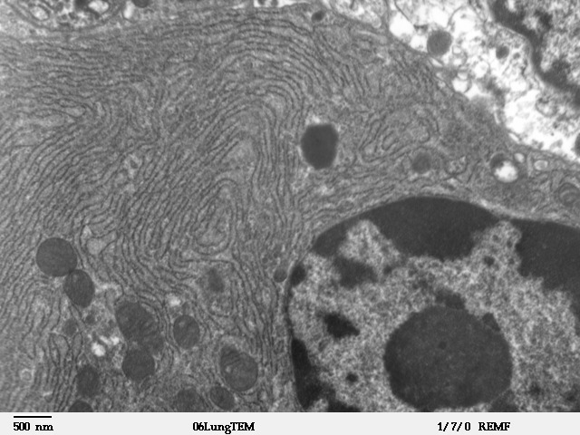
The endoplasmic reticulum (ER) is, in essence, the transportation system of the eukaryotic cell, and has many other important functions such as protein folding. It is a type of organelle made up of two subunits – rough endoplasmic reticulum (RER), and smooth endoplasmic reticulum (SER). The endoplasmic reticulum is found in most eukaryotic cells and forms an interconnected network of flattened, membrane-enclosed sacs known as cisternae (in the RER), and tubular structures in the SER. The membranes of the ER are continuous with the outer nuclear membrane. The endoplasmic reticulum is not found in red blood cells, or spermatozoa. The two types of ER share many of the same proteins and engage in certain common activities such as the synthesis of certain lipids and cholesterol. Different types of cells contain different ratios of the two types of ER depending on the activities of the cell. RER is found mainly toward the nucleus of cell and SER towards the cell membrane or plasma ... [...More Info...] [...Related Items...] OR: [Wikipedia] [Google] [Baidu] |
Lysosomal Lipase
Lysosomal lipase is a form of lipase which functions intracellularly, in the lysosomes. Biochemical Significance The primary function of lysosomal lipase is to hydrolyze lipids such as triglycerides and cholesterol. These fats are transported and degraded into free fatty acids. Lysosomal lipases function optimally at an acidic pH which are complementary with the environment found in the lysosomal lumen. These enzymes were believed to only hydrolyze the lipids found in organelle membranes and extracellular lipids. However, recent studies suggest that lysosomal lipases also play a significant role in the degradation of cytosolic lipids, a characteristic that was previously limited to neutral lipases. The ability of the lysosome to degrade a diverse set of cargo is attributed to the lysosomal lipase and other soluble hydrolases. These enzymes include sulphatases, phosphatases, peptidases, glycosidases, and nucleases. The biochemical role of these enzymes are observed in various path ... [...More Info...] [...Related Items...] OR: [Wikipedia] [Google] [Baidu] |
Cholesteryl Ester
300px, Cholesterol oleate, a member of the cholesteryl ester family Cholesteryl ester, a dietary lipid, is an ester of cholesterol. The ester bond is formed between the carboxylate group of a fatty acid and the hydroxyl group of cholesterol. Cholesteryl esters have a lower solubility in water due to their increased hydrophobicity. Esters are formed by replacing at least one –OH (hydroxyl) group with an –O–alkyl (alkoxy) group. They are hydrolyzed by pancreatic enzymes, cholesterol esterase, to produce cholesterol and free fatty acids. They are associated with atherosclerosis. Cholesteryl ester is found in human brains as lipid droplets which store and transport cholesterol. Increased levels of cholesteryl ester have been found in certain parts of the brain of people with Huntington disease. Higher concentrations of cholesteryl ester have been found in the caudate and putamen, but not the cerebellum, of people with Huntington Disease compared with levels in controls."Illawar ... [...More Info...] [...Related Items...] OR: [Wikipedia] [Google] [Baidu] |
Lysosome
A lysosome () is a membrane-bound organelle found in many animal cells. They are spherical vesicles that contain hydrolytic enzymes that can break down many kinds of biomolecules. A lysosome has a specific composition, of both its membrane proteins, and its lumenal proteins. The lumen's pH (~4.5–5.0) is optimal for the enzymes involved in hydrolysis, analogous to the activity of the stomach. Besides degradation of polymers, the lysosome is involved in various cell processes, including secretion, plasma membrane repair, apoptosis, cell signaling, and energy metabolism. Lysosomes act as the waste disposal system of the cell by digesting used materials in the cytoplasm, from both inside and outside the cell. Material from outside the cell is taken up through endocytosis, while material from the inside of the cell is digested through autophagy. The sizes of the organelles vary greatly—the larger ones can be more than 10 times the size of the smaller ones. They were discov ... [...More Info...] [...Related Items...] OR: [Wikipedia] [Google] [Baidu] |
Endosome
Endosomes are a collection of intracellular sorting organelles in eukaryotic cells. They are parts of endocytic membrane transport pathway originating from the trans Golgi network. Molecules or ligands internalized from the plasma membrane can follow this pathway all the way to lysosomes for degradation or can be recycled back to the cell membrane in the endocytic cycle. Molecules are also transported to endosomes from the trans Golgi network and either continue to lysosomes or recycle back to the Golgi apparatus. Endosomes can be classified as early, sorting, or late depending on their stage post internalization. Endosomes represent a major sorting compartment of the endomembrane system in cells. Function Endosomes provide an environment for material to be sorted before it reaches the degradative lysosome. For example, low-density lipoprotein (LDL) is taken into the cell by binding to the LDL receptor at the cell surface. Upon reaching early endosomes, the LDL dissociates ... [...More Info...] [...Related Items...] OR: [Wikipedia] [Google] [Baidu] |
Pinocytosis
In cellular biology, pinocytosis, otherwise known as fluid endocytosis and bulk-phase pinocytosis, is a mode of endocytosis in which small molecules dissolved in extracellular fluid are brought into the cell through an invagination of the cell membrane, resulting in their containment within a small vesicle inside the cell. These pinocytotic vesicles then typically fuse with early endosomes to hydrolyze (break down) the particles. Pinocytosis is variably subdivided into categories depending on the molecular mechanism and the fate of the internalized molecules. Function In humans, this process occurs primarily for absorption of fat droplets. In endocytosis the cell plasma membrane extends and folds around desired extracellular material, forming a pouch that pinches off creating an internalized vesicle. The invaginated pinocytosis vesicles are much smaller than those generated by phagocytosis. The vesicles eventually fuse with the lysosome, whereupon the vesicle contents are digeste ... [...More Info...] [...Related Items...] OR: [Wikipedia] [Google] [Baidu] |
Phagocytosis
Phagocytosis () is the process by which a cell uses its plasma membrane to engulf a large particle (≥ 0.5 μm), giving rise to an internal compartment called the phagosome. It is one type of endocytosis. A cell that performs phagocytosis is called a phagocyte. In a multicellular organism's immune system, phagocytosis is a major mechanism used to remove pathogens and cell debris. The ingested material is then digested in the phagosome. Bacteria, dead tissue cells, and small mineral particles are all examples of objects that may be phagocytized. Some protozoa use phagocytosis as means to obtain nutrients. History Phagocytosis was first noted by Canadian physician William Osler (1876), and later studied and named by Élie Metchnikoff (1880, 1883). In immune system Phagocytosis is one main mechanisms of the innate immune defense. It is one of the first processes responding to infection, and is also one of the initiating branches of an adaptive immune response. Although mo ... [...More Info...] [...Related Items...] OR: [Wikipedia] [Google] [Baidu] |
Endocytosis
Endocytosis is a cellular process in which substances are brought into the cell. The material to be internalized is surrounded by an area of cell membrane, which then buds off inside the cell to form a vesicle containing the ingested material. Endocytosis includes pinocytosis (cell drinking) and phagocytosis (cell eating). It is a form of active transport. History The term was proposed by De Duve in 1963. Phagocytosis was discovered by Élie Metchnikoff in 1882. Pathways Endocytosis pathways can be subdivided into four categories: namely, receptor-mediated endocytosis (also known as clathrin-mediated endocytosis), caveolae, pinocytosis, and phagocytosis Phagocytosis () is the process by which a cell uses its plasma membrane to engulf a large particle (≥ 0.5 μm), giving rise to an internal compartment called the phagosome. It is one type of endocytosis. A cell that performs phagocytosis is .... *Clathrin-mediated endocytosis is mediated by the production of smal ... [...More Info...] [...Related Items...] OR: [Wikipedia] [Google] [Baidu] |
Pattern Recognition Receptor
Pattern recognition receptors (PRRs) play a crucial role in the proper function of the innate immune system. PRRs are germline-encoded host sensors, which detect molecules typical for the pathogens. They are proteins expressed, mainly, by cells of the innate immune system, such as dendritic cells, macrophages, monocytes, neutrophils and epithelial cells, to identify two classes of molecules: pathogen-associated molecular patterns (PAMPs), which are associated with microbial pathogens, and damage-associated molecular patterns (DAMPs), which are associated with components of host's cells that are released during cell damage or death. They are also called primitive pattern recognition receptors because they evolved before other parts of the immune system, particularly before adaptive immunity. PRRs also mediate the initiation of antigen-specific adaptive immune response and release of inflammatory cytokines. The microbe-specific molecules that are recognized by a given PRR are called p ... [...More Info...] [...Related Items...] OR: [Wikipedia] [Google] [Baidu] |
CD36
CD36 (cluster of differentiation 36), also known as platelet glycoprotein 4, fatty acid translocase (FAT), scavenger receptor class B member 3 (SCARB3), and glycoproteins 88 (GP88), IIIb (GPIIIB), or IV (GPIV) is a protein that in humans is encoded by the CD36 gene. The CD36 antigen is an integral membrane protein found on the surface of many cell types in vertebrate animals. It imports fatty acids inside cells and is a member of the class B scavenger receptor family of cell surface proteins. CD36 binds many ligands including collagen, thrombospondin, erythrocytes parasitized with ''Plasmodium falciparum'', oxidized low density lipoprotein, native lipoproteins, oxidized phospholipids, and long-chain fatty acids. Work in genetically modified rodents suggest a role for CD36 in fatty acid metabolism,heart disease, taste, and dietary fat processing in the intestine. It may be involved in glucose intolerance, atherosclerosis, arterial hypertension, diabetes, cardiomyopathy, Alzheime ... [...More Info...] [...Related Items...] OR: [Wikipedia] [Google] [Baidu] |
.png)


