|
Fibularis Muscles
The fibularis muscles (also called peroneus muscles or peroneals) are a group of muscles in the lower leg. Description The muscle group is normally composed of three muscles: fibularis longus, fibularis brevis, and fibularis tertius. The fibularis longus and fibularis brevis are located in the lateral compartment of the leg and are supplied by the fibular artery and the superficial fibular nerve. The fibularis tertius is located in the anterior compartment of the leg and is supplied by the anterior tibial artery and the deep fibular nerve. While all three muscles move the sole of the foot outward, away from the midline of the body ( eversion), the longus and brevis extend the foot downward away from the body ( plantar flexion), whereas the tertius muscle pulls the foot upward toward the body (dorsiflexion). The fibularis muscles are highly variable. Several variants are occasionally present, including the peroneus digiti minimi and the peroneus quartus. The quartus is mor ... [...More Info...] [...Related Items...] OR: [Wikipedia] [Google] [Baidu] |
Human Leg
The human leg, in the general word sense, is the entire lower limb of the human body, including the foot, thigh or sometimes even the hip or gluteal region. However, the definition in human anatomy refers only to the section of the lower limb extending from the knee to the ankle, also known as the crus or, especially in non-technical use, the shank. Legs are used for standing, and all forms of locomotion including recreational such as dancing, and constitute a significant portion of a person's mass. Female legs generally have greater hip anteversion and tibiofemoral angles, but shorter femur and tibial lengths than those in males. Structure In human anatomy, the lower leg is the part of the lower limb that lies between the knee and the ankle. Anatomists restrict the term ''leg'' to this use, rather than to the entire lower limb. The thigh is between the hip and knee and makes up the rest of the lower limb. The term ''lower limb'' or ''lower extremity'' is commonly u ... [...More Info...] [...Related Items...] OR: [Wikipedia] [Google] [Baidu] |
Plantarflexion
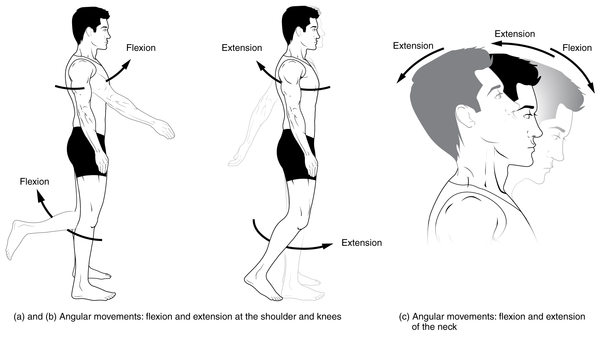
Motion, the process of movement, is described using specific anatomical terms. Motion includes movement of organs, joints, limbs, and specific sections of the body. The terminology used describes this motion according to its direction relative to the anatomical position of the body parts involved. Anatomists and others use a unified set of terms to describe most of the movements, although other, more specialized terms are necessary for describing unique movements such as those of the hands, feet, and eyes. In general, motion is classified according to the anatomical plane it occurs in. ''Flexion'' and ''extension'' are examples of ''angular'' motions, in which two axes of a joint are brought closer together or moved further apart. ''Rotational'' motion may occur at other joints, for example the shoulder, and are described as ''internal'' or ''external''. Other terms, such as ''elevation'' and ''depression'', describe movement above or below the horizontal plane. Many anatomic ... [...More Info...] [...Related Items...] OR: [Wikipedia] [Google] [Baidu] |
Terminologia Anatomica
''Terminologia Anatomica'' is the international standard for human anatomical terminology. It is developed by the Federative International Programme on Anatomical Terminology, a program of the International Federation of Associations of Anatomists (IFAA). The second edition was released in 2019 and approved and adopted by the IFAA General Assembly in 2020. ''Terminologia Anatomica'' supersedes the previous standard, ''Nomina Anatomica''. It contains terminology for about 7500 human anatomical structures. Categories of anatomical structures ''Terminologia Anatomica'' is divided into 16 chapters grouped into five parts. The official terms are in Latin. Although equivalent English-language terms are provided, as shown below, only the official Latin terms are used as the basis for creating lists of equivalent terms in other languages. Part I Chapter 1: General anatomy # General terms # Reference planes # Reference lines # Human body positions # Movements # Parts of human bod ... [...More Info...] [...Related Items...] OR: [Wikipedia] [Google] [Baidu] |
Thieme Medical Publishers
Thieme Medical Publishers is a German medical and science publisher in the Thieme Publishing Group. It produces professional journals, textbooks, atlases, monographs and reference books in both German and English covering a variety of medical specialties, including neurosurgery, orthopaedics, endocrinology, urology, radiology, anatomy, chemistry, otolaryngology, ophthalmology, audiology and speech-language pathology, complementary and alternative medicine. Thieme has more than 1,000 employees and maintains offices in seven cities worldwide, including New York City, Beijing, Delhi, Stuttgart, and three other cities in Germany. History Georg Thieme Verlag was founded in 1886 in Leipzig, Germany, by Georg Thieme when he was 26 years old. Thieme remains privately held and family-owned. The company received some early success in 1896 by publishing Wilhelm Röntgen's famous picture of his wife's hand in what is still one of Thieme's and Germany's oldest journals, the '' Deut ... [...More Info...] [...Related Items...] OR: [Wikipedia] [Google] [Baidu] |
Extensor Digitorum Longus Muscle
The extensor digitorum longus is a pennate muscle, situated at the lateral part of the front of the leg. Origin and insertion It arises from the lateral condyle of the tibia; from the upper three-quarters of the anterior surface of the body of the fibula; from the upper part of the interosseous membrane; from the deep surface of the fascia; and from the intermuscular septa between it and the tibialis anterior on the medial, and the peroneal muscles on the lateral side. Between it and the tibialis anterior are the upper portions of the anterior tibial vessels and deep peroneal nerve. The muscle passes under the superior and inferior extensor retinaculum of foot in company with the fibularis tertius, and divides into four slips, which run forward on the dorsum of the foot, and are inserted into the second and third phalanges of the four lesser toes. The tendons to the second, third, and fourth toes are each joined, opposite the metatarsophalangeal articulations, on the later ... [...More Info...] [...Related Items...] OR: [Wikipedia] [Google] [Baidu] |
Fibularis Tertius
In human anatomy, the fibularis tertius (also known as the peroneus tertius) is a muscle in the anterior compartment of the leg. It acts to tilt the sole of the foot away from the midline of the body ( eversion) and to pull the foot upward toward the body (dorsiflexion). Structure The fibularis tertius arises from the lower third of the front surface of the fibula, the lower part of the interosseous membrane, and septum, or connective tissue, between it and the fibularis brevis. The septum is sometimes called the intermuscular septum of Otto. The muscle passes downward and ends in a tendon that passes under the superior extensor retinaculum and the inferior extensor retinaculum of the foot in the same canal as the extensor digitorum longus muscle. It may be mistaken as a fifth tendon of the extensor digitorum longus. The tendon inserts into the medial part of the posterior surface of the shaft of the fifth metatarsal bone. The fibularis tertius is supplied by the deep fibular ... [...More Info...] [...Related Items...] OR: [Wikipedia] [Google] [Baidu] |
Fibularis Brevis
In human anatomy, the fibularis brevis (or peroneus brevis) is a muscle that lies underneath the fibularis longus within the lateral compartment of the leg. It acts to tilt the sole of the foot away from the midline of the body (eversion) and to extend the foot downward away from the body at the ankle (plantar flexion). Structure The fibularis brevis arises from the lower two-thirds of the lateral, or outward, surface of the fibula (inward in relation to the fibularis longus) and from the connective tissue between it and the muscles on the front and back of the leg. The muscle passes downward and ends in a tendon that runs behind the lateral malleolus of the ankle in a groove that it shares with the tendon of the fibularis longus; the groove is converted into a canal by the superior fibular retinaculum, and the tendons in it are contained in a common mucous sheath. The tendon then runs forward along the lateral side of the calcaneus, above the calcaneal tubercle and the tendon ... [...More Info...] [...Related Items...] OR: [Wikipedia] [Google] [Baidu] |
Lateral Compartment
The lateral compartment of the leg is a fascial compartment of the lower leg. It contains muscles which make eversion and plantarflexion of the foot. Muscles The lateral compartment of the leg contains: * Fibularis longus * Fibularis brevis Action * Foot evertors * Foot plantarflexion Nerve Supply The lateral compartment of the leg is supplied by the superficial fibular nerve (superficial peroneal nerve). Blood Supply Its proximal and distal arterial supply consists of perforating branches of the anterior tibial artery and fibular artery. Additional images File:Lateral compartment of leg - animation.gif, Animation. Fibularis longus (blue) and fibularis brevis (red). See also *Fascial compartments of leg The fascial compartments of the leg are the four fascial compartments that separate and contain the muscles of the lower leg (from the knee to the ankle). The compartments are divided by septa formed from the fascia. The compartments usually hav ... Refer ... [...More Info...] [...Related Items...] OR: [Wikipedia] [Google] [Baidu] |
Fibularis Longus
In human anatomy, the fibularis longus (also known as peroneus longus) is a superficial muscle in the lateral compartment of the leg. It acts to tilt the sole of the foot away from the midline of the body ( eversion) and to extend the foot downward away from the body (plantar flexion) at the ankle. The fibularis longus is the longest and most superficial of the three fibularis (peroneus) muscles. At its upper end, it is attached to the head of the fibula, and its "belly" runs down along most of this bone. The muscle becomes a tendon that wraps around and behind the lateral malleolus of the ankle, then continues under the foot to attach to the medial cuneiform and first metatarsal. It is supplied by the superficial fibular nerve. Structure The fibularis longus arises from the head and upper two-thirds of the lateral, or outward, surface of the fibula, from the deep surface of the fascia, and from the connective tissue between it and the muscles on the front and back of the leg. ... [...More Info...] [...Related Items...] OR: [Wikipedia] [Google] [Baidu] |
Dorsiflexion
Motion, the process of movement, is described using specific anatomical terms. Motion includes movement of organs, joints, limbs, and specific sections of the body. The terminology used describes this motion according to its direction relative to the anatomical position of the body parts involved. Anatomists and others use a unified set of terms to describe most of the movements, although other, more specialized terms are necessary for describing unique movements such as those of the hands, feet, and eyes. In general, motion is classified according to the anatomical plane it occurs in. ''Flexion'' and ''extension'' are examples of ''angular'' motions, in which two axes of a joint are brought closer together or moved further apart. ''Rotational'' motion may occur at other joints, for example the shoulder, and are described as ''internal'' or ''external''. Other terms, such as ''elevation'' and ''depression'', describe movement above or below the horizontal plane. Many anatomic ... [...More Info...] [...Related Items...] OR: [Wikipedia] [Google] [Baidu] |
Eversion (kinesiology)
Motion, the process of movement, is described using specific anatomical terms. Motion includes movement of organs, joints, limbs, and specific sections of the body. The terminology used describes this motion according to its direction relative to the anatomical position of the body parts involved. Anatomists and others use a unified set of terms to describe most of the movements, although other, more specialized terms are necessary for describing unique movements such as those of the hands, feet, and eyes. In general, motion is classified according to the anatomical plane it occurs in. ''Flexion'' and ''extension'' are examples of ''angular'' motions, in which two axes of a joint are brought closer together or moved further apart. ''Rotational'' motion may occur at other joints, for example the shoulder, and are described as ''internal'' or ''external''. Other terms, such as ''elevation'' and ''depression'', describe movement above or below the horizontal plane. Many anatomi ... [...More Info...] [...Related Items...] OR: [Wikipedia] [Google] [Baidu] |
Fibularis Longus
In human anatomy, the fibularis longus (also known as peroneus longus) is a superficial muscle in the lateral compartment of the leg. It acts to tilt the sole of the foot away from the midline of the body ( eversion) and to extend the foot downward away from the body (plantar flexion) at the ankle. The fibularis longus is the longest and most superficial of the three fibularis (peroneus) muscles. At its upper end, it is attached to the head of the fibula, and its "belly" runs down along most of this bone. The muscle becomes a tendon that wraps around and behind the lateral malleolus of the ankle, then continues under the foot to attach to the medial cuneiform and first metatarsal. It is supplied by the superficial fibular nerve. Structure The fibularis longus arises from the head and upper two-thirds of the lateral, or outward, surface of the fibula, from the deep surface of the fascia, and from the connective tissue between it and the muscles on the front and back of the leg. ... [...More Info...] [...Related Items...] OR: [Wikipedia] [Google] [Baidu] |


