|
Fat Pad
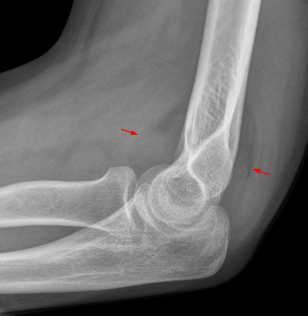
A fat pad (aka haversian gland) is a mass of closely packed fat cells surrounded by fibrous tissue septa.TheFreeDictionary > Fat padCiting: Mosby's Medical Dictionary, 8th edition. 2009 They may be extensively supplied with capillaries and nerve endings. Examples are: * ''Intraarticular fat pads''. These are also covered by a layer of synovial cells. A fat pad sign is an elevation of the anterior and posterior fat pads of the elbow joint, and suggests the presence of an occult fracture. * Buccal fat pad can be seen in nursing babies. * The fat pad of the labia majora, which can be used as a graft Graft or grafting may refer to: *Graft (politics), a form of political corruption * Graft, Netherlands, a village in the municipality of Graft-De Rijp Science and technology *Graft (surgery), a surgical procedure *Grafting, the joining of plant t ..., often as a so-called "Martius labial fat pad graft", which can be used, for example, in urethrolysis. *Fat pads within the heels which w ... [...More Info...] [...Related Items...] OR: [Wikipedia] [Google] [Baidu] |
Fat Cell
Adipocytes, also known as lipocytes and fat cells, are the cells that primarily compose adipose tissue, specialized in storing energy as fat. Adipocytes are derived from mesenchymal stem cells which give rise to adipocytes through adipogenesis. In cell culture, adipocyte progenitors can also form osteoblasts, myocytes and other cell types. There are two types of adipose tissue, white adipose tissue (WAT) and brown adipose tissue (BAT), which are also known as white and brown fat, respectively, and comprise two types of fat cells. Structure White fat cells White fat cells contain a single large lipid droplet surrounded by a layer of cytoplasm, and are known as unilocular. The nucleus is flattened and pushed to the periphery. A typical fat cell is 0.1 mm in diameter with some being twice that size, and others half that size. However, these numerical estimates of fat cell size depend largely on the measurement method and the location of the adipose tissue. The fat stored is ... [...More Info...] [...Related Items...] OR: [Wikipedia] [Google] [Baidu] |
Fat Pad Sign
The fat pad sign, also known as the sail sign, is a potential finding on elbow radiography which suggests a fracture (bone), fracture of one or more bones at the elbow. It is may indicate an occult fracture that is not directly visible. Its name derives from the fact that it has the shape of a Spinnaker, spinnaker (sail). It is caused by displacement of the fat pad around the elbow joint. Both ''anterior'' and ''posterior'' fat pad signs exist, and both can be found on the same X-ray. In children, a posterior fat pad sign suggests a condylar fracture of the humerus. In adults it suggests a radius (bone), radial head fracture. In addition to fracture, any process resulting in an elbow joint effusion may also demonstrate an abnormal fat pad sign. Increased intracapsular fluid is also seen in several conditions other than fracture and this produces the abnormal fat pad sign. (toxic synovitis, septic arthritis, Juvenile Rheumatoid Arthritis, osteomyelitis of the distal humeral ph ... [...More Info...] [...Related Items...] OR: [Wikipedia] [Google] [Baidu] |
Buccal Fat Pad
The buccal fat pad (also called Bichat’s fat pad, after Xavier Bichat, and the buccal pad of fat) is one of several encapsulated fat masses in the cheek. It is a deep fat pad located on either side of the face between the buccinator muscle and several more superficial muscles (including the masseter, the zygomaticus major, and the zygomaticus minor). The inferior portion of the buccal fat pad is contained within the buccal space. It should not be confused with the malar fat pad, which is directly below the skin of the cheek. It should also not be confused with jowl fat pads. It is implicated in the formation of hollow cheeks and the nasolabial fold, but not in the formation of jowls. Nomenclature and structure The buccal fat pad is composed of several parts, although exactly how many parts seems to be a point of disagreement and no single consistent nomenclature of these parts has been observed. It was described as being divided into three lobes, the anterior, intermediate, and ... [...More Info...] [...Related Items...] OR: [Wikipedia] [Google] [Baidu] |
Labia Majora
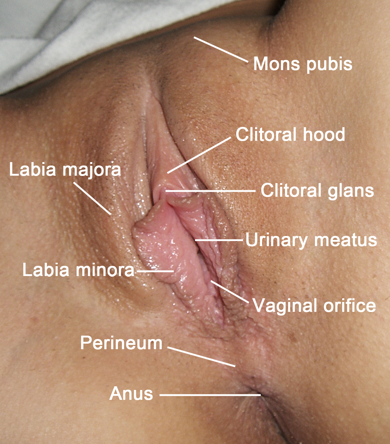
The labia majora (singular: ''labium majus'') are two prominent longitudinal cutaneous folds that extend downward and backward from the mons pubis to the perineum. Together with the labia minora they form the labia of the vulva. The labia majora are homologous to the male scrotum. Etymology ''Labia majora'' is the Latin plural for big ("major") lips; the singular is ''labium majus.'' The Latin term ''labium/labia'' is used in anatomy for a number of usually paired parallel structures, but in English it is mostly applied to two pairs of parts of female external genitals (vulva)—labia majora and labia minora. Labia majora are commonly known as the outer lips, while labia minora (Latin for ''small lips''), which run alongside between them, are referred to as the inner lips. Traditionally, to avoid confusion with other lip-like structures of the body, the labia of female genitals were termed by anatomists in Latin as ''labia majora (''or ''minora) pudendi.'' Embryology Embryolo ... [...More Info...] [...Related Items...] OR: [Wikipedia] [Google] [Baidu] |
Graft (surgery)
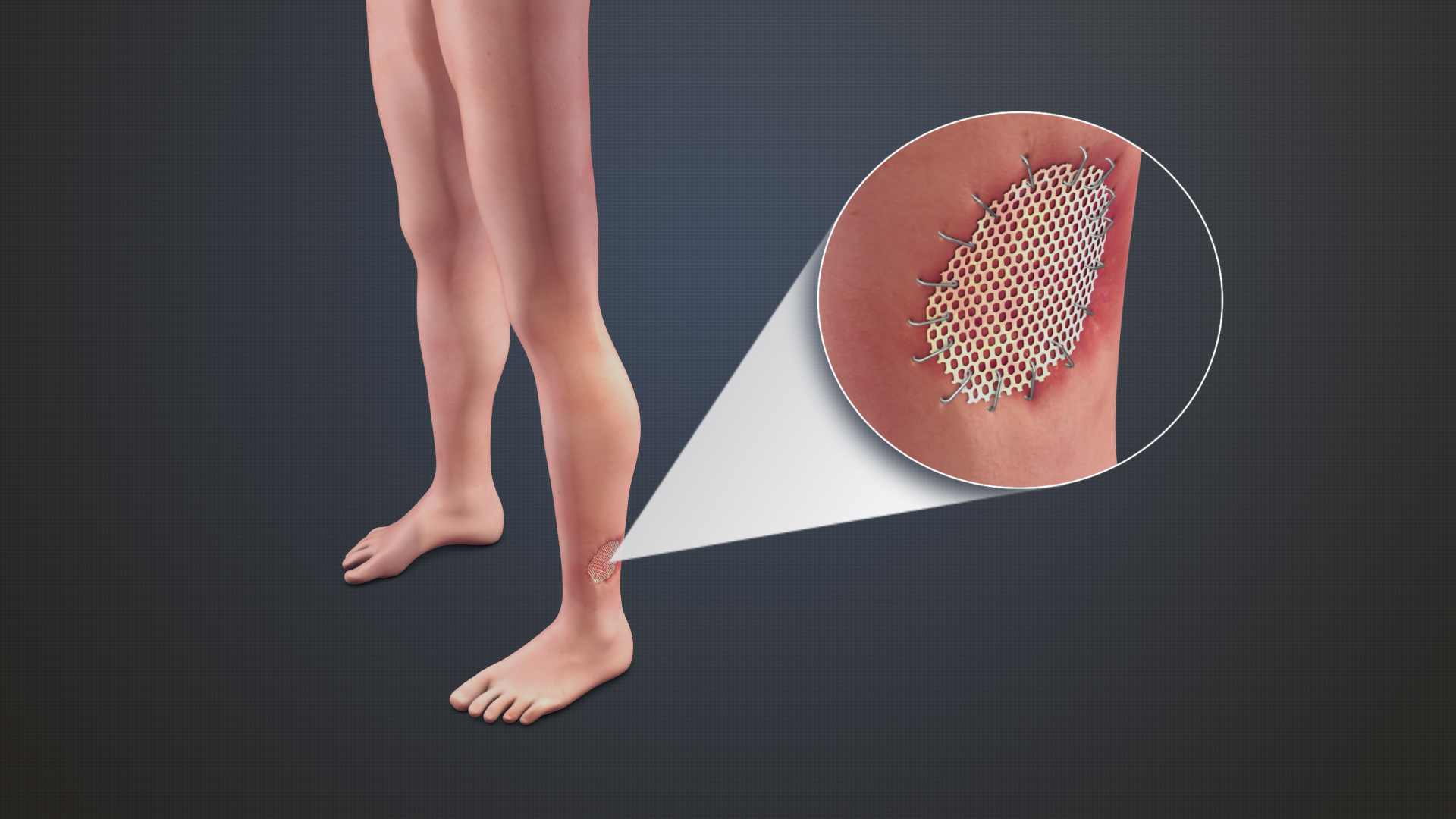
Grafting refers to a surgical procedure to move tissue from one site to another on the body, or from another creature, without bringing its own blood supply with it. Instead, a new blood supply grows in after it is placed. A similar technique where tissue is transferred with the blood supply intact is called a flap. In some instances, a graft can be an artificially manufactured device. Examples of this are a tube to carry blood flow across a defect or from an artery to a vein for use in hemodialysis. Classification Autografts and isografts are usually not considered as foreign and, therefore, do not elicit rejection. Allografts and xenografts may be recognized as foreign by the recipient and rejected. * Autograft: graft taken from one part of the body of an individual and transplanted onto another site in the same individual, e.g., skin graft. * Isograft: graft taken from one individual and placed on another individual of the same genetic constitution, e.g., grafts between ... [...More Info...] [...Related Items...] OR: [Wikipedia] [Google] [Baidu] |
Heel Pad Syndrome
Heel pad syndrome is a pain that occurs in the center of the heel. It is typically due to atrophy of the fat pad which makes up the heel. Risk factors include obesity. Other conditions with similar symptoms include plantar fasciitis. Treatment includes rest, pain medication, and heel cups. It becomes more common with age. Signs and symptoms * Pain in the heel, usually on the middle of the heel. This is in direct contrast to plantar fascia pain or heel spur pain which is present at the front of the heel, not the middle. * Pain is usually a deep, dull ache that feels like a bruise. * Pressing with the thumb into the centre of the heel should re-create the pain. * Condition can often be attributed to a blow to the heel – landing hard while barefoot on a hard surface, jumping in dress shoes with a hard heel, stepping on a stone while running. * Pain is aggravated by walking barefoot on hard surfaces like ceramic tile, concrete, hardwood floors, etc. Diagnosis The main differential ... [...More Info...] [...Related Items...] OR: [Wikipedia] [Google] [Baidu] |
Ball (anatomy)
The ball of the foot is the padded portion of the sole between the toes and the arch, underneath the heads of the metatarsal bones. In comparative foot morphology, the ball is most analogous to the metacarpal (forepaw) or metatarsal (hindpaw) pad in many mammals with paws, and serves mostly the same functions. The ball of the foot is of utmost importance when playing sports. Many sports, such as tennis, requires the player to stand on the balls of their feet for increased agility. The ball is a common area in which people develop pain, known as metatarsalgia. People who frequently wear high heels often develop pain in the balls of their feet from the immense amount of pressure that is placed on them for long periods of time, due to the inclination of the shoes. To remedy this, there is a market for ball-of-foot or general foot cushions that are placed into shoes to relieve some of the pressure. Alternately, people can have a procedure done in which a dermal filler is injected ... [...More Info...] [...Related Items...] OR: [Wikipedia] [Google] [Baidu] |


