|
Fascial Compartments Of Arm
The fascial compartments of arm refers to the specific anatomical term of the compartments within the upper segment of the upper limb (the arm) of the body. The upper limb is divided into two segments, the arm and the forearm. Each of these segments is further divided into two compartments which are formed by deep fascia – tough connective tissue septa (walls). Each compartment encloses specific muscles and nerves. The compartments of the arm are the anterior compartment of the arm and the posterior compartment of the arm, divided by the lateral and the medial intermuscular septa. The compartments of the forearm are the anterior compartment of the forearm and posterior compartment of the forearm. Intermuscular septa The lateral intermuscular septum extends from the lower part of the crest of the greater tubercle of the humerus, along the lateral supracondylar ridge, to the lateral epicondyle; it is blended with the tendon of the deltoid muscle, gives attachment to the tricep ... [...More Info...] [...Related Items...] OR: [Wikipedia] [Google] [Baidu] |
Radial Nerve
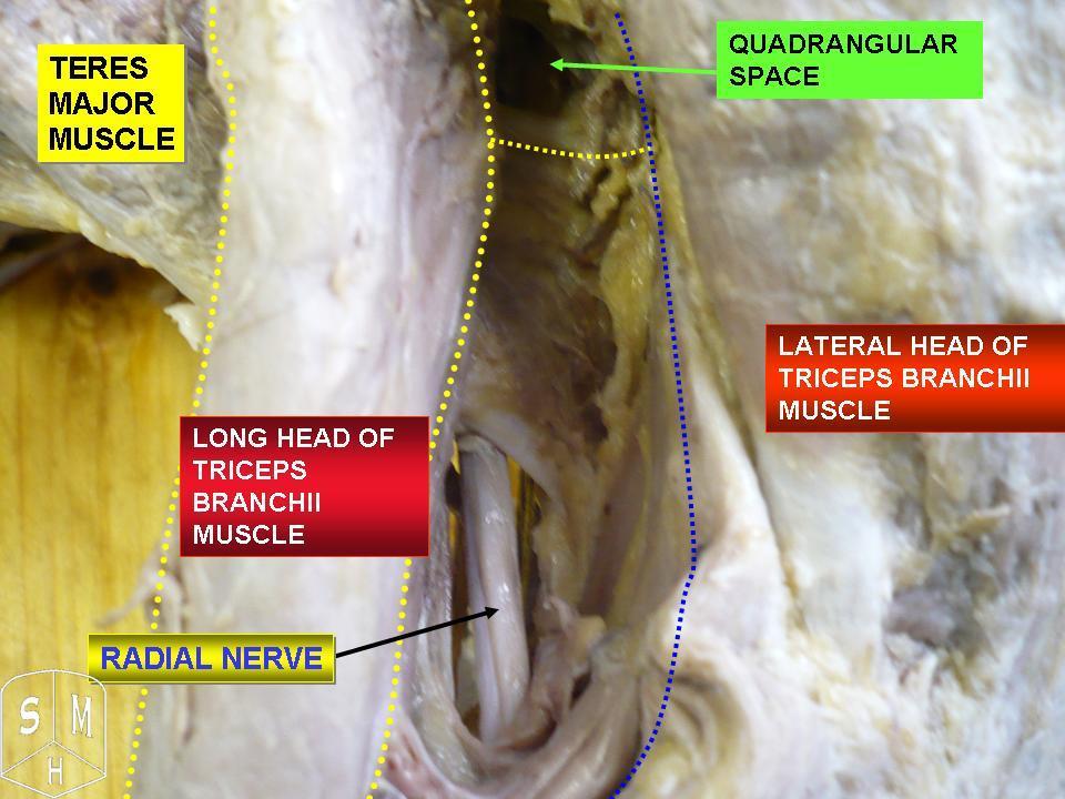
The radial nerve is a nerve in the human body that supplies the posterior portion of the upper limb. It innervates the medial and lateral heads of the triceps brachii muscle of the arm, as well as all 12 muscles in the posterior osteofascial compartment of the forearm and the associated joints and overlying skin. It originates from the brachial plexus, carrying fibers from the ventral roots of spinal nerves C5, C6, C7, C8 & T1. The radial nerve and its branches provide motor innervation to the dorsal arm muscles (the triceps brachii and the anconeus) and the extrinsic extensors of the wrists and hands; it also provides cutaneous sensory innervation to most of the back of the hand, except for the back of the little finger and adjacent half of the ring finger (which are innervated by the ulnar nerve). The radial nerve divides into a deep branch, which becomes the posterior interosseous nerve, and a superficial branch, which goes on to innervate the dorsum (back) of the hand. Th ... [...More Info...] [...Related Items...] OR: [Wikipedia] [Google] [Baidu] |
Cervical Spinal Nerve 6
The cervical spinal nerve 6 (C6) is a spinal nerve of the cervical segment. It originates from the spinal column from above the cervical vertebra 6 (C6). The C6 nerve root shares a common branch from C5, and has a role in innervating many muscles of the rotator cuff and distal arm, including: *Subclavius *Supraspinatus *Infraspinatus * Biceps Brachii * Brachialis * Deltoid *Teres Minor *Brachioradialis *Serratus Anterior *Subscapularis *Pectoralis Major *Coracobrachialis *Teres Major * Supinator *Extensor Carpi Radialis Brevis * Extensor Carpi Radialis Longus * Latissimus Dorsi Damage to the C6 motor neuron, by way of impingement, ischemia, trauma, or degeneration of nerve tissue, can cause denervation Denervation is any loss of nerve supply regardless of the cause. If the nerves lost to denervation are part of the neuronal communication to a specific function in the body then altered or a loss of physiological functioning can occur. Denervati ... of one or more of the asso ... [...More Info...] [...Related Items...] OR: [Wikipedia] [Google] [Baidu] |
Cervical Spinal Nerve 5
The cervical spinal nerve 5 (C5) is a spinal nerve of the cervical segment. Nervous System -- Groups of Nerves It originates from the spinal column from above the (C5). It contributes to the phrenic nerve, , and before joining |
Musculocutaneous Nerve
The musculocutaneous nerve arises from the lateral cord of the brachial plexus, opposite the lower border of the pectoralis major, its fibers being derived from C5, C6 and C7. Structure The musculocutaneous nerve arises from the lateral cord of the brachial plexus, courses through the anterior part of the arm, and terminates at 2 cm above elbow as the lateral cutaneous nerve of the forearm. Musculocutaneous nerve arises from the lateral cord of the brachial plexus with root value of C5 to C7 of the spinal cord. It follows the course of the third part of the axillary artery (part of the axillary artery distal to the pectoralis minor) laterally and enters the frontal aspect of the arm where it penetrates the coracobrachialis muscle. It then passes downwards and laterally between the biceps brachii (above) and the brachialis muscles (below), to the lateral side of the arm; at 2 cm above the elbow it pierces the deep fascia lateral to the tendon of the biceps brachii ... [...More Info...] [...Related Items...] OR: [Wikipedia] [Google] [Baidu] |
Biceps Brachii
The biceps or biceps brachii ( la, musculus biceps brachii, "two-headed muscle of the arm") is a large muscle that lies on the front of the upper arm between the shoulder and the elbow. Both heads of the muscle arise on the scapula and join to form a single muscle belly which is attached to the upper forearm. While the biceps crosses both the shoulder and elbow joints, its main function is at the elbow where it flexes the forearm and supinates the forearm. Both these movements are used when opening a bottle with a corkscrew: first biceps screws in the cork (supination), then it pulls the cork out (flexion). Structure The biceps is one of three muscles in the anterior compartment of the upper arm, along with the brachialis muscle and the coracobrachialis muscle, with which the biceps shares a nerve supply. The biceps muscle has two heads, the short head and the long head, distinguished according to their origin at the coracoid process and supraglenoid tubercle of the sca ... [...More Info...] [...Related Items...] OR: [Wikipedia] [Google] [Baidu] |
Inferior Ulnar Collateral Artery
The inferior ulnar collateral artery (anastomotica magna artery) is an artery in the arm. It arises about 5 cm. above the elbow from the brachial artery. Course It passes medialward upon the Brachialis, and piercing the medial intermuscular septum, winds around the back of the humerus between the Triceps brachii and the bone, forming, by its junction with the profunda brachii, an arch above the olecranon fossa. Branches and anastomoses As the vessel lies on the brachialis, it gives off branches which ascend to join the superior ulnar collateral: others descend in front of the medial epicondyle, to anastomose with the anterior ulnar recurrent. Behind the medial epicondyle a branch anastomoses with the superior ulnar collateral and posterior ulnar recurrent The posterior ulnar recurrent artery is an artery in the forearm. It is one of two recurrent arteries that arises from the ulnar artery, the other being the anterior ulnar recurrent artery. The posterior ulnar recurrent ... [...More Info...] [...Related Items...] OR: [Wikipedia] [Google] [Baidu] |
Superior Ulnar Collateral Artery
The superior ulnar collateral artery (inferior profunda artery), of small size, arises from the brachial artery a little below the middle of the arm; it frequently springs from the upper part of the a. profunda brachii. It pierces the medial intermuscular septum, and descends on the surface of the medial head of the Triceps brachii to the space between the medial epicondyle and olecranon, accompanied by the ulnar nerve, and ends under the Flexor carpi ulnaris by anastomosing with the posterior ulnar recurrent, and inferior ulnar collateral. It sometimes sends a branch in front of the medial epicondyle, to anastomose with the anterior ulnar recurrent The anterior ulnar recurrent artery is an artery in the forearm. It is one of two recurrent arteries that arises from the ulnar artery, the other being the posterior ulnar recurrent artery. It arises from the ulnar artery immediately below the el .... Additional images File:Gray413_color.png, Cross-section through the middle ... [...More Info...] [...Related Items...] OR: [Wikipedia] [Google] [Baidu] |
Ulnar Nerve
In human anatomy, the ulnar nerve is a nerve that runs near the ulna bone. The ulnar collateral ligament of elbow joint is in relation with the ulnar nerve. The nerve is the largest in the human body unprotected by muscle or bone, so injury is common. This nerve is directly connected to the little finger, and the adjacent half of the ring finger, innervating the palmar aspect of these fingers, including both front and back of the tips, perhaps as far back as the fingernail beds. This nerve can cause an electric shock-like sensation by striking the medial epicondyle of the humerus posteriorly, or inferiorly with the elbow flexed. The ulnar nerve is trapped between the bone and the overlying skin at this point. This is commonly referred to as bumping one's "funny bone". This name is thought to be a pun, based on the sound resemblance between the name of the bone of the upper arm, the humerus, and the word "humorous". Alternatively, according to the Oxford English Dictionary, i ... [...More Info...] [...Related Items...] OR: [Wikipedia] [Google] [Baidu] |
Coracobrachialis
The coracobrachialis muscle is the smallest of the three muscles that attach to the coracoid process of the scapula. (The other two muscles are pectoralis minor and the short head of the biceps brachii.) It is situated at the upper and medial part of the arm. Structure Coracobrachialis muscle arises from the apex of the coracoid process, in common with the short head of the biceps brachii, and from the intermuscular septum between the two muscles. It is inserted by means of a flat tendon into an impression at the middle of the medial surface and border of the body of the humerus (shaft of the humerus) between the origins of the triceps brachii and brachialis. Innervation Coracobrachialis muscle is perforated by and innervated by the musculocutaneous nerve, which arises from the anterior division of the upper trunk ( C5, C6) and middle trunk ( C7) of the brachial plexus. Development Variation Function The action of the coracobrachialis is to flex and adduct the arm at the ... [...More Info...] [...Related Items...] OR: [Wikipedia] [Google] [Baidu] |
Medial Epicondyle Of The Humerus
The medial epicondyle of the humerus is an epicondyle of the humerus bone of the upper arm in humans. It is larger and more prominent than the lateral epicondyle and is directed slightly more posteriorly in the anatomical position. In birds, where the arm is somewhat rotated compared to other tetrapods, it is called the ventral epicondyle of the humerus. In comparative anatomy, the more neutral term entepicondyle is used. The medial epicondyle gives attachment to the ulnar collateral ligament of elbow joint, to the pronator teres, and to a common tendon of origin (the common flexor tendon) of some of the flexor muscles of the forearm: the flexor carpi radialis, the flexor carpi ulnaris, the flexor digitorum superficialis, and the palmaris longus. The medial epicondyle is located on the distal end of the humerus. Additionally, the medial epicondyle is inferior to the medial supracondylar ridge. It is also proximal to the olecranon fossa. The medial epicondyle protects the uln ... [...More Info...] [...Related Items...] OR: [Wikipedia] [Google] [Baidu] |
Medial Supracondylar Ridge
The inferior third of the medial border of the humerus is raised into a slight ridge, the medial supracondylar ridge (or medial supracondylar line), which becomes very prominent below; it presents an anterior lip for the origins of the Brachialis and Pronator teres, a posterior lip for the medial head of the Triceps brachii The triceps, or triceps brachii (Latin for "three-headed muscle of the arm"), is a large muscle on the back of the upper limb of many vertebrates Vertebrates () comprise all animal taxa within the subphylum Vertebrata () ( chordates with ba ..., and an intermediate ridge for the attachment of the medial intermuscular septum. References External links * Image at u-szeged.hu Humerus {{musculoskeletal-stub ... [...More Info...] [...Related Items...] OR: [Wikipedia] [Google] [Baidu] |


