|
Foramen Rotundum
The foramen rotundum is a circular hole in the sphenoid bone of the skull. It connects the middle cranial fossa and the pterygopalatine fossa. It allows for the passage of the maxillary nerve (V2), a branch of the trigeminal nerve. Structure The foramen rotundum is one of the several circular apertures (the foramina) located in the base of the skull, in the anterior and medial part of the sphenoid bone. The mean area of the foramina rotunda is not considerable, which may suggest that they play a minor role in the dynamics of blood circulation in the venous system of the head. Development The foramen rotundum evolves in shape throughout the fetal period, and from birth to adolescence. It achieves a perfect ring-shaped formation in the fetus after the 4th fetal month. It is mostly oval-shaped in the fetal period, and round-shaped after birth (generally speaking). After birth, the rotundum is about 2.5 mm and in 15- to 17-year-olds about 3 mm in length. The average di ... [...More Info...] [...Related Items...] OR: [Wikipedia] [Google] [Baidu] |
Sphenoid Bone
The sphenoid bone is an unpaired bone of the neurocranium. It is situated in the middle of the skull towards the front, in front of the basilar part of occipital bone, basilar part of the occipital bone. The sphenoid bone is one of the seven bones that articulate to form the orbit (anatomy), orbit. Its shape somewhat resembles that of a butterfly or bat with its wings extended. Structure It is divided into the following parts: * a median portion, known as the body of sphenoid bone, containing the sella turcica, which houses the pituitary gland as well as the paired paranasal sinuses, the sphenoidal sinuses * two Greater wing of sphenoid bone, greater wings on the lateral side of the body and two Lesser wing of sphenoid bone, lesser wings from the anterior side. * Pterygoid processes of the sphenoides, directed downwards from the junction of the body and the greater wings. Two sphenoidal conchae are situated at the anterior and inferior part of the body. Intrinsic ligaments of ... [...More Info...] [...Related Items...] OR: [Wikipedia] [Google] [Baidu] |
Base Of The Skull
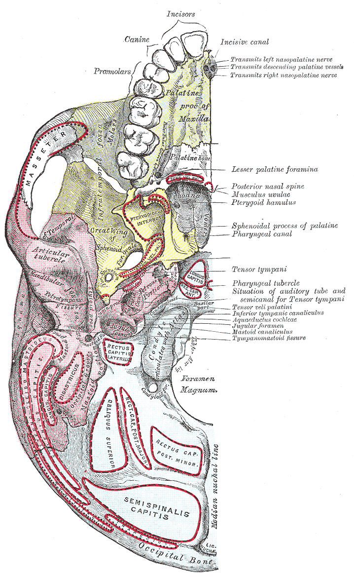
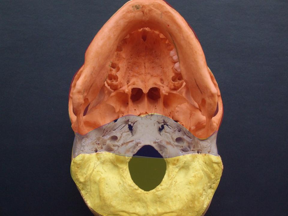
The base of skull, also known as the cranial base or the cranial floor, is the most inferior area of the skull. It is composed of the endocranium and the lower parts of the calvaria. Structure Structures found at the base of the skull are for example: Bones There are five bones that make up the base of the skull: *Ethmoid bone * Sphenoid bone * Occipital bone *Frontal bone *Temporal bone Sinuses *Occipital sinus * Superior sagittal sinus *Superior petrosal sinus Foramina of the skull * Foramen cecum *Optic foramen *Foramen lacerum *Foramen rotundum * Foramen magnum * Foramen ovale *Jugular foramen *Internal auditory meatus *Mastoid foramen *Sphenoidal emissary foramen *Foramen spinosum Sutures *Frontoethmoidal suture *Sphenofrontal suture *Sphenopetrosal suture *Sphenoethmoidal suture * Petrosquamous suture *Sphenosquamosal suture Other *Sphenoidal lingula *Subarcuate fossa *Dorsum sellae *Jugular process *Petro-occipital fissure *Condylar canal * Jugular tubercle * ... [...More Info...] [...Related Items...] OR: [Wikipedia] [Google] [Baidu] |
Sphenoid Bone
The sphenoid bone is an unpaired bone of the neurocranium. It is situated in the middle of the skull towards the front, in front of the basilar part of occipital bone, basilar part of the occipital bone. The sphenoid bone is one of the seven bones that articulate to form the orbit (anatomy), orbit. Its shape somewhat resembles that of a butterfly or bat with its wings extended. Structure It is divided into the following parts: * a median portion, known as the body of sphenoid bone, containing the sella turcica, which houses the pituitary gland as well as the paired paranasal sinuses, the sphenoidal sinuses * two Greater wing of sphenoid bone, greater wings on the lateral side of the body and two Lesser wing of sphenoid bone, lesser wings from the anterior side. * Pterygoid processes of the sphenoides, directed downwards from the junction of the body and the greater wings. Two sphenoidal conchae are situated at the anterior and inferior part of the body. Intrinsic ligaments of ... [...More Info...] [...Related Items...] OR: [Wikipedia] [Google] [Baidu] |
Skeletal System
A skeleton is the structural frame that supports the body of an animal. There are several types of skeletons, including the exoskeleton, which is the stable outer shell of an organism, the endoskeleton, which forms the support structure inside the body, and the hydroskeleton, a flexible internal skeleton supported by fluid pressure. Vertebrates are animals with a vertebral column, and their skeletons are typically composed of bone and cartilage. Invertebrates are animals that lack a vertebral column. The skeletons of invertebrates vary, including hard exoskeleton shells, plated endoskeletons, or Sponge spicule, spicules. Cartilage is a rigid connective tissue that is found in the skeletal systems of vertebrates and invertebrates. Etymology The term ''skeleton'' comes . ''Sceleton'' is an archaic form of the word. Classification Skeletons can be defined by several attributes. Solid skeletons consist of hard substances, such as bone, cartilage, or cuticle. These can be further ... [...More Info...] [...Related Items...] OR: [Wikipedia] [Google] [Baidu] |
Middle Cranial Fossa
The middle cranial fossa, deeper than the anterior cranial fossa, is narrow medially and widens laterally to the sides of the skull. It is separated from the posterior fossa by the clivus and the petrous crest. It is bounded in front by the posterior margins of the lesser wings of the sphenoid bone, the anterior clinoid processes, and the ridge forming the anterior margin of the chiasmatic groove; behind, by the superior angles of the petrous portions of the temporal bones and the dorsum sellæ; laterally by the temporal squamæ, sphenoidal angles of the parietals, and greater wings of the sphenoid. It is traversed by the squamosal, sphenoparietal, sphenosquamosal, and sphenopetrosal sutures. It houses the temporal lobes of the brain and the pituitary gland. A middle fossa craniotomy is one means to surgically remove acoustic neuromas (vestibular schwannoma) growing within the internal auditory canal of the temporal bone. Middle part The middle part of the fossa presents, i ... [...More Info...] [...Related Items...] OR: [Wikipedia] [Google] [Baidu] |
Pterygopalatine Fossa
In human anatomy, the pterygopalatine fossa (sphenopalatine fossa) is a fossa in the skull. A human skull contains two pterygopalatine fossae—one on the left side, and another on the right side. Each fossa is a cone-shaped paired depression deep to the infratemporal fossa and posterior to the maxilla on each side of the skull, located between the pterygoid process and the maxillary tuberosity close to the apex of the orbit. It is the indented area medial to the pterygomaxillary fissure leading into the sphenopalatine foramen. It communicates with the nasal and oral cavities, infratemporal fossa, orbit, pharynx, and middle cranial fossa through eight foramina. Structure Boundaries It has the following boundaries: * ''anterior'': superomedial part of the infratemporal surface of maxilla * ''posterior'': root of the pterygoid process and adjoining anterior surface of the greater wing of sphenoid bone * ''medial'': perpendicular plate of the palatine bone and its orbital and sph ... [...More Info...] [...Related Items...] OR: [Wikipedia] [Google] [Baidu] |
Maxillary Nerve
In neuroanatomy, the maxillary nerve (V) is one of the three branches or divisions of the trigeminal nerve, the fifth (CN V) cranial nerve. It comprises the principal functions of sensation from the maxilla, nasal cavity, sinuses, the palate and subsequently that of the mid-face, and is intermediate, both in position and size, between the ophthalmic nerve and the mandibular nerve.Illustrated Anatomy of the Head and Neck, Fehrenbach and Herring, Elsevier, 2012, page 180 Structure It begins at the middle of the trigeminal ganglion as a flattened plexiform band then it passes through the lateral wall of the cavernous sinus. It leaves the skull through the foramen rotundum, where it becomes more cylindrical in form, and firmer in texture. After leaving foramen rotundum it gives two branches to the pterygopalatine ganglion. It then crosses the pterygopalatine fossa, inclines lateralward on the back of the maxilla, and enters the orbit through the inferior orbital fissure. It then r ... [...More Info...] [...Related Items...] OR: [Wikipedia] [Google] [Baidu] |
Trigeminal Nerve
In neuroanatomy, the trigeminal nerve ( lit. ''triplet'' nerve), also known as the fifth cranial nerve, cranial nerve V, or simply CN V, is a cranial nerve responsible for sensation in the face and motor functions such as biting and chewing; it is the most complex of the cranial nerves. Its name ("trigeminal", ) derives from each of the two nerves (one on each side of the pons) having three major branches: the ophthalmic nerve (V), the maxillary nerve (V), and the mandibular nerve (V). The ophthalmic and maxillary nerves are purely sensory, whereas the mandibular nerve supplies motor as well as sensory (or "cutaneous") functions. Adding to the complexity of this nerve is that autonomic nerve fibers as well as special sensory fibers (taste) are contained within it. The motor division of the trigeminal nerve derives from the basal plate of the embryonic pons, and the sensory division originates in the cranial neural crest. Sensory information from the face and body is proc ... [...More Info...] [...Related Items...] OR: [Wikipedia] [Google] [Baidu] |
Foramina Of The Skull
This article lists foramina that occur in the human body. __TOC__ Skull The human skull has numerous openings (foramina), through which cranial nerves, arteries, veins, and other structures pass. These foramina vary in size and number, with age. Gray193.png , Base of the skull, upper surface Gray187.png , Base of the skull, inferior surface, attachment of muscles marked in red Spine Within the vertebral column (spine) of vertebrates, including the human spine, each bone has an opening at both its top and bottom to allow nerves, arteries, veins, etc. to pass through. Other * Apical foramen, the opening at the tip of the root of a tooth * Foramen ovale (heart), an opening between the venous and arterial sides of the fetal heart * Foramen transversarium, one of a pair of openings in each cervical vertebra, in which the vertebral artery travels * Greater sciatic foramen, a major foramen of the pelvis * Interventricular foramen, channels connecting ventricles in ... [...More Info...] [...Related Items...] OR: [Wikipedia] [Google] [Baidu] |
Base Of The Skull
The base of skull, also known as the cranial base or the cranial floor, is the most inferior area of the skull. It is composed of the endocranium and the lower parts of the calvaria. Structure Structures found at the base of the skull are for example: Bones There are five bones that make up the base of the skull: *Ethmoid bone * Sphenoid bone * Occipital bone *Frontal bone *Temporal bone Sinuses *Occipital sinus * Superior sagittal sinus *Superior petrosal sinus Foramina of the skull * Foramen cecum *Optic foramen *Foramen lacerum *Foramen rotundum * Foramen magnum * Foramen ovale *Jugular foramen *Internal auditory meatus *Mastoid foramen *Sphenoidal emissary foramen *Foramen spinosum Sutures *Frontoethmoidal suture *Sphenofrontal suture *Sphenopetrosal suture *Sphenoethmoidal suture * Petrosquamous suture *Sphenosquamosal suture Other *Sphenoidal lingula *Subarcuate fossa *Dorsum sellae *Jugular process *Petro-occipital fissure *Condylar canal * Jugular tubercle * ... [...More Info...] [...Related Items...] OR: [Wikipedia] [Google] [Baidu] |
Birth
Birth is the act or process of bearing or bringing forth offspring, also referred to in technical contexts as parturition. In mammals, the process is initiated by hormones which cause the muscular walls of the uterus to contract, expelling the fetus at a developmental stage when it is ready to feed and breathe. In some species the offspring is precocial and can move around almost immediately after birth but in others it is altricial and completely dependent on parenting. In marsupials, the fetus is born at a very immature stage after a short gestation and develops further in its mother's womb pouch. It is not only mammals that give birth. Some reptiles, amphibians, fish and invertebrates carry their developing young inside them. Some of these are ovoviviparous, with the eggs being hatched inside the mother's body, and others are viviparous, with the embryo developing inside her body, as in the case of mammals. Mammals Large mammals, such as primates, cattle, horses, some ... [...More Info...] [...Related Items...] OR: [Wikipedia] [Google] [Baidu] |


.jpg)