|
Fleck Corneal Dystrophy
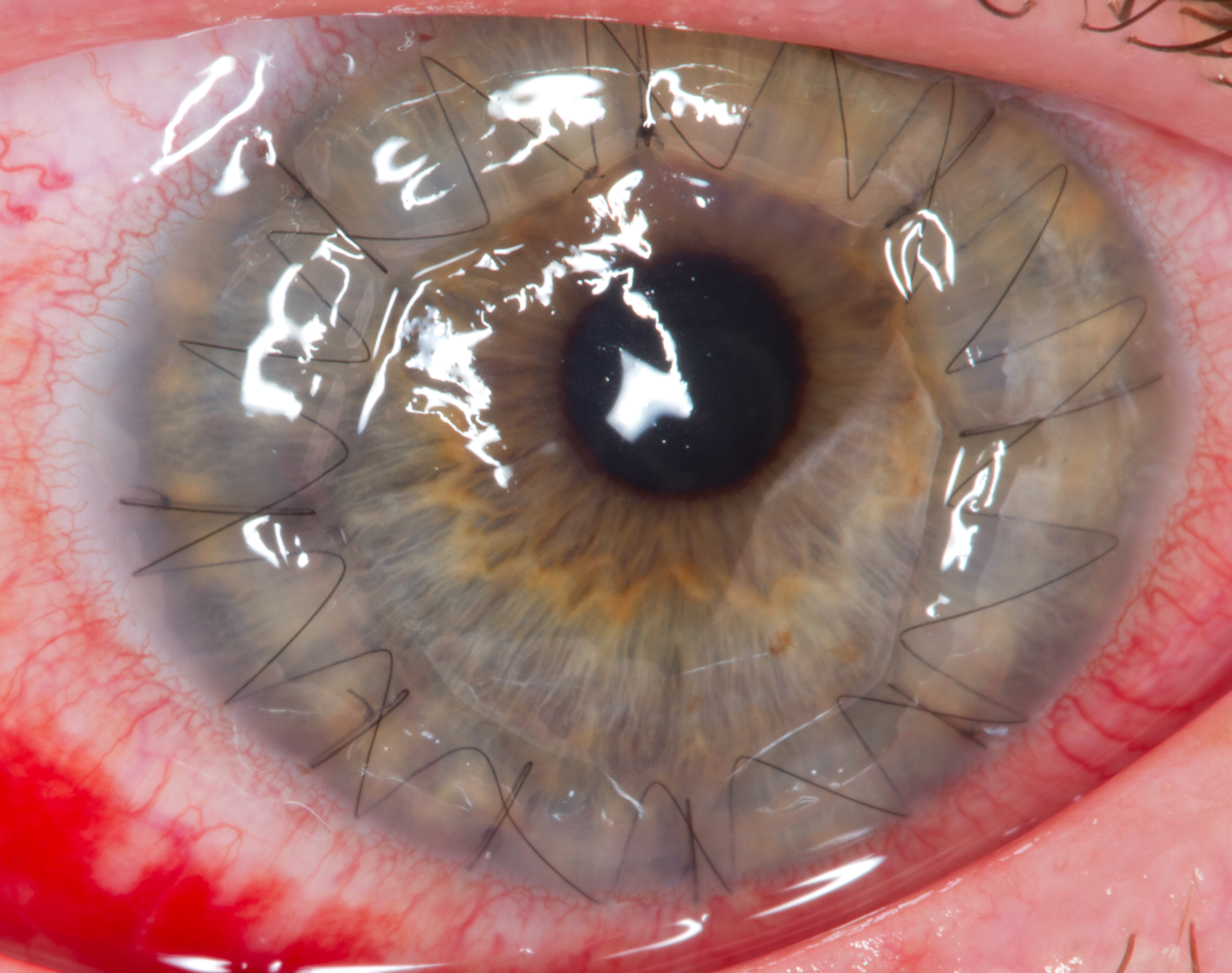
Fleck corneal dystrophy, also known as ''Francois-Neetens speckled corneal dystrophy'', is a rare form of corneal dystrophy. It is caused by mutations in PIKFYVE gene. Small opacities, some of which resemble "flecks", are scattered in the stroma of the patients. Other opacities look more like snowflakes or clouds. The disease is non-progressive and in most cases asymptomatic, with mild photophobia reported by some patients. In a single case report, a corneal transplantation was performed for concurrent keratoconus Keratoconus (KC) is a disorder of the eye that results in progressive thinning of the cornea. This may result in blurry vision, double vision, nearsightedness, irregular astigmatism, and light sensitivity leading to poor quality-of-life. Usu ..., and at 10 years follow-up there was still no evidence of the inclusions in the stroma. References External links Disorders of sclera and cornea {{Genetic-disorder-stub} ... [...More Info...] [...Related Items...] OR: [Wikipedia] [Google] [Baidu] |
Cornea
The cornea is the transparent front part of the eye that covers the iris, pupil, and anterior chamber. Along with the anterior chamber and lens, the cornea refracts light, accounting for approximately two-thirds of the eye's total optical power. In humans, the refractive power of the cornea is approximately 43 dioptres. The cornea can be reshaped by surgical procedures such as LASIK. While the cornea contributes most of the eye's focusing power, its focus is fixed. Accommodation (the refocusing of light to better view near objects) is accomplished by changing the geometry of the lens. Medical terms related to the cornea often start with the prefix "'' kerat-''" from the Greek word κέρας, ''horn''. Structure The cornea has unmyelinated nerve endings sensitive to touch, temperature and chemicals; a touch of the cornea causes an involuntary reflex to close the eyelid. Because transparency is of prime importance, the healthy cornea does not have or need blood vess ... [...More Info...] [...Related Items...] OR: [Wikipedia] [Google] [Baidu] |
Slit Lamp
A slit lamp is an instrument consisting of a high-intensity light source that can be focused to shine a thin sheet of light into the eye. It is used in conjunction with a biomicroscope. The lamp facilitates an examination of the anterior segment and posterior segment of the human eye, which includes the eyelid, sclera, conjunctiva, iris (anatomy), iris, natural crystalline lens, and cornea. The binocular slit-lamp examination provides a stereoscopic magnified view of the eye structures in detail, enabling anatomical diagnoses to be made for a variety of eye conditions. A second, hand-held lens is used to examine the retina. History Two conflicting trends emerged in the development of the slit lamp. One trend originated from clinical research and aimed to apply the increasingly complex and advanced technology of the time. [...More Info...] [...Related Items...] OR: [Wikipedia] [Google] [Baidu] |
Confocal Microscopy
Confocal microscopy, most frequently confocal laser scanning microscopy (CLSM) or laser confocal scanning microscopy (LCSM), is an optical imaging technique for increasing optical resolution and contrast of a micrograph by means of using a spatial pinhole to block out-of-focus light in image formation. Capturing multiple two-dimensional images at different depths in a sample enables the reconstruction of three-dimensional structures (a process known as optical sectioning) within an object. This technique is used extensively in the scientific and industrial communities and typical applications are in life sciences, semiconductor inspection and materials science. Light travels through the sample under a conventional microscope as far into the specimen as it can penetrate, while a confocal microscope only focuses a smaller beam of light at one narrow depth level at a time. The CLSM achieves a controlled and highly limited depth of field. Basic concept The principle of ... [...More Info...] [...Related Items...] OR: [Wikipedia] [Google] [Baidu] |
Corneal Dystrophy
Corneal dystrophy is a group of rare hereditary disorders characterised by bilateral abnormal deposition of substances in the transparent front part of the eye called the cornea. Signs and symptoms Corneal dystrophy may not significantly affect vision in the early stages. However, it does require proper evaluation and treatment for restoration of optimal vision. Corneal dystrophies usually manifest themselves during the first or second decade but sometimes later. It appears as grayish white lines, circles, or clouding of the cornea. Corneal dystrophy can also have a crystalline appearance. There are over 20 corneal dystrophies that affect all parts of the cornea. These diseases share many traits: * They are usually inherited. * They affect the right and left eyes equally. * They are not caused by outside factors, such as injury or diet. * Most progress gradually. * Most usually begin in one of the five corneal layers and may later spread to nearby layers. * Most do not affect oth ... [...More Info...] [...Related Items...] OR: [Wikipedia] [Google] [Baidu] |
Stroma Of Cornea
The stroma of the cornea (or substantia propria) is a fibrous, tough, unyielding, perfectly transparent and the thickest layer of the cornea of the eye. It is between Bowman's membrane anteriorly, and Descemet's membrane posteriorly. At its centre, human corneal stroma is composed of about 200 flattened ''lamellæ'' (layers of collagen fibrils), superimposed one on another. They are each about 1.5-2.5 μm in thickness. The anterior lamellæ interweave more than posterior lamellæ. The fibrils of each lamella are parallel with one another, but at different angles to those of adjacent lamellæ. The lamellæ are produced by keratocytes (corneal connective tissue cells), which occupy about 10% of the substantia propria. Apart from the cells, the major non-aqueous constituents of the stroma are collagen fibrils and proteoglycans. The collagen fibrils are made of a mixture of type I and type V collagens. These molecules are tilted by about 15 degrees to the fibril axis, and because ... [...More Info...] [...Related Items...] OR: [Wikipedia] [Google] [Baidu] |
Corneal Transplantation
Corneal transplantation, also known as corneal grafting, is a surgical procedure where a damaged or diseased cornea is replaced by donated corneal tissue (the graft). When the entire cornea is replaced it is known as penetrating keratoplasty and when only part of the cornea is replaced it is known as lamellar keratoplasty. Keratoplasty simply means surgery to the cornea. The graft is taken from a recently deceased individual with no known diseases or other factors that may affect the chance of survival of the donated tissue or the health of the recipient. The cornea is the transparent front part of the eye that covers the iris, pupil and anterior chamber. The surgical procedure is performed by ophthalmologists, physicians who specialize in eyes, and is often done on an outpatient basis. Donors can be of any age, as is shown in the case of Janis Babson, who donated her eyes after dying at the age of 10. Corneal transplantation is performed when medicines, keratoconus conse ... [...More Info...] [...Related Items...] OR: [Wikipedia] [Google] [Baidu] |
Keratoconus
Keratoconus (KC) is a disorder of the eye that results in progressive thinning of the cornea. This may result in blurry vision, double vision, nearsightedness, irregular astigmatism, and light sensitivity leading to poor quality-of-life. Usually both eyes are affected. In more severe cases a scarring or a circle may be seen within the cornea. While the cause is unknown, it is believed to occur due to a combination of genetic, environmental, and hormonal factors. Patients with a parent, sibling, or child who has keratoconus have 15 to 67 times higher risk in developing corneal ectasia compared to patients with no affected relatives. Proposed environmental factors include rubbing the eyes and allergies. The underlying mechanism involves changes of the cornea to a cone shape. Diagnosis is most often by topography. Topography measures the curvature of the cornea and creates a colored "map" of the cornea. Keratoconus causes very distinctive changes in the appearance of thes ... [...More Info...] [...Related Items...] OR: [Wikipedia] [Google] [Baidu] |
Orphanet J Rare Dis
The ''Orphanet Journal of Rare Diseases'' is a peer-reviewed open access medical journal covering research on rare diseases. It was established in 2006 and the editor-in-chief is Francesc Palau (Hospital Sant Joan de Déu Barcelona and CIBERER, Spain). It is an official journal of Orphanet and is published by BioMed Central, which is part of Springer Nature. Aims, scope and content By its own definition, Orphanet Journal of Rare Diseases is an open access, peer-reviewed journal that encompasses all aspects of rare diseases and orphan drugs. The journal publishes reviews on specific rare diseases, as well as articles on clinical trial outcome reports (positive or negative), and articles on public health issues in the field of rare diseases and orphan drugs. Readers can find contributions which are directly from patients affected by a rare disease or about events, such as the Rare Disease Day. Readers also have the possibility to search articles according to their subject or have t ... [...More Info...] [...Related Items...] OR: [Wikipedia] [Google] [Baidu] |


