|
Fetal Pig
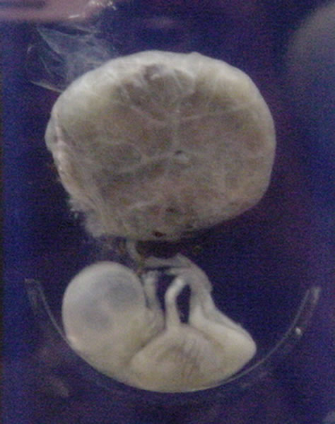
Fetal pigs are unborn pigs used in elementary as well as advanced biology classes as objects for dissection. Pigs, as a mammalian species, provide a good specimen for the study of physiological systems and processes due to the similarities between many pig and human organs. Use in biology labs Along with frogs and earthworms, fetal pigs are among the most common animals used in classroom dissection. There are several reasons for this, including that pigs, like humans, are mammals. Shared traits include common hair, mammary glands, live birth, similar organ systems, metabolic levels, and basic body form. They also allow for the study of fetal circulation, which differs from that of an adult. Fetal pigs are easy to obtain at a relatively low price because they are by-products of the meat-packing industry. These pigs are not bred and killed for this purpose, but are extracted from the deceased sow's uterus. Fetal pigs not used in classroom dissections are often used in fertilizer or s ... [...More Info...] [...Related Items...] OR: [Wikipedia] [Google] [Baidu] |
Fetal Pig - Forensics Class (MxCC)
A fetus or foetus (; : fetuses, foetuses, rarely feti or foeti) is the unborn offspring of a viviparous animal that develops from an embryo. Following the embryonic stage, the fetal stage of development takes place. Prenatal development is a continuum, with no clear defining feature distinguishing an embryo from a fetus. However, in general a fetus is characterized by the presence of all the major body organs, though they will not yet be fully developed and functional, and some may not yet be situated in their final anatomical location. In human prenatal development, fetal development begins from the ninth week after fertilization (which is the eleventh week of gestational age) and continues until the birth of a newborn. Etymology The word ''fetus'' (plural ''fetuses'' or rarely, the solecism '' feti''''Oxford English Dictionary'', 2013''s.v.'' 'fetus') comes from Latin '' fētus'' 'offspring, bringing forth, hatching of young'. The Latin plural ''fetūs'' is not used in ... [...More Info...] [...Related Items...] OR: [Wikipedia] [Google] [Baidu] |
Periarteriolar Lymphoid Sheaths
Periarteriolar lymphoid sheaths (or periarterial lymphatic sheaths, or PALS) are a portion of the white pulp of the spleen. They are populated largely by T cells and surround central arteries within the spleen; the PALS T-cells are presented with blood borne antigens via myeloid dendritic cells. In contrast, the lymphoid portions of the white pulp are dominated by B cell B cells, also known as B lymphocytes, are a type of the lymphocyte subtype. They function in the humoral immunity component of the adaptive immune system. B cells produce antibody molecules which may be either secreted or inserted into the plasm ...s. External links * * Diagram at okstate.edu {{lymphatic-stub Lymphatic organ anatomy ... [...More Info...] [...Related Items...] OR: [Wikipedia] [Google] [Baidu] |
Mesenteries
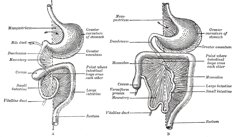
In human anatomy, the mesentery is an organ that attaches the intestines to the posterior abdominal wall, consisting of a double fold of the peritoneum. It helps (among other functions) in storing fat and allowing blood vessels, lymphatics, and nerves to supply the intestines. The (the part of the mesentery that attaches the colon to the abdominal wall) was formerly thought to be a fragmented structure, with all named parts—the ascending, transverse, descending, and sigmoid mesocolons, the mesoappendix, and the mesorectum—separately terminating their insertion into the posterior abdominal wall. However, in 2012, new microscopic and electron microscopic examinations showed the mesocolon to be a single structure derived from the duodenojejunal flexure and extending to the distal mesorectal layer. Thus the mesentery is an internal organ. Structure The mesentery of the small intestine arises from the root of the mesentery (or mesenteric root) and is the part connected wit ... [...More Info...] [...Related Items...] OR: [Wikipedia] [Google] [Baidu] |
Large Intestines
The large intestine, also known as the large bowel, is the last part of the gastrointestinal tract and of the digestive system in tetrapods. Water is absorbed here and the remaining waste material is stored in the rectum as feces before being removed by defecation. The colon (progressing from the ascending colon to the transverse, the descending and finally the sigmoid colon) is the longest portion of the large intestine, and the terms "large intestine" and "colon" are often used interchangeably, but most sources define the large intestine as the combination of the cecum, colon, rectum, and anal canal. Some other sources exclude the anal canal. In humans, the large intestine begins in the right iliac region of the pelvis, just at or below the waist, where it is joined to the end of the small intestine at the cecum, via the ileocecal valve. It then continues as the colon ascending the abdomen, across the width of the abdominal cavity as the transverse colon, and then descending ... [...More Info...] [...Related Items...] OR: [Wikipedia] [Google] [Baidu] |
Stomach
The stomach is a muscular, hollow organ in the upper gastrointestinal tract of Human, humans and many other animals, including several invertebrates. The Ancient Greek name for the stomach is ''gaster'' which is used as ''gastric'' in medical terms related to the stomach. The stomach has a dilated structure and functions as a vital organ in the digestive system. The stomach is involved in the gastric phase, gastric phase of digestion, following the cephalic phase in which the sight and smell of food and the act of chewing are stimuli. In the stomach a chemical breakdown of food takes place by means of secreted digestive enzymes and gastric acid. It also plays a role in regulating gut microbiota, influencing digestion and overall health. The stomach is located between the esophagus and the small intestine. The pyloric sphincter controls the passage of partially digested food (chyme) from the stomach into the duodenum, the first and shortest part of the small intestine, where p ... [...More Info...] [...Related Items...] OR: [Wikipedia] [Google] [Baidu] |
Esophagus
The esophagus (American English), oesophagus (British English), or œsophagus (Œ, archaic spelling) (American and British English spelling differences#ae and oe, see spelling difference) all ; : ((o)e)(œ)sophagi or ((o)e)(œ)sophaguses), colloquially known also as the food pipe, food tube, or gullet, is an Organ (anatomy), organ in vertebrates through which food passes, aided by Peristalsis, peristaltic contractions, from the Human pharynx, pharynx to the stomach. The esophagus is a :wiktionary:fibromuscular, fibromuscular tube, about long in adults, that travels behind the trachea and human heart, heart, passes through the Thoracic diaphragm, diaphragm, and empties into the uppermost region of the stomach. During swallowing, the epiglottis tilts backwards to prevent food from going down the larynx and lungs. The word ''esophagus'' is from Ancient Greek οἰσοφάγος (oisophágos), from οἴσω (oísō), future form of φέρω (phérō, "I carry") + ἔφαγον ( ... [...More Info...] [...Related Items...] OR: [Wikipedia] [Google] [Baidu] |
Monogastric
A monogastric organism defines one of the many types of digestive tracts found among different species of animals. The defining feature of a monogastric is that it has a simple single-chambered stomach (one stomach). A monogastric can be classified as an herbivore, an omnivore (facultative carnivore), or a carnivore (obligate carnivore). Herbivores have a plant-based diet, omnivores have a plant and meat-based diet, and carnivores only eat meat. Examples of monogastric herbivores include horses, rabbits, and guinea pigs. Examples of monogastric omnivores include humans, pigs, and hamsters. Furthermore, there are monogastric carnivores such as cats and seals. A monogastric digestive tract is slightly different from other types of digestive tracts such as a ruminant and avian. Ruminant organisms have a four-chambered complex stomach and avian organisms have a two-chambered stomach. An example of a ruminant and avian are cattle and chickens. Digestive System The digestive system o ... [...More Info...] [...Related Items...] OR: [Wikipedia] [Google] [Baidu] |
Foramen Ovale (heart)
In the fetal heart, the foramen ovale (), also foramen Botalli or the ostium secundum of Born, allows blood to enter the left atrium from the right atrium. It is one of two fetal cardiac shunts, the other being the ductus arteriosus (which allows blood that still escapes to the right ventricle to bypass the pulmonary circulation). Another similar adaptation in the fetus is the ductus venosus. In most individuals, the foramen ovale closes at birth. It later forms the fossa ovalis. Development The foramen ovale () forms in the late fourth week of gestation, as a small passageway between the septum secundum and the ostium secundum. Initially the atria are separated from one another by the septum primum except for a small opening below the septum, the ostium primum. As the septum primum grows, the ostium primum narrows and eventually closes. Before it does so, bloodflow from the inferior vena cava wears down a portion of the septum primum, forming the ostium secundum. Some e ... [...More Info...] [...Related Items...] OR: [Wikipedia] [Google] [Baidu] |
Umbilical Arteries
The umbilical artery is a paired artery (with one for each half of the body) that is found in the abdominal and pelvic regions. In the fetus, it extends into the umbilical cord. Structure Development The umbilical arteries supply systemic arterial blood from the fetus to the placenta. Although this blood is sometimes referred to as deoxygenated blood it is not, and has the same oxygen saturation and nutrients as blood distributed to the other fetal tissues. There are usually two umbilical arteries present together with one umbilical vein in the umbilical cord. The umbilical arteries surround the urinary bladder and then carry all the deoxygenated blood out of the fetus through the umbilical cord. Inside the placenta, the umbilical arteries connect with each other at a distance of approximately 5 mm from the cord insertion in what is called the ''Hyrtl anastomosis''. Subsequently, they branch into chorionic arteries or ''intraplacental fetal arteries''. The umbilical art ... [...More Info...] [...Related Items...] OR: [Wikipedia] [Google] [Baidu] |
Muscular
MUSCULAR (DS-200B), located in the United Kingdom, is the name of a surveillance program jointly operated by Britain's Government Communications Headquarters (GCHQ) and the U.S. National Security Agency (NSA) that was revealed by documents released by Edward Snowden and interviews with knowledgeable officials. GCHQ is the primary operator of the program. GCHQ and the NSA have secretly broken into the main communications links that connect the data centers of Yahoo! and Google. Substantive information about the program was made public at the end of October 2013. Overview The programme is jointly run by: * – Government Communications Headquarters (GCHQ) (United Kingdom) * – U.S. National Security Agency (NSA) MUSCULAR is one of at least four other similar programs that rely on a trusted 2nd party, programs which together are known as WINDSTOP. In a 30-day period from December 2012 to January 2013, MUSCULAR was responsible for collecting 181 million records. It was however dw ... [...More Info...] [...Related Items...] OR: [Wikipedia] [Google] [Baidu] |
Respiratory
The respiratory system (also respiratory apparatus, ventilatory system) is a biological system consisting of specific organs and structures used for gas exchange in animals and plants. The anatomy and physiology that make this happen varies greatly, depending on the size of the organism, the environment in which it lives and its evolutionary history. In land animals, the respiratory surface is internalized as linings of the lungs. Gas exchange in the lungs occurs in millions of small air sacs; in mammals and reptiles, these are called alveoli, and in birds, they are known as atria. These microscopic air sacs have a very rich blood supply, thus bringing the air into close contact with the blood. These air sacs communicate with the external environment via a system of airways, or hollow tubes, of which the largest is the trachea, which branches in the middle of the chest into the two main bronchi. These enter the lungs where they branch into progressively narrower secondary an ... [...More Info...] [...Related Items...] OR: [Wikipedia] [Google] [Baidu] |
Skeletal
A skeleton is the structural frame that supports the body of most animals. There are several types of skeletons, including the exoskeleton, which is a rigid outer shell that holds up an organism's shape; the endoskeleton, a rigid internal frame to which the organs and soft tissues attach; and the hydroskeleton, a flexible internal structure supported by the hydrostatic pressure of body fluids. Vertebrates are animals with an endoskeleton centered around an axial vertebral column, and their skeletons are typically composed of bones and cartilages. Invertebrates are other animals that lack a vertebral column, and their skeletons vary, including hard-shelled exoskeleton (arthropods and most molluscs), plated internal shells (e.g. cuttlebones in some cephalopods) or rods (e.g. ossicles in echinoderms), hydrostatically supported body cavities (most), and spicules (sponges). Cartilage is a rigid connective tissue that is found in the skeletal systems of vertebrates and inverteb ... [...More Info...] [...Related Items...] OR: [Wikipedia] [Google] [Baidu] |


