|
Eye Of The Jaguar
Eyes are organs of the visual system. They provide living organisms with vision, the ability to receive and process visual detail, as well as enabling several photo response functions that are independent of vision. Eyes detect light and convert it into electro-chemical impulses in neurons (neurones). In higher organisms, the eye is a complex optical system which collects light from the surrounding environment, regulates its intensity through a diaphragm, focuses it through an adjustable assembly of lenses to form an image, converts this image into a set of electrical signals, and transmits these signals to the brain through complex neural pathways that connect the eye via the optic nerve to the visual cortex and other areas of the brain. Eyes with resolving power have come in ten fundamentally different forms, and 96% of animal species possess a complex optical system. Image-resolving eyes are present in molluscs, chordates and arthropods. The most simple eyes, pit ... [...More Info...] [...Related Items...] OR: [Wikipedia] [Google] [Baidu] |
Nervous System
In biology, the nervous system is the highly complex part of an animal that coordinates its actions and sensory information by transmitting signals to and from different parts of its body. The nervous system detects environmental changes that impact the body, then works in tandem with the endocrine system to respond to such events. Nervous tissue first arose in wormlike organisms about 550 to 600 million years ago. In vertebrates it consists of two main parts, the central nervous system (CNS) and the peripheral nervous system (PNS). The CNS consists of the brain and spinal cord. The PNS consists mainly of nerves, which are enclosed bundles of the long fibers or axons, that connect the CNS to every other part of the body. Nerves that transmit signals from the brain are called motor nerves or '' efferent'' nerves, while those nerves that transmit information from the body to the CNS are called sensory nerves or '' afferent''. Spinal nerves are mixed nerves that serve both fu ... [...More Info...] [...Related Items...] OR: [Wikipedia] [Google] [Baidu] |
Annual Review Of Neuroscience , in biology
{{disambiguation ...
Annual may refer to: * Annual publication, periodical publications appearing regularly once per year **Yearbook ** Literary annual *Annual plant * Annual report * Annual giving * Annual, Morocco, a settlement in northeastern Morocco * Annuals (band), a musical group See also * Annual Review (other) * Circannual cycle A circannual cycle is a biological process that occurs in living creatures over the period of approximately one year. This cycle was first discovered by Ebo Gwinner and Canadian biologist Ted Pengelley. It is classified as an Infradian rhythm, whi ... [...More Info...] [...Related Items...] OR: [Wikipedia] [Google] [Baidu] |
Depth Perception
Depth perception is the ability to perceive distance to objects in the world using the visual system and visual perception. It is a major factor in perceiving the world in three dimensions. Depth perception happens primarily due to stereopsis and accommodation of the eye. Depth sensation is the corresponding term for non-human animals, since although it is known that they can sense the distance of an object, it is not known whether they perceive it in the same way that humans do. Depth perception arises from a variety of depth cues. These are typically classified into binocular cues and monocular cues. Binocular cues are based on the receipt of sensory information in three dimensions from both eyes and monocular cues can be observed with just one eye. Binocular cues include retinal disparity, which exploits parallax and vergence. Stereopsis is made possible with binocular vision. Monocular cues include relative size (distant objects subtend smaller visual angles than near obj ... [...More Info...] [...Related Items...] OR: [Wikipedia] [Google] [Baidu] |
Binocular Vision
In biology, binocular vision is a type of vision in which an animal has two eyes capable of facing the same direction to perceive a single three-dimensional image of its surroundings. Binocular vision does not typically refer to vision where an animal has eyes on opposite sides of its head and shares no field of view between them, like in some animals. Neurological researcher Manfred Fahle has stated six specific advantages of having two eyes rather than just one: #It gives a creature a "spare eye" in case one is damaged. #It gives a wider field of view. For example, humans have a maximum horizontal field of view of approximately 190 degrees with two eyes, approximately 120 degrees of which makes up the binocular field of view (seen by both eyes) flanked by two uniocular fields (seen by only one eye) of approximately 40 degrees. #It can give stereopsis in which binocular disparity (or parallax) provided by the two eyes' different positions on the head gives precise depth per ... [...More Info...] [...Related Items...] OR: [Wikipedia] [Google] [Baidu] |
Colour
Color (American English) or colour (British English) is the visual perceptual property deriving from the spectrum of light interacting with the photoreceptor cells of the eyes. Color categories and physical specifications of color are associated with objects or materials based on their physical properties such as light absorption, reflection, or emission spectra. By defining a color space, colors can be identified numerically by their coordinates. Because perception of color stems from the varying spectral sensitivity of different types of cone cells in the retina to different parts of the spectrum, colors may be defined and quantified by the degree to which they stimulate these cells. These physical or physiological quantifications of color, however, do not fully explain the psychophysical perception of color appearance. Color science includes the perception of color by the eye and brain, the origin of color in materials, color theory in art, and the physics of electr ... [...More Info...] [...Related Items...] OR: [Wikipedia] [Google] [Baidu] |
Bison Bonasus Right Eye Close-up
Bison are large bovines in the genus ''Bison'' (Greek: "wild ox" (bison)) within the tribe Bovini. Two extant and numerous extinct species are recognised. Of the two surviving species, the American bison, ''B. bison'', found only in North America, is the more numerous. Although colloquially referred to as a buffalo in the United States and Canada, it is only distantly related to the true buffalo. The North American species is composed of two subspecies, the Plains bison, ''B. b. bison'', and the wood bison, ''B. b. athabascae'', which is the namesake of Wood Buffalo National Park in Canada. A third subspecies, the eastern bison (''B. b. pennsylvanicus'') is no longer considered a valid taxon, being a junior synonym of ''B. b. bison''. References to "woods bison" or "wood bison" from the eastern United States refer to this subspecies, not ''B. b. athabascae'', which was not found in the region. The European bison, ''B. bonasus'', or wisent, or zubr, or colloquially European bu ... [...More Info...] [...Related Items...] OR: [Wikipedia] [Google] [Baidu] |
Pupillary Light Reflex
The pupillary light reflex (PLR) or photopupillary reflex is a reflex that controls the diameter of the pupil, in response to the intensity (luminance) of light that falls on the retinal ganglion cells of the retina in the back of the eye, thereby assisting in adaptation of vision to various levels of lightness/darkness. A greater intensity of light causes the pupil to constrict ( miosis/myosis; thereby allowing less light in), whereas a lower intensity of light causes the pupil to dilate ( mydriasis, expansion; thereby allowing more light in). Thus, the pupillary light reflex regulates the intensity of light entering the eye. Light shone into one eye will cause both pupils to constrict. Terminology The pupil is the dark circular opening in the center of the iris and is where light enters the eye. By analogy with a camera, the pupil is equivalent to aperture, whereas the iris is equivalent to the diaphragm. It may be helpful to consider the ''Pupillary reflex'' as an Iris' refle ... [...More Info...] [...Related Items...] OR: [Wikipedia] [Google] [Baidu] |
Pretectal Area
In neuroanatomy, the pretectal area, or pretectum, is a midbrain structure composed of seven nuclei and comprises part of the subcortical visual system. Through reciprocal bilateral projections from the retina, it is involved primarily in mediating behavioral responses to acute changes in ambient light such as the pupillary light reflex, the optokinetic reflex, and temporary changes to the circadian rhythm. In addition to the pretectum's role in the visual system, the anterior pretectal nucleus has been found to mediate somatosensory and nociceptive information. Location and structure The pretectum is a bilateral group of highly interconnected nuclei located near the junction of the midbrain and forebrain. The pretectum is generally classified as a midbrain structure, although because of its proximity to the forebrain it is sometimes classified as part of the caudal diencephalon (forebrain). Within vertebrates, the pretectum is located directly anterior to the superior colliculus ... [...More Info...] [...Related Items...] OR: [Wikipedia] [Google] [Baidu] |
Suprachiasmatic Nucleus
The suprachiasmatic nucleus or nuclei (SCN) is a tiny region of the brain in the hypothalamus, situated directly above the optic chiasm. It is responsible for controlling circadian rhythms. The neuronal and hormonal activities it generates regulate many different body functions in a 24-hour cycle. The mouse SCN contains approximately 20,000 neurons. The SCN interacts with many other regions of the brain. It contains several cell types and several different peptides (including vasopressin and vasoactive intestinal peptide) and neurotransmitters. Neuroanatomy The SCN is situated in the anterior part of the hypothalamus immediately dorsal, or ''superior'' (hence supra) to the optic chiasm (CHO) bilateral to (on either side of) the third ventricle. The nucleus can be divided into ventrolateral and dorsolateral portions, also known as the core and shell, respectively. These regions differ in their expression of the clock genes, the core expresses them in response to stimuli whereas ... [...More Info...] [...Related Items...] OR: [Wikipedia] [Google] [Baidu] |
Retinohypothalamic Tract
In neuroanatomy, the retinohypothalamic tract (RHT) is a photic neural input pathway involved in the circadian rhythms of mammals. The origin of the retinohypothalamic tract is the intrinsically photosensitive retinal ganglion cells (ipRGC), which contain the photopigment melanopsin. The axons of the ipRGCs belonging to the retinohypothalamic tract project directly, monosynaptically, to the suprachiasmatic nuclei (SCN) via the optic nerve and the optic chiasm. The suprachiasmatic nuclei receive and interpret information on environmental light, dark and day length, important in the entrainment of the "body clock". They can coordinate peripheral "clocks" and direct the pineal gland to secrete the hormone melatonin. Structure The retinohypothalamic tract consists of retinal ganglion cells.http://www.ifc.unam.mx/curso_ritmos/capitulo12/Hannibal021.pdf A distinct population of ganglion cells, known as intrinsically photosensitive retinal ganglion cells (ipRGCs), is critically resp ... [...More Info...] [...Related Items...] OR: [Wikipedia] [Google] [Baidu] |
Photosensitive Ganglion Cell
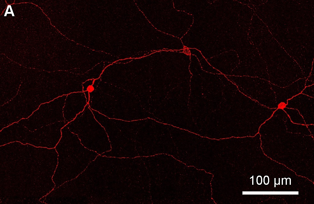
Intrinsically photosensitive retinal ganglion cells (ipRGCs), also called photosensitive retinal ganglion cells (pRGC), or melanopsin-containing retinal ganglion cells (mRGCs), are a type of neuron in the retina of the mammalian eye. The presence of (something like) ipRGCs was first suspected in 1927 when rodless, coneless mice still responded to a light stimulus through pupil constriction, This implied that rods and cones are not the only light-sensitive neurons in the retina. Yet research on these cells did not advance until the 1980s. Recent research has shown that these retinal ganglion cells, unlike other retinal ganglion cells, are intrinsically photosensitive due to the presence of melanopsin, a light-sensitive protein. Therefore they constitute a third class of photoreceptors, in addition to rod and cone cells. Overview Compared to the rods and cones, the ipRGCs respond more sluggishly and signal the presence of light over the long term. They represent a very small su ... [...More Info...] [...Related Items...] OR: [Wikipedia] [Google] [Baidu] |


