|
Extrafusal Muscle Fiber
Extrafusal muscle fibers are the standard skeletal muscle fibers that are innervated by alpha motor neurons and generate tension by contracting, thereby allowing for skeletal movement. They make up the large mass of skeletal striated muscle tissue and are attached to bone by fibrous tissue extensions (tendons). Each alpha motor neuron and the extrafusal muscle fibers innervated by it make up a motor unit. The connection between the alpha motor neuron and the extrafusal muscle fiber is a neuromuscular junction, where the neuron's signal, the action potential, is transduced to the muscle fiber by the neurotransmitter acetylcholine. Extrafusal muscle fibers are not to be confused with intrafusal muscle fibers, which are innervated by sensory nerve endings in central noncontractile parts and by gamma motor neurons in contractile ends and thus serve as a sensory proprioceptor. Extrafusal muscle fibers can be generated in vitro (in a dish) from pluripotent stem cells through dire ... [...More Info...] [...Related Items...] OR: [Wikipedia] [Google] [Baidu] |
Skeletal Muscle
Skeletal muscles (commonly referred to as muscles) are organs of the vertebrate muscular system and typically are attached by tendons to bones of a skeleton. The muscle cells of skeletal muscles are much longer than in the other types of muscle tissue, and are often known as muscle fibers. The muscle tissue of a skeletal muscle is striated – having a striped appearance due to the arrangement of the sarcomeres. Skeletal muscles are voluntary muscles under the control of the somatic nervous system. The other types of muscle are cardiac muscle which is also striated and smooth muscle which is non-striated; both of these types of muscle tissue are classified as involuntary, or, under the control of the autonomic nervous system. A skeletal muscle contains multiple muscle fascicle, fascicles – bundles of muscle fibers. Each individual fiber, and each muscle is surrounded by a type of connective tissue layer of fascia. Muscle fibers are formed from the cell fusion, fusion of ... [...More Info...] [...Related Items...] OR: [Wikipedia] [Google] [Baidu] |
Proprioceptor
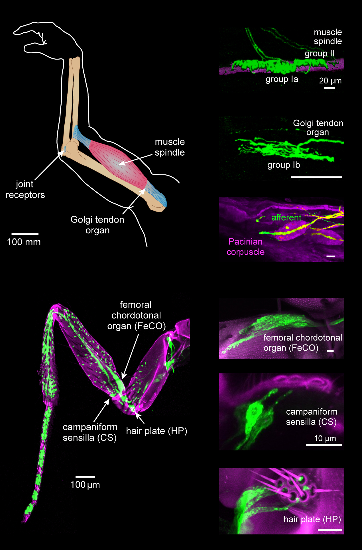
Proprioception ( ), also referred to as kinaesthesia (or kinesthesia), is the sense of self-movement, force, and body position. It is sometimes described as the "sixth sense". Proprioception is mediated by proprioceptors, mechanosensory neurons located within muscles, tendons, and joints. Most animals possess multiple subtypes of proprioceptors, which detect distinct kinematic parameters, such as joint position, movement, and load. Although all mobile animals possess proprioceptors, the structure of the sensory organs can vary across species. Proprioceptive signals are transmitted to the central nervous system, where they are integrated with information from other sensory systems, such as the visual system and the vestibular system, to create an overall representation of body position, movement, and acceleration. In many animals, sensory feedback from proprioceptors is essential for stabilizing body posture and coordinating body movement. System overview In vertebrates, limb v ... [...More Info...] [...Related Items...] OR: [Wikipedia] [Google] [Baidu] |
Gamma Motor Neuron
A gamma motor neuron (γ motor neuron), also called gamma motoneuron, or fusimotor neuron, is a type of lower motor neuron that takes part in the process of muscle contraction, and represents about 30% of ( Aγ) fibers going to the muscle. Like alpha motor neurons, their cell bodies are located in the anterior grey column of the spinal cord. They receive input from the reticular formation of the pons in the brainstem. Their axons are smaller than those of the alpha motor neurons, with a diameter of only 5 μm. Unlike the alpha motor neurons, gamma motor neurons do not directly adjust the lengthening or shortening of muscles. However, their role is important in keeping muscle spindles taut, thereby allowing the continued firing of alpha neurons, leading to muscle contraction. These neurons also play a role in adjusting the sensitivity of muscle spindles. The presence of myelination in gamma motor neurons allows a conduction velocity of 4 to 24 meters per second, significantl ... [...More Info...] [...Related Items...] OR: [Wikipedia] [Google] [Baidu] |
Alpha Motor Neuron
Alpha (α) motor neurons (also called alpha motoneurons), are large, multipolar lower motor neurons of the brainstem and spinal cord. They innervate extrafusal muscle fibers of skeletal muscle and are directly responsible for initiating their contraction. Alpha motor neurons are distinct from gamma motor neurons, which innervate intrafusal muscle fibers of muscle spindles. While their cell bodies are found in the central nervous system (CNS), α motor neurons are also considered part of the somatic nervous system—a branch of the peripheral nervous system (PNS)—because their axons extend into the periphery to innervate skeletal muscles. An alpha motor neuron and the muscle fibers it innervates is a motor unit. A motor neuron pool contains the cell bodies of all the alpha motor neurons involved in contracting a single muscle. Location Alpha motor neurons (α-MNs) innervating the head and neck are found in the brainstem; the remaining α-MNs innervate the rest of the body and ... [...More Info...] [...Related Items...] OR: [Wikipedia] [Google] [Baidu] |
Type II Sensory Fiber
Type II sensory fiber (group Aβ) is a type of sensory fiber, the second of the two main groups of touch receptors. The responses of different type Aβ fibers to these stimuli can be subdivided based on their adaptation properties, traditionally into rapidly adapting (RA) or slowly adapting (SA) neurons. Type II sensory fibers are slowly-adapting (SA), meaning that even when there is no change in touch, they keep respond to stimuli and fire action potentials. In the body, Type II sensory fibers belong to pseudounipolar neurons. The most notable example are neurons with Merkel cell-neurite complexes on their dendrites (sense static touch) and Ruffini endings (sense stretch on the skin and over-extension inside joints). Under pathological conditions they may become hyper-excitable leading to stimuli that would usually elicit sensations of tactile touch causing pain. These changes are in part induced by PGE2 which is produced by COX1, and type II fibers with free nerve endings ... [...More Info...] [...Related Items...] OR: [Wikipedia] [Google] [Baidu] |
Type Ia Sensory Fiber
A type Ia sensory fiber, or a primary afferent fiber is a type of afferent nerve fiber. It is the sensory fiber of a stretch receptor called the muscle spindle found in muscles, which constantly monitors the rate at which a muscle stretch changes. The information carried by type Ia fibers contributes to the sense of proprioception. Function of muscle spindles For the body to keep moving properly and with finesse, the nervous system has to have a constant input of sensory data coming from areas such as the muscles and joints. In order to receive a continuous stream of sensory data, the body has developed special sensory receptors called proprioceptors. Muscle spindles are a type of proprioceptor, and they are found inside the muscle itself. They lie parallel with the contractile fibers. This gives them the ability to monitor muscle length with precision. Types of sensory fibers This change in length of the spindle is transduced (transformed into electric membrane potentials ... [...More Info...] [...Related Items...] OR: [Wikipedia] [Google] [Baidu] |
Intrafusal Muscle Fiber
Intrafusal muscle fibers are skeletal muscle fibers that serve as specialized sensory organs (proprioceptors). They detect the amount and rate of change in length of a muscle.Casagrand, Janet (2008) ''Action and Movement: Spinal Control of Motor Units and Spinal Reflexes.'' University of Colorado, Boulder. They constitute the muscle spindle, and are innervated by both sensory (afferent) and motor (efferent) fibers. Intrafusal muscle fibers are not to be confused with extrafusal muscle fibers, which contract, generating skeletal movement and are innervated by alpha motor neurons. Structure Types There are two types of intrafusal muscle fibers: nuclear bag fibers and nuclear chain fibers. They bear two types of sensory ending, known as annulospiral and flower-spray endings. Both ends of these fibers contract, but the central region only stretches and does not contract. Intrafusal muscle fibers are walled off from the rest of the muscle by an outer connective tissue sheath ... [...More Info...] [...Related Items...] OR: [Wikipedia] [Google] [Baidu] |
Directed Differentiation
Directed differentiation is a bioengineering methodology at the interface of stem cell biology, developmental biology and tissue engineering. It is essentially harnessing the potential of stem cells by constraining their differentiation in vitro toward a specific cell type or tissue of interest. Stem cells are by definition pluripotent, able to differentiate into several cell types such as neurons, cardiomyocytes, hepatocytes, etc. Efficient ''directed differentiation'' requires a detailed understanding of the lineage and cell fate decision, often provided by developmental biology. Conceptual frame During differentiation, pluripotent cells make a number of developmental decisions to generate first the three germ layers (ectoderm, mesoderm and endoderm) of the embryo and intermediate progenitors, followed by subsequent decisions or check points, giving rise to all the body's mature tissues. The differentiation process can be modeled as sequence of binary decisions based on probabi ... [...More Info...] [...Related Items...] OR: [Wikipedia] [Google] [Baidu] |
Induced Pluripotent Stem Cells
Induced pluripotent stem cells (also known as iPS cells or iPSCs) are a type of pluripotent stem cell that can be generated directly from a somatic cell. The iPSC technology was pioneered by Shinya Yamanaka's lab in Kyoto, Japan, who showed in 2006 that the introduction of four specific genes (named Myc, Oct3/4, Sox2 and Klf4), collectively known as Yamanaka factors, encoding transcription factors could convert somatic cells into pluripotent stem cells. He was awarded the 2012 Nobel Prize along with Sir John Gurdon "for the discovery that mature cells can be reprogrammed to become pluripotent." Pluripotent stem cells hold promise in the field of regenerative medicine. Because they can propagate indefinitely, as well as give rise to every other cell type in the body (such as neurons, heart, pancreatic, and liver cells), they represent a single source of cells that could be used to replace those lost to damage or disease. The most well-known type of pluripotent stem cell is the ... [...More Info...] [...Related Items...] OR: [Wikipedia] [Google] [Baidu] |
Gamma Motor Neurons
A gamma motor neuron (γ motor neuron), also called gamma motoneuron, or fusimotor neuron, is a type of lower motor neuron that takes part in the process of muscle contraction, and represents about 30% of ( Aγ) fibers going to the muscle. Like alpha motor neurons, their cell bodies are located in the anterior grey column of the spinal cord. They receive input from the reticular formation of the pons in the brainstem. Their axons are smaller than those of the alpha motor neurons, with a diameter of only 5 μm. Unlike the alpha motor neurons, gamma motor neurons do not directly adjust the lengthening or shortening of muscles. However, their role is important in keeping muscle spindles taut, thereby allowing the continued firing of alpha neurons, leading to muscle contraction. These neurons also play a role in adjusting the sensitivity of muscle spindles. The presence of myelination in gamma motor neurons allows a conduction velocity of 4 to 24 meters per second, significantl ... [...More Info...] [...Related Items...] OR: [Wikipedia] [Google] [Baidu] |
Alpha Motor Neuron
Alpha (α) motor neurons (also called alpha motoneurons), are large, multipolar lower motor neurons of the brainstem and spinal cord. They innervate extrafusal muscle fibers of skeletal muscle and are directly responsible for initiating their contraction. Alpha motor neurons are distinct from gamma motor neurons, which innervate intrafusal muscle fibers of muscle spindles. While their cell bodies are found in the central nervous system (CNS), α motor neurons are also considered part of the somatic nervous system—a branch of the peripheral nervous system (PNS)—because their axons extend into the periphery to innervate skeletal muscles. An alpha motor neuron and the muscle fibers it innervates is a motor unit. A motor neuron pool contains the cell bodies of all the alpha motor neurons involved in contracting a single muscle. Location Alpha motor neurons (α-MNs) innervating the head and neck are found in the brainstem; the remaining α-MNs innervate the rest of the body and ... [...More Info...] [...Related Items...] OR: [Wikipedia] [Google] [Baidu] |
Intrafusal Muscle Fiber
Intrafusal muscle fibers are skeletal muscle fibers that serve as specialized sensory organs (proprioceptors). They detect the amount and rate of change in length of a muscle.Casagrand, Janet (2008) ''Action and Movement: Spinal Control of Motor Units and Spinal Reflexes.'' University of Colorado, Boulder. They constitute the muscle spindle, and are innervated by both sensory (afferent) and motor (efferent) fibers. Intrafusal muscle fibers are not to be confused with extrafusal muscle fibers, which contract, generating skeletal movement and are innervated by alpha motor neurons. Structure Types There are two types of intrafusal muscle fibers: nuclear bag fibers and nuclear chain fibers. They bear two types of sensory ending, known as annulospiral and flower-spray endings. Both ends of these fibers contract, but the central region only stretches and does not contract. Intrafusal muscle fibers are walled off from the rest of the muscle by an outer connective tissue sheath ... [...More Info...] [...Related Items...] OR: [Wikipedia] [Google] [Baidu] |


.jpg)