|
Excretory System
The excretory system is a passive biological system that removes excess, unnecessary materials from the body fluids of an organism, so as to help maintain internal chemical homeostasis and prevent damage to the body. The dual function of excretory systems is the elimination of the waste products of metabolism and to drain the body of used up and broken down components in a liquid and gaseous state. In humans and other amniotes ( mammals, birds and reptiles) most of these substances leave the body as urine and to some degree exhalation, mammals also expel them through sweating. Only the organs specifically used for the excretion are considered a part of the excretory system. In the narrow sense, the term refers to the urinary system. However, as excretion involves several functions that are only superficially related, it is not usually used in more formal classifications of anatomy or function. As most healthy functioning organs produce metabolic and other wastes, the entire or ... [...More Info...] [...Related Items...] OR: [Wikipedia] [Google] [Baidu] |
Body Fluid
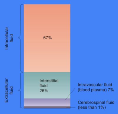
Body fluids, bodily fluids, or biofluids, sometimes body liquids, are liquids within the human body. In lean healthy adult men, the total body water is about 60% (60–67%) of the total Human body weight, body weight; it is usually slightly lower in women (52-55%). The exact percentage of fluid relative to body weight is inversely proportional to the percentage of body fat. A lean 70 kg (160 pound) man, for example, has about 42 (42–47) liters of water in his body. The total body of water is divided into fluid compartments, between the Fluid compartments#Intracellular compartment, intracellular fluid (ICF) compartment (also called space, or volume) and the extracellular fluid (ECF) compartment (space, volume) in a ''two-to-one ratio'': 28 (28–32) liters are inside cells and 14 (14–15) liters are outside cells. The ECF compartment is divided into the interstitial fluid volume – the fluid outside both the cells and the blood vessels – and the Blood vessel, intravascular vo ... [...More Info...] [...Related Items...] OR: [Wikipedia] [Google] [Baidu] |
Renal Artery
The renal arteries are paired arteries that supply the kidneys with blood. Each is directed across the crus of the diaphragm, so as to form nearly a right angle. The renal arteries carry a large portion of total blood flow to the kidneys. Up to a third of total cardiac output can pass through the renal arteries to be filtered by the kidneys. Structure The renal arteries normally arise at a 90° angle off of the left interior side of the abdominal aorta, immediately below the superior mesenteric artery. They have a radius of approximately 0.25 cm, 0.26 cm at the root. The measured mean diameter can differ depending on the imaging method used. For example, the diameter was found to be 5.04 ± 0.74 mm using ultrasound but 5.68 ± 1.19 mm using angiography. Due to the anatomical position of the aorta, the inferior vena cava, and the kidneys, the right renal artery is normally longer than the left renal artery. * The right passes behind the inferior vena cava, ... [...More Info...] [...Related Items...] OR: [Wikipedia] [Google] [Baidu] |
Prostate
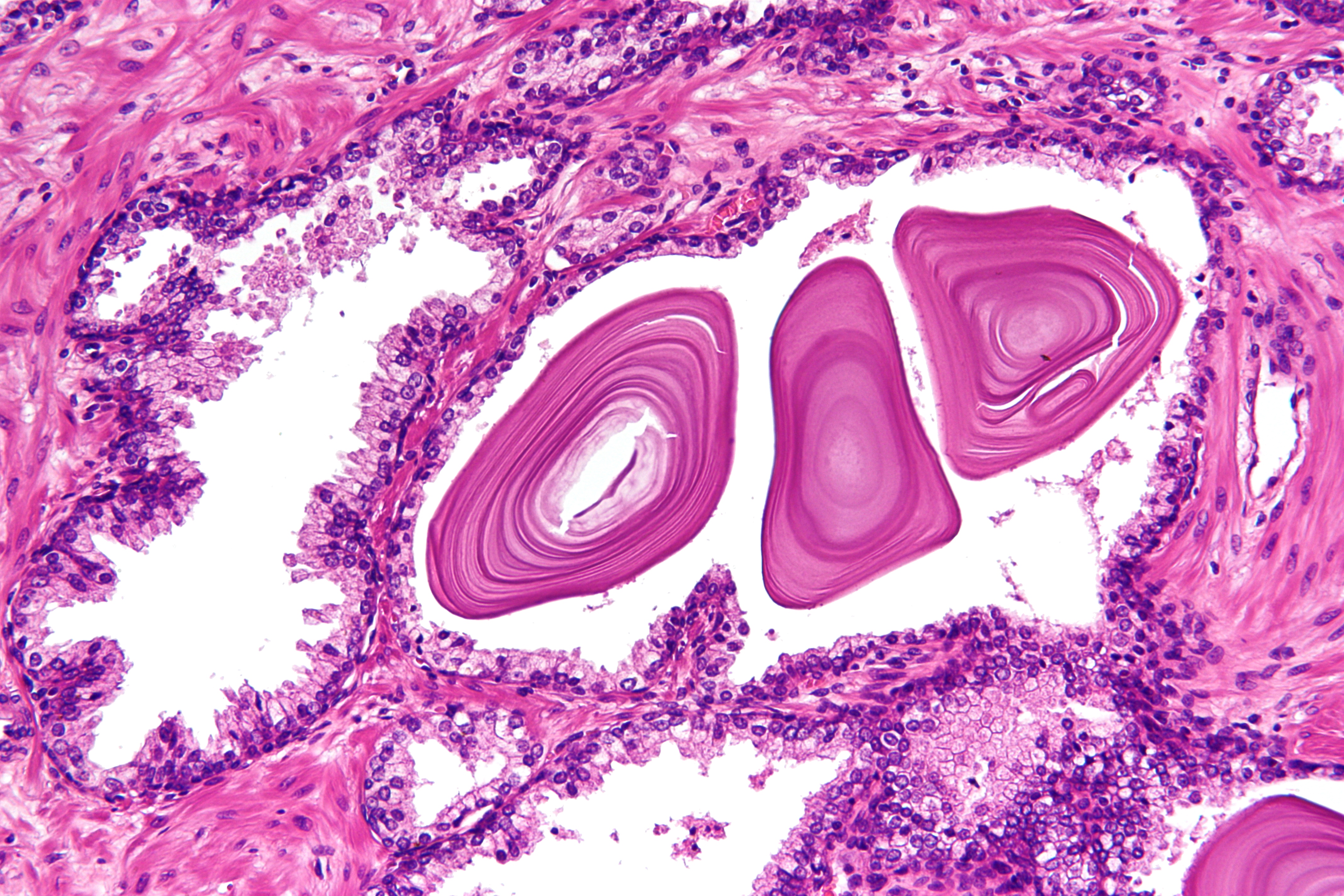
The prostate is both an Male accessory gland, accessory gland of the male reproductive system and a muscle-driven mechanical switch between urination and ejaculation. It is found only in some mammals. It differs between species anatomically, chemically, and physiologically. Anatomically, the prostate is found below the Urinary bladder, bladder, with the urethra passing through it. It is described in gross anatomy as consisting of lobes and in microanatomy by zone. It is surrounded by an elastic, fibromuscular capsule and contains glandular tissue as well as connective tissue. The prostate glands produce and contain fluid that forms part of semen, the substance emitted during ejaculation as part of the male Human sexual response cycle, sexual response. This prostatic fluid is slightly alkaline, milky or white in appearance. The alkalinity of semen helps neutralize the acidity of the vagina, vaginal tract, prolonging the lifespan of sperm. The prostatic fluid is expelled in the ... [...More Info...] [...Related Items...] OR: [Wikipedia] [Google] [Baidu] |
Allantois
The allantois (plural ''allantoides'' or ''allantoises'') is a hollow sac-like structure filled with clear fluid that forms part of a developing amniote's conceptus (which consists of all embryonic and extraembryonic tissues). It helps the embryo exchange gases and handle liquid waste. The allantois, along with the amnion, chorion, and yolk sac (other extraembryonic membranes), identify humans and other mammals, birds, and other reptiles as amniotes. Fish and amphibians are anamniotes, and lack the allantois. In mammals the extraembryonic membranes are known as the fetal membranes. Function This sac-like structure, whose name is the New Latin equivalent of "sausage" (in reference to its shape when first formed) is primarily involved in nutrition and excretion, and is webbed with blood vessels. The function of the allantois is to collect liquid waste from the embryo, as well as to exchange gases used by the embryo. In mammals In mammals excluding egg-laying monotremes, the all ... [...More Info...] [...Related Items...] OR: [Wikipedia] [Google] [Baidu] |
Urogenital Sinus
The urogenital sinus is a part of the human body only present in the development of the urinary and reproductive organs. It is the ventral part of the cloaca, formed after the cloaca separates from the anal canal during the fourth to seventh weeks of development. In males, the UG sinus is divided into three regions: upper, pelvic, and phallic. The upper part gives rise to the urinary bladder and the pelvic part gives rise to the prostatic and membranous parts of the urethra, the prostate and the bulbourethral gland (Cowper's). The phallic portion gives rise to the spongy (bulbar) part of the urethra and the urethral glands (Littre's). Note that the penile part of the urethra originates from urogenital fold. In females, the pelvic part of the UG sinus gives rise to the sinovaginal bulbs, structures that will eventually form the inferior two thirds of the vagina. This process begins when the lower tip of the paramesonephric ducts, the structures that will eventually form the ... [...More Info...] [...Related Items...] OR: [Wikipedia] [Google] [Baidu] |
Urethra
The urethra (from Greek οὐρήθρα – ''ourḗthrā'') is a tube that connects the urinary bladder to the urinary meatus for the removal of urine from the body of both females and males. In human females and other primates, the urethra connects to the urinary meatus above the vagina, whereas in marsupials, the female's urethra empties into the urogenital sinus. Females use their urethra only for urinating, but males use their urethra for both urination and ejaculation. The external urethral sphincter is a striated muscle that allows voluntary control over urination. The internal sphincter, formed by the involuntary smooth muscles lining the bladder neck and urethra, receives its nerve supply by the sympathetic division of the autonomic nervous system. The internal sphincter is present both in males and females. Structure The urethra is a fibrous and muscular tube which connects the urinary bladder to the external urethral meatus. Its length differs between the sexes, ... [...More Info...] [...Related Items...] OR: [Wikipedia] [Google] [Baidu] |
Pelvic Floor
The pelvic floor or pelvic diaphragm is composed of muscle fibers of the levator ani, the coccygeus muscle, and associated connective tissue which span the area underneath the pelvis. The pelvic diaphragm is a muscular partition formed by the levatores ani and coccygei, with which may be included the parietal pelvic fascia on their upper and lower aspects. The pelvic floor separates the pelvic cavity above from the perineal region (including perineum) below. Both males and females have a pelvic floor. To accommodate the birth canal, a female's pelvic cavity is larger than a male's. Structure The right and left levator ani lie almost horizontally in the floor of the pelvis, separated by a narrow gap that transmits the urethra, vagina, and anal canal. The levator ani is usually considered in three parts: pubococcygeus, puborectalis, and iliococcygeus. The pubococcygeus, the main part of the levator, runs backward from the body of the pubis toward the coccyx and may be damaged during ... [...More Info...] [...Related Items...] OR: [Wikipedia] [Google] [Baidu] |
Muscular
Skeletal muscles (commonly referred to as muscles) are organs of the vertebrate muscular system and typically are attached by tendons to bones of a skeleton. The muscle cells of skeletal muscles are much longer than in the other types of muscle tissue, and are often known as muscle fibers. The muscle tissue of a skeletal muscle is striated – having a striped appearance due to the arrangement of the sarcomeres. Skeletal muscles are voluntary muscles under the control of the somatic nervous system. The other types of muscle are cardiac muscle which is also striated and smooth muscle which is non-striated; both of these types of muscle tissue are classified as involuntary, or, under the control of the autonomic nervous system. A skeletal muscle contains multiple fascicles – bundles of muscle fibers. Each individual fiber, and each muscle is surrounded by a type of connective tissue layer of fascia. Muscle fibers are formed from the fusion of developmental myoblasts in a pro ... [...More Info...] [...Related Items...] OR: [Wikipedia] [Google] [Baidu] |
Urination
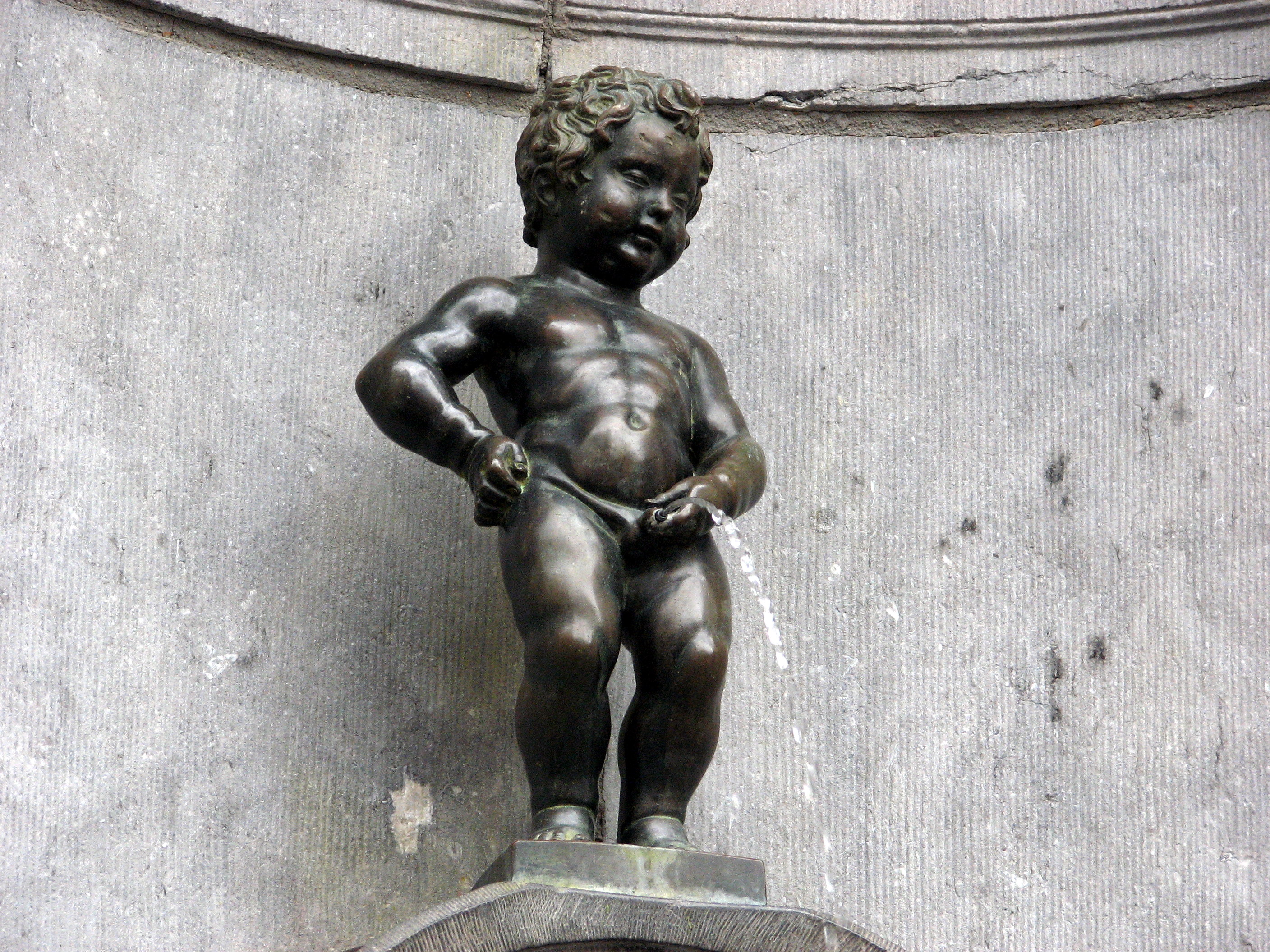
Urination, also known as micturition, is the release of urine from the urinary bladder through the urethra to the outside of the body. It is the urinary system's form of excretion. It is also known medically as micturition, voiding, uresis, or, rarely, emiction, and known colloquially by various names including peeing, weeing, and pissing. In healthy humans (and many other animals), the process of urination is under voluntary control. In infants, some elderly individuals, and those with neurological injury, urination may occur as a reflex. It is normal for adult humans to urinate up to seven times during the day. In some animals, in addition to expelling waste material, urination can mark territory or express submissiveness. Physiologically, urination involves coordination between the central, autonomic, and somatic nervous systems. Brain centres that regulate urination include the pontine micturition center, periaqueductal gray, and the cerebral cortex. In placental ma ... [...More Info...] [...Related Items...] OR: [Wikipedia] [Google] [Baidu] |
Kidney Stones
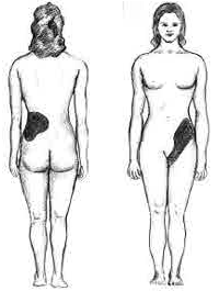
Kidney stone disease, also known as nephrolithiasis or urolithiasis, is a crystallopathy where a solid piece of material (kidney stone) develops in the urinary tract. Kidney stones typically form in the kidney and leave the body in the urine stream. A small stone may pass without causing symptoms. If a stone grows to more than , it can cause blockage of the ureter, resulting in sharp and severe pain in the lower back or abdomen. A stone may also result in blood in the urine, vomiting, or painful urination. About half of people who have had a kidney stone will have another within ten years. Most stones form by a combination of genetics and environmental factors. Risk factors include high urine calcium levels, obesity, certain foods, some medications, calcium supplements, hyperparathyroidism, gout and not drinking enough fluids. Stones form in the kidney when minerals in urine are at high concentration. The diagnosis is usually based on symptoms, urine testing, and medical ... [...More Info...] [...Related Items...] OR: [Wikipedia] [Google] [Baidu] |
Psoas Major
The psoas major ( or ; from grc, ψόᾱ, psóā, muscles of the loins) is a long fusiform muscle located in the lateral lumbar region between the vertebral column and the brim of the lesser pelvis. It joins the iliacus muscle to form the iliopsoas. In animals, this muscle is equivalent to the tenderloin. Structure The psoas major is divided into a superficial and a deep part. The deep part originates from the transverse processes of lumbar vertebrae L1–L5. The superficial part originates from the lateral surfaces of the last thoracic vertebra, lumbar vertebrae L1–L4, and the neighboring intervertebral discs. The lumbar plexus lies between the two layers. Together, the iliacus muscle and the psoas major form the iliopsoas, which is surrounded by the iliac fascia. The iliopsoas runs across the iliopubic eminence through the muscular lacuna to its insertion on the lesser trochanter of the femur. The iliopectineal bursa separates the tendon of the iliopsoas muscle from th ... [...More Info...] [...Related Items...] OR: [Wikipedia] [Google] [Baidu] |
Urinary Bladder
The urinary bladder, or simply bladder, is a hollow organ in humans and other vertebrates that stores urine from the kidneys before disposal by urination. In humans the bladder is a distensible organ that sits on the pelvic floor. Urine enters the bladder via the ureters and exits via the urethra. The typical adult human bladder will hold between 300 and (10.14 and ) before the urge to empty occurs, but can hold considerably more. The Latin phrase for "urinary bladder" is ''vesica urinaria'', and the term ''vesical'' or prefix ''vesico -'' appear in connection with associated structures such as vesical veins. The modern Latin word for "bladder" – ''cystis'' – appears in associated terms such as cystitis (inflammation of the bladder). Structure In humans, the bladder is a hollow muscular organ situated at the base of the pelvis. In gross anatomy, the bladder can be divided into a broad , a body, an apex, and a neck. The apex (also called the vertex) is directed forward ... [...More Info...] [...Related Items...] OR: [Wikipedia] [Google] [Baidu] |


.png)


