|
Erector Spinae Muscles
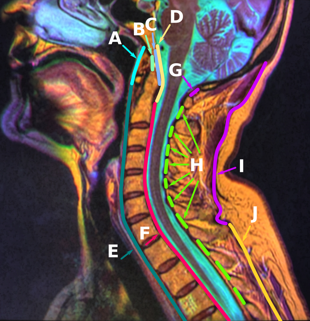
The erector spinae ( ) or spinal erectors is a set of muscles that straighten and rotate the back. The spinal erectors work together with the glutes (gluteus maximus, gluteus medius and gluteus minimus) to maintain stable posture standing or sitting. Structure The erector spinae is not just one muscle, but a group of muscles and tendons which run more or less the length of the spine on the left and the right, from the sacrum, or sacral region, and hips to the base of the skull. They are also known as the sacrospinalis group of muscles. These muscles lie on either side of the spinous processes of the vertebrae and extend throughout the lumbar, thoracic, and cervical regions. The erector spinae is covered in the lumbar and thoracic regions by the thoracolumbar fascia, and in the cervical region by the nuchal ligament. This large muscular and tendinous mass varies in size and structure at different parts of the vertebral column. In the sacral region, it is narrow and pointed, and at ... [...More Info...] [...Related Items...] OR: [Wikipedia] [Google] [Baidu] |
Thoracic Vertebrae
In vertebrates, thoracic vertebrae compose the middle segment of the vertebral column, between the cervical vertebrae and the lumbar vertebrae. In humans, there are twelve thoracic vertebra (anatomy), vertebrae and they are intermediate in size between the cervical and lumbar vertebrae; they increase in size going towards the lumbar vertebrae, with the lower ones being much larger than the upper. They are distinguished by the presence of Zygapophysial joint, facets on the sides of the bodies for Articulation (anatomy), articulation with the head of rib, heads of the ribs, as well as facets on the transverse processes of all, except the eleventh and twelfth, for articulation with the tubercle (rib), tubercles of the ribs. By convention, the human thoracic vertebrae are numbered T1–T12, with the first one (T1) located closest to the skull and the others going down the spine toward the lumbar region. General characteristics These are the general characteristics of the second throu ... [...More Info...] [...Related Items...] OR: [Wikipedia] [Google] [Baidu] |
Vertebra
The spinal column, a defining synapomorphy shared by nearly all vertebrates,Hagfish are believed to have secondarily lost their spinal column is a moderately flexible series of vertebrae (singular vertebra), each constituting a characteristic irregular bone whose complex structure is composed primarily of bone, and secondarily of hyaline cartilage. They show variation in the proportion contributed by these two tissue types; such variations correlate on one hand with the cerebral/caudal rank (i.e., location within the backbone), and on the other with phylogenetic differences among the vertebrate taxa. The basic configuration of a vertebra varies, but the bone is its ''body'', with the central part of the body constituting the ''centrum''. The upper (closer to) and lower (further from), respectively, the cranium and its central nervous system surfaces of the vertebra body support attachment to the intervertebral discs. The posterior part of a vertebra forms a vertebral arch ... [...More Info...] [...Related Items...] OR: [Wikipedia] [Google] [Baidu] |
Iliocostalis Cervicis
Iliocostalis muscle is the muscle immediately lateral to the longissimus that is the nearest to the furrow that separates the epaxial muscles from the hypaxial. It lies very deep to the fleshy portion of the serratus posterior muscle. It laterally flexes the vertebral column to the same side. Structure Iliocostalis muscle has a common origin from the iliac crest, the sacrum, the thoracolumbar fascia, and the spinous processes of the vertebrae from T11 to L5. Iliocostalis cervicis (cervicalis ascendens) arises from the angles of the third, fourth, fifth, and sixth ribs, and is inserted into the posterior tubercles of the transverse processes of the fourth, fifth, and sixth cervical vertebrae. Iliocostalis thoracis (musculus accessorius; iliocostalis thoracis) arises by flattened tendons from the upper borders of the angles of the lower six ribs medial to the tendons of insertion of the iliocostalis lumborum; these become muscular, and are inserted into the upper borders of the ... [...More Info...] [...Related Items...] OR: [Wikipedia] [Google] [Baidu] |
Iliocostalis Thoracis
Iliocostalis muscle is the muscle immediately lateral to the longissimus that is the nearest to the furrow that separates the epaxial muscles from the hypaxial. It lies very deep to the fleshy portion of the serratus posterior muscle. It laterally flexes the vertebral column to the same side. Structure Iliocostalis muscle has a common origin from the iliac crest, the sacrum, the thoracolumbar fascia, and the spinous processes of the vertebrae from T11 to L5. Iliocostalis cervicis (cervicalis ascendens) arises from the angles of the third, fourth, fifth, and sixth ribs, and is inserted into the posterior tubercles of the transverse processes of the fourth, fifth, and sixth cervical vertebrae. Iliocostalis thoracis (musculus accessorius; iliocostalis thoracis) arises by flattened tendons from the upper borders of the angles of the lower six ribs medial to the tendons of insertion of the iliocostalis lumborum; these become muscular, and are inserted into the upper borders of th ... [...More Info...] [...Related Items...] OR: [Wikipedia] [Google] [Baidu] |
Iliocostalis Lumborum
Iliocostalis muscle is the muscle immediately lateral to the longissimus that is the nearest to the furrow that separates the epaxial muscles from the hypaxial. It lies very deep to the fleshy portion of the serratus posterior muscle. It laterally flexes the vertebral column to the same side. Structure Iliocostalis muscle has a common origin from the iliac crest, the sacrum, the thoracolumbar fascia, and the spinous processes of the vertebrae from T11 to L5. Iliocostalis cervicis (cervicalis ascendens) arises from the angles of the third, fourth, fifth, and sixth ribs, and is inserted into the posterior tubercles of the transverse processes of the fourth, fifth, and sixth cervical vertebrae. Iliocostalis thoracis (musculus accessorius; iliocostalis thoracis) arises by flattened tendons from the upper borders of the angles of the lower six ribs medial to the tendons of insertion of the iliocostalis lumborum; these become muscular, and are inserted into the upper borders of t ... [...More Info...] [...Related Items...] OR: [Wikipedia] [Google] [Baidu] |
Iliac Crest
The crest of the ilium (or iliac crest) is the superior border of the wing of ilium and the superiolateral margin of the greater pelvis. Structure The iliac crest stretches posteriorly from the anterior superior iliac spine (ASIS) to the posterior superior iliac spine (PSIS). Behind the ASIS, it divides into an outer and inner lip separated by the intermediate zone. The outer lip bulges laterally into the iliac tubercle. Platzer (2004), p 186 Palpable in its entire length, the crest is convex superiorly but is sinuously curved, being concave inward in front, concave outward behind. Palastanga (2006), p 243 It is thinner at the center than at the extremities. Development The iliac crest is derived from endochondral bone. Function To the external lip are attached the ''Tensor fasciae latae'', '' Obliquus externus abdominis'', and '' Latissimus dorsi'', and along its whole length the '' fascia lata''; to the intermediate line, the '' Obliquus internus abdominis''. To the inter ... [...More Info...] [...Related Items...] OR: [Wikipedia] [Google] [Baidu] |
Erector Spinae Aponeurosis
Erector Set (trademark styled as "ERECTOR") was a brand of metal toy construction sets which were originally patented by Alfred Carlton Gilbert and first sold by his company, the Mysto Manufacturing Company of New Haven, Connecticut in 1913. In 1916, the company was reorganized as the A.C. Gilbert Company. The brand continued its independent existence under various corporate ownerships until 2000, when Meccano bought the Erector brand and consolidated its worldwide marketing with its own brand. The coverage here focuses on the historical legacy of the classic Erector Set; for current developments under the "Erector by Meccano" brand name, see the Meccano article. Basic Erector parts included various metal beams with regularly spaced holes for assembly using nuts and bolts. A frequently promoted patented feature was the ability to fabricate a strong but lightweight hollow structural girder from four long flat pieces of stamped sheet steel, held together by bolts and nuts (US Pate ... [...More Info...] [...Related Items...] OR: [Wikipedia] [Google] [Baidu] |
Posterior Sacroiliac Ligament
The posterior sacroiliac ligament is situated in a deep depression between the sacrum and ilium behind; it is strong and forms the chief bond of union between the bones. It consists of numerous fasciculi, which pass between the bones in various directions. * The ''upper part'' (''short posterior sacroiliac ligament'') is nearly horizontal in direction, and pass from the first and second transverse tubercles on the back of the sacrum to the tuberosity of the ilium. * The ''lower part'' (''long posterior sacroiliac ligament'') is oblique in direction; it is attached by one extremity to the third transverse tubercle of the back of the sacrum, and by the other to the posterior superior spine of the ilium. See also *Anterior sacroiliac ligament The anterior sacroiliac ligament consists of numerous thin bands, which connect the anterior surface of the lateral part of the sacrum to the margin of the auricular surface of the ilium and to the preauricular sulcus. See also *Posterior s ... [...More Info...] [...Related Items...] OR: [Wikipedia] [Google] [Baidu] |
Sacrotuberous
The sacrotuberous ligament (great or posterior sacrosciatic ligament) is situated at the lower and back part of the pelvis. It is flat, and triangular in form; narrower in the middle than at the ends. Structure It runs from the sacrum (the lower transverse sacral tubercles, the inferior margins sacrum and the upper coccyx) to the tuberosity of the ischium. It is a remnant of part of Biceps femoris muscle. The sacrotuberous ligament is attached by its broad base to the posterior superior iliac spine, the posterior sacroiliac ligaments (with which it is partly blended), to the lower transverse sacral tubercles and the lateral margins of the lower sacrum and upper coccyx. Its oblique fibres descend laterally, converging to form a thick, narrow band that widens again below and is attached to the medial margin of the ischial tuberosity. It then spreads along the ischial ramus as the falciform process, whose concave edge blends with the fascial sheath of the internal pudendal vessels and ... [...More Info...] [...Related Items...] OR: [Wikipedia] [Google] [Baidu] |
Supraspinous Ligament
The supraspinous ligament, also known as the supraspinal ligament, is a ligament found along the vertebral column. Structure The supraspinous ligament connects the tips of the spinous processes from the seventh cervical vertebra to the sacrum. Above the seventh cervical vertebra, the supraspinous ligament is continuous with the nuchal ligament. Between the spinous processes it is continuous with the interspinous ligaments. It is thicker and broader in the lumbar than in the thoracic region, and intimately blended, in both situations, with the neighboring fascia. The most superficial fibers of this ligament extend over three or four vertebrae; those more deeply seated pass between two or three vertebrae while the deepest connect the spinous processes of neighboring vertebrae. Development Function The supraspinous ligament, along with the posterior longitudinal ligament, interspinous ligaments and ligamentum flavum, help to limit hyperflexion of the vertebral column. Clinical ... [...More Info...] [...Related Items...] OR: [Wikipedia] [Google] [Baidu] |
Nuchal Ligament
The nuchal ligament is a ligament at the back of the neck that is continuous with the supraspinous ligament. Structure The nuchal ligament extends from the external occipital protuberance on the skull and median nuchal line to the spinous process of the seventh cervical vertebra in the lower part of the neck. From the anterior border of the nuchal ligament, a fibrous lamina is given off. This is attached to the posterior tubercle of the atlas, and to the spinous processes of the cervical vertebrae, and forms a septum between the muscles on either side of the neck. The trapezius and splenius capitis muscle attach to the nuchal ligament. Function It is a tendon-like structure that has developed independently in humans and other animals well adapted for running. In some four-legged animals, particularly ungulates, the nuchal ligament serves to sustain the weight of the head. Clinical significance In Chiari malformation treatment, decompression and duraplasty with a harvested n ... [...More Info...] [...Related Items...] OR: [Wikipedia] [Google] [Baidu] |
Thoracolumbar Fascia
The thoracolumbar fascia (lumbodorsal fascia or thoracodorsal fascia) is a deep investing membrane throughout most of the posterior thorax and abdomen although it is a thin fibrous lamina in the thoracic region. Above, it is continuous with a similar investing layer on the back of the neck—the nuchal fascia. It is formed of longitudinal and transverse fibers that bridge the aponeuroses of internal oblique and transversus, costal angles and iliac crest laterally, to the vertebral column and sacrum medially. In doing so, they cover the paravertebral muscles. It is made up of three layers, anterior, middle, and posterior. The anterior and middle layers insert onto the transverse processes of the vertebral column while the posterior layer inserts onto the tips of the spinous processes, hence it is indirectly continuous with the interspinous ligaments. The anterior layer is the thinnest and the posterior layer is the thickest. Two spaces are formed between these three layers of the ... [...More Info...] [...Related Items...] OR: [Wikipedia] [Google] [Baidu] |


