|
Epiploic Appendix
The epiploic appendices (or appendices epiploicae, or epiploic appendages, or appendix epiploica, or omental appendices) are small pouches of the peritoneum filled with fat and situated along the colon, but are absent in the rectum. They are chiefly appended to the transverse and sigmoid parts of the colon, however, their function is unknown. The appendages can become inflamed in a painful process known as epiploic appendagitis which can mimic acute appendicitis Appendicitis is inflammation of the appendix. Symptoms commonly include right lower abdominal pain, nausea, vomiting, and decreased appetite. However, approximately 40% of people do not have these typical symptoms. Severe complications of a rupt ... and other conditions. External links * - "Intestines and Pancreas: Large Intestine" * * "Michael Sand et al. Epiploic appendagitis – clinical characteristics of an uncommon surgical diagnosisBMC Surgery 2007, 7:11 (1 July 2007)". {{Authority control Pelvis ... [...More Info...] [...Related Items...] OR: [Wikipedia] [Google] [Baidu] |
Epiploic Appendages 0002
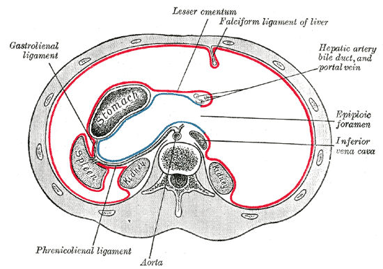
The greater omentum (also the great omentum, omentum majus, gastrocolic omentum, epiploon, or, especially in animals, caul) is a large apron-like fold of visceral peritoneum that hangs down from the stomach. It extends from the greater curvature of the stomach, passing in front of the small intestines and doubles back to ascend to the transverse colon before reaching to the posterior abdominal wall. The greater omentum is larger than the lesser omentum, which hangs down from the liver to the lesser curvature. The common anatomical term "epiploic" derives from "epiploon", from the Greek ''epipleein'', meaning to float or sail on, since the greater omentum appears to float on the surface of the intestines. It is the first structure observed when the abdominal cavity is opened anteriorly (from the front). Structure The greater omentum is the larger of the two peritoneal folds. It consists of a double sheet of peritoneum, folded on itself so that it has four layers. The two layers o ... [...More Info...] [...Related Items...] OR: [Wikipedia] [Google] [Baidu] |
Greater Omentum
The greater omentum (also the great omentum, omentum majus, gastrocolic omentum, epiploon, or, especially in animals, caul) is a large apron-like fold of visceral peritoneum that hangs down from the stomach. It extends from the greater curvature of the stomach, passing in front of the small intestines and doubles back to ascend to the transverse colon before reaching to the posterior abdominal wall. The greater omentum is larger than the lesser omentum, which hangs down from the liver to the lesser curvature. The common anatomical term "epiploic" derives from "epiploon", from the Greek ''epipleein'', meaning to float or sail on, since the greater omentum appears to float on the surface of the intestines. It is the first structure observed when the abdominal cavity is opened anteriorly (from the front). Structure The greater omentum is the larger of the two peritoneal folds. It consists of a double sheet of peritoneum, folded on itself so that it has four layers. The two layers o ... [...More Info...] [...Related Items...] OR: [Wikipedia] [Google] [Baidu] |
Peritoneum
The peritoneum is the serous membrane forming the lining of the abdominal cavity or coelom in amniotes and some invertebrates, such as annelids. It covers most of the intra-abdominal (or coelomic) organs, and is composed of a layer of mesothelium supported by a thin layer of connective tissue. This peritoneal lining of the cavity supports many of the abdominal organs and serves as a conduit for their blood vessels, lymphatic vessels, and nerves. The abdominal cavity (the space bounded by the vertebrae, abdominal muscles, diaphragm, and pelvic floor) is different from the intraperitoneal space (located within the abdominal cavity but wrapped in peritoneum). The structures within the intraperitoneal space are called "intraperitoneal" (e.g., the stomach and intestines), the structures in the abdominal cavity that are located behind the intraperitoneal space are called "retroperitoneal" (e.g., the kidneys), and those structures below the intraperitoneal space are called "subp ... [...More Info...] [...Related Items...] OR: [Wikipedia] [Google] [Baidu] |
Colon (anatomy)
The large intestine, also known as the large bowel, is the last part of the gastrointestinal tract and of the digestive system in tetrapods. Water is absorbed here and the remaining waste material is stored in the rectum as feces before being removed by defecation. The colon is the longest portion of the large intestine, and the terms are often used interchangeably but most sources define the large intestine as the combination of the cecum, colon, rectum, and anal canal. Some other sources exclude the anal canal. In humans, the large intestine begins in the right iliac region of the pelvis, just at or below the waist, where it is joined to the end of the small intestine at the cecum, via the ileocecal valve. It then continues as the colon ascending the abdomen, across the width of the abdominal cavity as the transverse colon, and then descending to the rectum and its endpoint at the anal canal. Overall, in humans, the large intestine is about long, which is about one-fifth o ... [...More Info...] [...Related Items...] OR: [Wikipedia] [Google] [Baidu] |
Epiploic Appendagitis
Epiploic appendagitis (EA) is an uncommon, benign, self-limiting inflammatory process of the epiploic appendices. Other, older terms for the process include appendicitis epiploica and appendagitis, but these terms are used less now in order to avoid confusion with acute appendicitis. Epiploic appendices are small, fat-filled sacs or finger-like projections along the surface of the upper and lower colon and rectum. They may become acutely inflamed as a result of torsion (twisting) or venous thrombosis. The inflammation causes pain, often described as sharp or stabbing, located on the left, right, or central regions of the abdomen. There is sometimes nausea and vomiting. The symptoms may mimic those of acute appendicitis, diverticulitis, or cholecystitis. The pain is characteristically intense during/after defecation or micturition (espec. in the sigmoid type) due to the effect of traction on the pedicle of the lesion caused by straining and emptying of the bowel and bladder. Ini ... [...More Info...] [...Related Items...] OR: [Wikipedia] [Google] [Baidu] |
Appendicitis
Appendicitis is inflammation of the appendix. Symptoms commonly include right lower abdominal pain, nausea, vomiting, and decreased appetite. However, approximately 40% of people do not have these typical symptoms. Severe complications of a ruptured appendix include widespread, painful inflammation of the inner lining of the abdominal wall and sepsis. Appendicitis is caused by a blockage of the hollow portion of the appendix. This is most commonly due to a calcified "stone" made of feces. Inflamed lymphoid tissue from a viral infection, parasites, gallstone, or tumors may also cause the blockage. This blockage leads to increased pressures in the appendix, decreased blood flow to the tissues of the appendix, and bacterial growth inside the appendix causing inflammation. The combination of inflammation, reduced blood flow to the appendix and distention of the appendix causes tissue injury and tissue death. If this process is left untreated, the appendix may burst, releasing ba ... [...More Info...] [...Related Items...] OR: [Wikipedia] [Google] [Baidu] |


