|
Endoscopic Ear Surgery
Endoscopic ear surgery (EES) is a minimally invasive alternative to traditional ear surgery and is defined as the use of the rigid endoscope, as opposed to a surgical microscope, to visualize the middle and inner ear during otologic surgery. During endoscopic ear surgery the surgeon holds the endoscope in one hand while working in the ear with the other. To allow this kind of single-handed surgery, different surgical instruments have to be used. Endoscopic visualization has improved due to high-definition video imaging and wide-field endoscopy, and being less invasive, EES is gaining importance as an adjunct to microscopic ear surgery. History Endoscopic Ear Surgery was first described in 1992 by Professor Ahmed El-Guindy and pioneered by Dr Muaaz Tarabichi in Dubai during the late 90s. His contributions to the field have led to him being recognized globally as the father of endoscopic ear surgery. He now lectures extensively on the topic worldwide. Similar to the early years ... [...More Info...] [...Related Items...] OR: [Wikipedia] [Google] [Baidu] |
Endoscope
An endoscope is an inspection instrument composed of image sensor, optical lens, light source and mechanical device, which is used to look deep into the body by way of openings such as the mouth or anus. A typical endoscope applies several modern technologies including optics, ergonomics, precision mechanics, electronics, and software engineering. With an endoscope, it is possible to observe lesions that cannot be detected by X-ray, making it useful in medical diagnosis. Endoscopes use tubes which are only a few millimeters thick to transfer illumination in one direction and high-resolution images in real time in the other direction, resulting in minimally invasive surgeries. It is used to examine the internal organs like the throat or esophagus. Specialized instruments are named after their target organ. Examples include the cystoscope (bladder), nephroscope (kidney), bronchoscope (bronchus), arthroscope (joints) and colonoscope (colon), and laparoscope (abdomen or pelvis). They c ... [...More Info...] [...Related Items...] OR: [Wikipedia] [Google] [Baidu] |
Cholesteatoma
Cholesteatoma is a destructive and expanding growth consisting of keratinizing squamous epithelium in the middle ear and/or mastoid process. Cholesteatomas are not cancerous as the name may suggest, but can cause significant problems because of their erosive and expansile properties. This can result in the destruction of the bones of the middle ear ( ossicles), as well as growth through the base of the skull into the brain. They often become infected and can result in chronically draining ears. Treatment almost always consists of surgical removal. Signs and symptoms Other more common conditions (e.g. otitis externa) may also present with these symptoms, but cholesteatoma is much more serious and should not be overlooked. If a patient presents to a doctor with ear discharge and hearing loss, the doctor should consider cholesteatoma until the disease is definitely excluded. Other less common symptoms (all less than 15%) of cholesteatoma may include pain, balance disruption, tinnitu ... [...More Info...] [...Related Items...] OR: [Wikipedia] [Google] [Baidu] |
Endoscopy
An endoscopy is a procedure used in medicine to look inside the body. The endoscopy procedure uses an endoscope to examine the interior of a hollow organ or cavity of the body. Unlike many other medical imaging techniques, endoscopes are inserted directly into the organ. There are many types of endoscopies. Depending on the site in the body and type of procedure, an endoscopy may be performed by either a doctor or a surgeon. A patient may be fully conscious or anaesthesia, anaesthetised during the procedure. Most often, the term ''endoscopy'' is used to refer to an examination of the upper part of the human gastrointestinal tract, gastrointestinal tract, known as an esophagogastroduodenoscopy. For nonmedical use, similar instruments are called borescopes. History Adolf Kussmaul was fascinated by sword swallowers who would insert a sword down their throat without gagging. This drew inspiration to insert a camera, the next problem to solve was how to insert a source of light, as ... [...More Info...] [...Related Items...] OR: [Wikipedia] [Google] [Baidu] |
Eustachian Tube
In anatomy, the Eustachian tube, also known as the auditory tube or pharyngotympanic tube, is a tube that links the nasopharynx to the middle ear, of which it is also a part. In adult humans, the Eustachian tube is approximately long and in diameter. It is named after the sixteenth-century Italian anatomist Bartolomeo Eustachi. In humans and other tetrapods, both the middle ear and the ear canal are normally filled with air. Unlike the air of the ear canal, however, the air of the middle ear is not in direct contact with the atmosphere outside the body; thus, a pressure difference can develop between the atmospheric pressure of the ear canal and the middle ear. Normally, the Eustachian tube is collapsed, but it gapes open with swallowing and with positive pressure, allowing the middle ear's pressure to adjust to the atmospheric pressure. When taking off in an aircraft, the ambient air pressure goes from higher (on the ground) to lower (in the sky). The air in the middle ear ... [...More Info...] [...Related Items...] OR: [Wikipedia] [Google] [Baidu] |
Stapedectomy
A stapedectomy is a surgical procedure of the middle ear performed in order to improve hearing. If the stapes footplate is fixed in position, rather than being normally mobile, the result is a conductive hearing loss. There are two major causes of stapes fixation. The first is a disease process of abnormal mineralization of the temporal bone called otosclerosis. The second is a congenital malformation of the stapes. In both of these situations, it is possible to improve hearing by removing the stapes bone and replacing it with a micro prosthesis - a stapedectomy, or creating a fenestra, small hole in the fixed stapes footplate and inserting a tiny, piston-like prosthesis - a stapedotomy. The results of this surgery are generally most reliable in patients whose stapes has lost mobility because of otosclerosis. Nine out of ten patients who undergo the procedure will come out with significantly improved hearing while less than 1% will experience worsened hearing acuity or deafness. Su ... [...More Info...] [...Related Items...] OR: [Wikipedia] [Google] [Baidu] |
Stapes
The ''stapes'' or stirrup is a bone in the middle ear of humans and other animals which is involved in the conduction of sound vibrations to the inner ear. This bone is connected to the oval window by its annular ligament, which allows the footplate to transmit sound energy through the oval window into the inner ear. The ''stapes'' is the smallest and lightest bone in the human body, and is so-called because of its resemblance to a stirrup ( la, Stapes). Structure The ''stapes'' is the third bone of the three ossicles in the middle ear and the smallest in the human body. It measures roughly , greater along the head-base span. It rests on the oval window, to which it is connected by an annular ligament and articulates with the ''incus'', or anvil through the incudostapedial joint. They are connected by anterior and posterior limbs ( la, crura). Development The ''stapes'' develops from the second pharyngeal arch during the sixth to eighth week of embryological life. The ce ... [...More Info...] [...Related Items...] OR: [Wikipedia] [Google] [Baidu] |
Otosclerosis
Otosclerosis is a condition of the middle ear where portions of the dense enchondral layer of the bony labyrinth remodel into one or more lesions of irregularly-laid spongy bone. As the lesions reach the stapes the bone is resorbed, then hardened ( sclerotized), which limits its movement and results in hearing loss, tinnitus, vertigo or a combination of symptoms. The term otosclerosis is something of a misnomer: much of the clinical course is characterized by lucent rather than sclerotic bony changes, so the disease is also known as otospongiosis. Etymology The word ''otosclerosis'' derives from Greek ὠτός (''ōtos''), genitive of οὖς (''oûs'') "ear" + σκλήρωσις (''sklērōsis''), "hardening". Presentation The primary form of hearing loss in otosclerosis is conductive hearing loss (CHL) whereby sounds reach the ear drum but are incompletely transferred via the ossicular chain in the middle ear, and thus partly fail to reach the inner ear (cochlea). This can af ... [...More Info...] [...Related Items...] OR: [Wikipedia] [Google] [Baidu] |
Eardrum
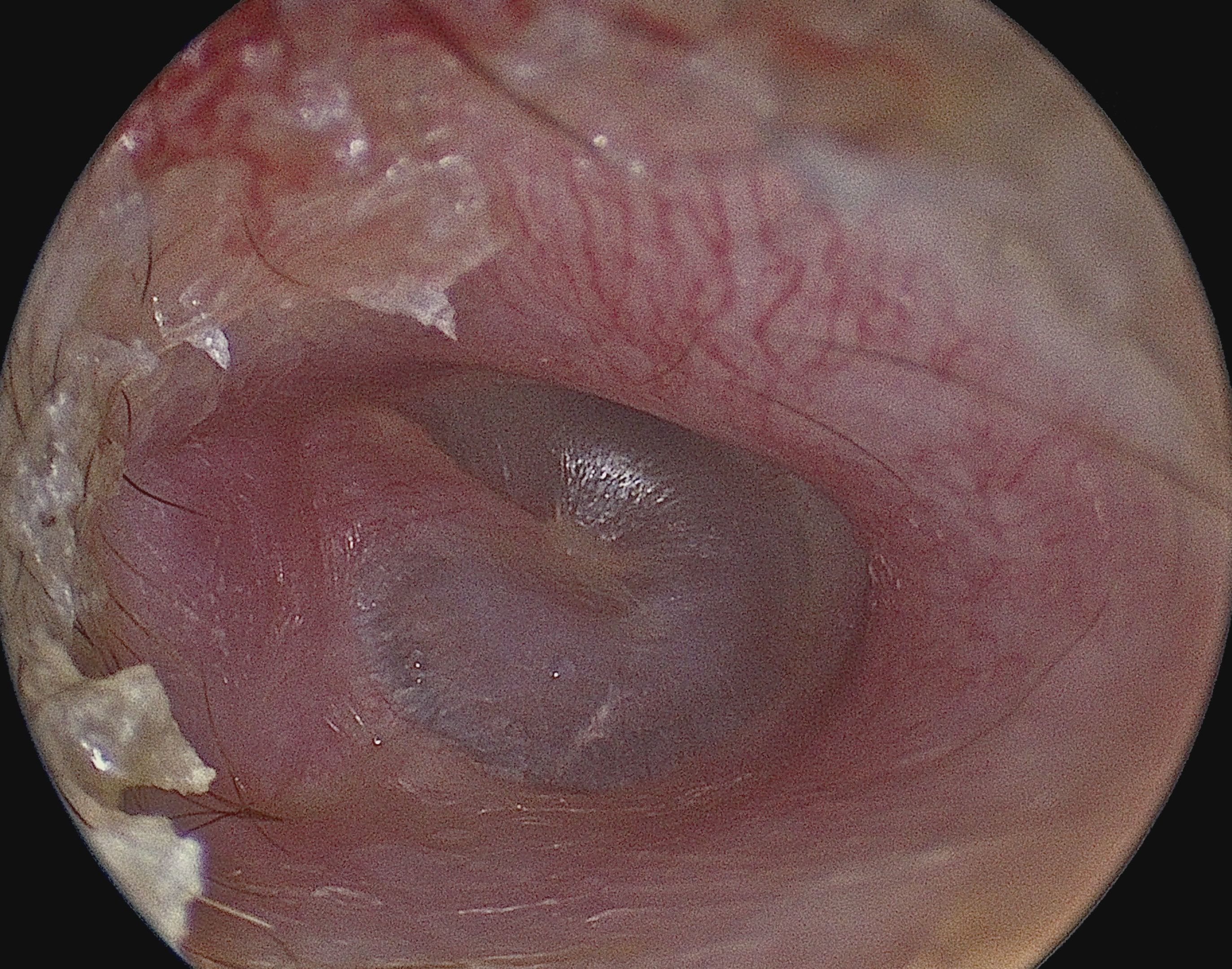
In the anatomy of humans and various other tetrapods, the eardrum, also called the tympanic membrane or myringa, is a thin, cone-shaped membrane that separates the external ear The outer ear, external ear, or auris externa is the external part of the ear, which consists of the auricle (also pinna) and the ear canal. It gathers sound energy and focuses it on the eardrum (tympanic membrane). Structure Auricle The ... from the middle ear. Its function is to transmit sound from the air to the ossicles inside the middle ear, and then to the oval window in the fluid-filled cochlea. Hence, it ultimately converts and amplifies vibration in the air to vibration in cochlear fluid. The malleus bone bridges the gap between the eardrum and the other ossicles. Rupture or perforation of the eardrum can lead to conductive hearing loss. Collapse or tympanic membrane retraction, retraction of the eardrum can cause conductive hearing loss or cholesteatoma. Structure Orientation and r ... [...More Info...] [...Related Items...] OR: [Wikipedia] [Google] [Baidu] |
Perforated Eardrum
A perforated eardrum (tympanic membrane perforation) is a hole in the eardrum. It can be caused by infection (otitis media), trauma, overpressure (loud noise), inappropriate ear clearing, and changes in middle ear pressure. An otoscope can be used to view the eardrum to diagnose a perforation. Perforations may heal naturally, or require surgery. Presentation A perforated eardrum leads to conductive hearing loss, which is usually temporary. Other symptoms may include tinnitus, ear pain, vertigo, or a discharge of mucus. Nausea and/or vomiting secondary to vertigo may occur. Causes A perforated eardrum can have one of many causes, such as: * infection (otitis media). This infection may then spread through the middle ear, and may reoccur. * trauma. This may be caused by trying to clean ear wax with sharp instruments. It may also occur due to surgical complications. * overpressure (loud noise or shockwave from an explosion). * inappropriate ear clearing. * flying with a seve ... [...More Info...] [...Related Items...] OR: [Wikipedia] [Google] [Baidu] |
Sinus Tympani
Sinus may refer to: Anatomy * Sinus (anatomy), a sac or cavity in any organ or tissue ** Paranasal sinuses, air cavities in the cranial bones, especially those near the nose, including: *** Maxillary sinus, is the largest of the paranasal sinuses, under the eyes, in the maxillary bones *** Frontal sinus, superior to the eyes, in the frontal bone, which forms the hard part of the forehead *** Ethmoid sinus, formed from several discrete air cells within the ethmoid bone between the eyes and under the nose *** Sphenoidal sinus, in the sphenoid bone at the center of the skull base under the pituitary gland ** Anal sinuses, the furrows which separate the columns in the rectum ** Dural venous sinuses, venous channels found between layers of dura mater in the brain * Sinus (botany), a space or indentation, usually on a leaf Heart * Sinus node, a structure in the superior part of the right atrium * Sinus rhythm, normal beating on an ECG * Coronary sinus, a vein collecting blood from ... [...More Info...] [...Related Items...] OR: [Wikipedia] [Google] [Baidu] |
Operating Microscope
An operating microscope or surgical microscope is an optical microscope specifically designed to be used in a surgical setting, typically to perform microsurgery. Design features of an operating microscope are: magnification typically in the range from 4x-40x, components that are easy to sterilize or disinfect in order to ensure cross-infection control. There is often a prism that allows splitting of the light beam in order that assistants may also visualize the procedure or to allow photography or video to be taken of the operating field. Typically an operating microscope might cost several thousand dollars for a basic model, more advanced models may be much more expensive. Additionally specialized microsurgical instruments may be required to make full use of the improved vision the microscope affords. It can take time to master use of an operating microscope. Fields of medicine that make significant use of the operating microscope include plastic surgery, dentistry (especia ... [...More Info...] [...Related Items...] OR: [Wikipedia] [Google] [Baidu] |


