|
Ectasis
Ectasia (), also called ectasis (), is dilation or distention of a tubular structure, either normal or Pathophysiology, pathophysiologic but usually the latter (except in atelectasis, where absence of ectasis is the problem). Specific conditions * Bronchiectasis, chronic dilatation of the bronchi * Duct ectasia of breast, a dilated milk duct. Duct ectasia syndrome is a synonym for nonpuerperal (unrelated to pregnancy and breastfeeding) mastitis. * Dural ectasia, dilation of the dural sac surrounding the spinal cord, usually in the very low back. * Pyelectasis, dilation of a part of the kidney, most frequently seen in prenatal ultrasounds. It usually resolves on its own. * Rete tubular ectasia, dilation of tubular structures in the testicles. It is usually found in older men. * Acral arteriolar ectasia * Keratoconus, Corneal ectasia (secondary keratoconus), a bulging of the cornea. ;Vascular ectasias * Most broadly, any abnormal dilatation of a blood vessel, including aneurysms * ... [...More Info...] [...Related Items...] OR: [Wikipedia] [Google] [Baidu] |
Bronchiectasis
Bronchiectasis is a disease in which there is permanent enlargement of parts of the bronchi, airways of the lung. Symptoms typically include a chronic cough with sputum, mucus production. Other symptoms include shortness of breath, hemoptysis, coughing up blood, and chest pain. Wheezing and nail clubbing may also occur. Those with the disease often get lung infections. Bronchiectasis may result from a number of infection, infectious and acquired causes, including measles, pneumonia, tuberculosis, immune system problems, as well as the genetic disorder cystic fibrosis. Cystic fibrosis eventually results in severe bronchiectasis in nearly all cases. The cause in 10–50% of those without cystic fibrosis is unknown. The mechanism of disease is breakdown of the airways due to an excessive inflammatory response. Involved airways (bronchi) become enlarged and thus less able to clear secretions. These secretions increase the amount of bacteria in the lungs, resulting in airway blockage ... [...More Info...] [...Related Items...] OR: [Wikipedia] [Google] [Baidu] |
Atelectasis
Atelectasis is the collapse or closure of a lung resulting in reduced or absent gas exchange. It is usually unilateral, affecting part or all of one lung. It is a condition where the alveoli are deflated down to little or no volume, as distinct from pulmonary consolidation, in which they are filled with liquid. It is often called a ''collapsed lung'', although that term may also refer to pneumothorax. It is a very common finding in chest X-rays and other radiological studies, and may be caused by normal exhalation or by various medical conditions. Although frequently described as a ''collapse of lung tissue'', atelectasis is not synonymous with a pneumothorax, which is a more specific condition that can cause atelectasis. Acute atelectasis may occur as a post-operative complication or as a result of surfactant deficiency. In premature babies, this leads to infant respiratory distress syndrome. The term uses combining forms of ''atel-'' + ''ectasis'', from el, ἀτελής, ... [...More Info...] [...Related Items...] OR: [Wikipedia] [Google] [Baidu] |
Pyelectasis
Pyelectasis is a dilation of the renal pelvis. It is a relatively common ultrasound finding in fetuses and is three times more common in male fetuses. In most cases pyelectasis resolves normally, having no ill effects on the baby. The significance of pyelectasis in fetuses is not clear. It was thought to be a marker for obstruction, but in most cases it resolves spontaneously. In some studies it has been shown to appear and disappear several times throughout the course of pregnancy. There is some discussion about what degree of pyelectasis is considered severe enough to warrant further investigation and most authorities use 6mm as the cut-off point. Pyelectasis is considered to be a "soft marker” for Down syndrome. This, along with other factors such as age and abnormal maternal serum screening (exa, Integrated or Quad screen), may be grounds for a prenatal diagnostic test such as an amniocentesis to rule out Down syndrome. Babies with unresolved pyelectasis may experience urol ... [...More Info...] [...Related Items...] OR: [Wikipedia] [Google] [Baidu] |
Marfan Syndrome
Marfan syndrome (MFS) is a multi-systemic genetic disorder that affects the connective tissue. Those with the condition tend to be tall and thin, with long arms, legs, fingers, and toes. They also typically have exceptionally flexible joints and abnormally curved spines. The most serious complications involve the heart and aorta, with an increased risk of mitral valve prolapse and aortic aneurysm. The lungs, eyes, bones, and the covering of the spinal cord are also commonly affected. The severity of the symptoms is variable. MFS is caused by a mutation in ''FBN1'', one of the genes that makes fibrillin, which results in abnormal connective tissue. It is an autosomal dominant disorder. In about 75% of cases, it is inherited from a parent with the condition, while in about 25% it is a new mutation. Diagnosis is often based on the Ghent criteria. There is no known cure for MFS. Many of those with the disorder have a normal life expectancy with proper treatment. Management of ... [...More Info...] [...Related Items...] OR: [Wikipedia] [Google] [Baidu] |
Pathophysiology
Pathophysiology ( physiopathology) – a convergence of pathology with physiology – is the study of the disordered physiological processes that cause, result from, or are otherwise associated with a disease or injury. Pathology is the medical discipline that describes conditions typically ''observed'' during a disease state, whereas physiology is the biological discipline that describes processes or mechanisms ''operating'' within an organism. Pathology describes the abnormal or undesired condition, whereas pathophysiology seeks to explain the functional changes that are occurring within an individual due to a disease or pathologic state. History Etymology The term ''pathophysiology'' comes from the Ancient Greek πάθος (''pathos'') and φυσιολογία (''phusiologia''). Nineteenth century Reductionism In Germany in the 1830s, Johannes Müller led the establishment of physiology research autonomous from medical research. In 1843, the Berlin Physical Socie ... [...More Info...] [...Related Items...] OR: [Wikipedia] [Google] [Baidu] |
Aorta
The aorta ( ) is the main and largest artery in the human body, originating from the left ventricle of the heart and extending down to the abdomen, where it splits into two smaller arteries (the common iliac arteries). The aorta distributes oxygenated blood to all parts of the body through the systemic circulation. Structure Sections In anatomical sources, the aorta is usually divided into sections. One way of classifying a part of the aorta is by anatomical compartment, where the thoracic aorta (or thoracic portion of the aorta) runs from the heart to the diaphragm. The aorta then continues downward as the abdominal aorta (or abdominal portion of the aorta) from the diaphragm to the aortic bifurcation. Another system divides the aorta with respect to its course and the direction of blood flow. In this system, the aorta starts as the ascending aorta, travels superiorly from the heart, and then makes a hairpin turn known as the aortic arch. Following the aortic arch ... [...More Info...] [...Related Items...] OR: [Wikipedia] [Google] [Baidu] |
Jugular Vein Ectasia
Jugular vein ectasia is a venous anomaly that commonly presents itself as a unilateral neck swelling in children and adults. It is rare to have bilateral neck swelling due to internal jugular vein ectasia. References External links Vascular diseases {{circulatory-stub ... [...More Info...] [...Related Items...] OR: [Wikipedia] [Google] [Baidu] |
Chronic Venous Insufficiency
Chronic venous insufficiency (CVI) is a medical condition in which blood pools in the veins, straining the walls of the vein. The most common cause of CVI is superficial venous reflux which is a treatable condition. As functional venous valves are required to provide for efficient blood return from the lower extremities, this condition typically affects the legs. If the impaired vein function causes significant symptoms, such as swelling and ulcer formation, it is referred to as chronic venous disease. It is sometimes called ''chronic peripheral venous insufficiency'' and should not be confused with post-thrombotic syndrome in which the deep veins have been damaged by previous deep vein thrombosis. Most cases of CVI can be improved with treatments to the superficial venous system or stenting the deep system. Varicose veins for example can now be treated by local anesthetic endovenous surgery. Rates of CVI are higher in women than in men. Other risk factors include genetics, smok ... [...More Info...] [...Related Items...] OR: [Wikipedia] [Google] [Baidu] |
Vein
Veins are blood vessels in humans and most other animals that carry blood towards the heart. Most veins carry deoxygenated blood from the tissues back to the heart; exceptions are the pulmonary and umbilical veins, both of which carry oxygenated blood to the heart. In contrast to veins, arteries carry blood away from the heart. Veins are less muscular than arteries and are often closer to the skin. There are valves (called ''pocket valves'') in most veins to prevent backflow. Structure Veins are present throughout the body as tubes that carry blood back to the heart. Veins are classified in a number of ways, including superficial vs. deep, pulmonary vs. systemic, and large vs. small. * Superficial veins are those closer to the surface of the body, and have no corresponding arteries. *Deep veins are deeper in the body and have corresponding arteries. *Perforator veins drain from the superficial to the deep veins. These are usually referred to in the lower limbs and feet. *Communic ... [...More Info...] [...Related Items...] OR: [Wikipedia] [Google] [Baidu] |
Telangiectasia
Telangiectasias, also known as spider veins, are small dilated blood vessels that can occur near the surface of the skin or mucous membranes, measuring between 0.5 and 1 millimeter in diameter. These dilated blood vessels can develop anywhere on the body, but are commonly seen on the face around the nose, cheeks and chin. Dilated blood vessels can also develop on the legs, although when they occur on the legs, they often have underlying venous reflux or "hidden varicose veins" (see Venous hypertension section below). When found on the legs, they are found specifically on the upper thigh, below the knee joint and around the ankles. Many patients with spider veins seek the assistance of physicians who specialize in vein care or peripheral vascular disease. These physicians are called vascular surgeons or phlebologists. More recently, interventional radiologists have started treating venous problems. Some telangiectasias are due to developmental abnormalities that can closely mimic ... [...More Info...] [...Related Items...] OR: [Wikipedia] [Google] [Baidu] |
Stomach
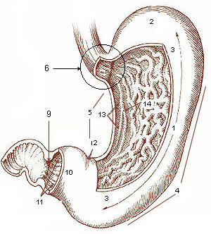
The stomach is a muscular, hollow organ in the gastrointestinal tract of humans and many other animals, including several invertebrates. The stomach has a dilated structure and functions as a vital organ in the digestive system. The stomach is involved in the gastric phase of digestion, following chewing. It performs a chemical breakdown by means of enzymes and hydrochloric acid. In humans and many other animals, the stomach is located between the oesophagus and the small intestine. The stomach secretes digestive enzymes and gastric acid to aid in food digestion. The pyloric sphincter controls the passage of partially digested food ( chyme) from the stomach into the duodenum, where peristalsis takes over to move this through the rest of intestines. Structure In the human digestive system, the stomach lies between the oesophagus and the duodenum (the first part of the small intestine). It is in the left upper quadrant of the abdominal cavity. The top of the stomach lies ag ... [...More Info...] [...Related Items...] OR: [Wikipedia] [Google] [Baidu] |
Gastric Antral Vascular Ectasia
Gastric antral vascular ectasia (GAVE) is an uncommon cause of chronic gastrointestinal bleeding or iron deficiency anemia. The condition is associated with dilated small blood vessels in the pyloric antrum, which is a distal part of the stomach. The dilated vessels result in intestinal bleeding. It is also called watermelon stomach because streaky long red areas that are present in the stomach may resemble the markings on watermelon. The condition was first discovered in 1952, and reported in the literature in 1953. Watermelon disease was first diagnosed by Wheeler ''et al.'' in 1979, and definitively described in four living patients by Jabbari ''et al.'' only in 1984. As of 2011, the cause and pathogenesis are still not known. However, there are several competing hypotheses as to various causes. Signs and symptoms Most patients who are eventually diagnosed with watermelon stomach come to a physician complaining of anemia and blood loss. Sometimes, a patient may come to the physi ... [...More Info...] [...Related Items...] OR: [Wikipedia] [Google] [Baidu] |


