|
Extraembryonic Membrane
The extraembryonic membranes are four membranes which assist in the development of an animal's embryo. Such membranes occur in a range of animals from humans to insects. They originate from the embryo, but are not considered part of it. They typically perform roles in nutrition, gas exchange, and waste removal. There are four standard extraembryonic membranes in birds, reptiles, and mammals: the yolk sac which surrounds the yolk, the amnion which surrounds and cushions the embryo, the allantois which among avians stores embryonic waste and assists with the exchange of carbon dioxide with oxygen as well as the resorption of calcium from the shell, and the chorion which surrounds all of these and in avians successively merges with the allantois in the later stages of egg development to form a combined respiratory and excretory organ called the chorioallantois. The extraembryonic membranes in insects include a serous membrane originating from blastoderm cells, an amnion or amniot ... [...More Info...] [...Related Items...] OR: [Wikipedia] [Google] [Baidu] |
Fetal Membranes
The fetal membranes are the four extraembryonic membranes, associated with the developing embryo, and fetus in humans and other mammals.. They are the amnion, chorion, allantois, and yolk sac. The amnion and the chorion are the chorioamniotic membranes that make up the amniotic sac which surrounds and protects the embryo. The fetal membranes are four of six accessory organs developed by the conceptus that are not part of the embryo itself, the other two are the placenta, and the umbilical cord. Structure The fetal membranes surround the developing embryo and form the fetal-maternal interface. The fetal membranes are derived from the trophoblast layer (outer layer of cells) of the implanting blastocyst. The trophoblast layer differentiates into amnion and the chorion, which then comprise the fetal membranes. The amnion is the innermost layer and, therefore, contacts the amniotic fluid, the fetus and the umbilical cord. The internal pressure of the amniotic fluid causes the amn ... [...More Info...] [...Related Items...] OR: [Wikipedia] [Google] [Baidu] |
Membrane
A membrane is a selective barrier; it allows some things to pass through but stops others. Such things may be molecules, ions, or other small particles. Membranes can be generally classified into synthetic membranes and biological membranes. Biological membranes include cell membranes (outer coverings of cells or organelles that allow passage of certain constituents); nuclear membranes, which cover a cell nucleus; and tissue membranes, such as mucosae and serosae. Synthetic membranes are made by humans for use in laboratories and industry (such as chemical plants). This concept of a membrane has been known since the eighteenth century but was used little outside of the laboratory until the end of World War II. Drinking water supplies in Europe had been compromised by the war and membrane filters were used to test for water safety. However, due to the lack of reliability, slow operation, reduced selectivity and elevated costs, membranes were not widely exploited. The first us ... [...More Info...] [...Related Items...] OR: [Wikipedia] [Google] [Baidu] |
Embryo
An embryo is an initial stage of development of a multicellular organism. In organisms that reproduce sexually, embryonic development is the part of the life cycle that begins just after fertilization of the female egg cell by the male sperm cell. The resulting fusion of these two cells produces a single-celled zygote that undergoes many cell divisions that produce cells known as blastomeres. The blastomeres are arranged as a solid ball that when reaching a certain size, called a morula, takes in fluid to create a cavity called a blastocoel. The structure is then termed a blastula, or a blastocyst in mammals. The mammalian blastocyst hatches before implantating into the endometrial lining of the womb. Once implanted the embryo will continue its development through the next stages of gastrulation, neurulation, and organogenesis. Gastrulation is the formation of the three germ layers that will form all of the different parts of the body. Neurulation forms the nervous ... [...More Info...] [...Related Items...] OR: [Wikipedia] [Google] [Baidu] |
Yolk Sac
The yolk sac is a membranous sac attached to an embryo, formed by cells of the hypoblast layer of the bilaminar embryonic disc. This is alternatively called the umbilical vesicle by the Terminologia Embryologica (TE), though ''yolk sac'' is far more widely used. In humans, the yolk sac is important in early embryonic blood supply, and much of it is incorporated into the primordial gut during the fourth week of embryonic development. In humans The yolk sac is the first element seen within the gestational sac during pregnancy, usually at 3 days gestation. The yolk sac is situated on the front (ventral) part of the embryo; it is lined by extra-embryonic endoderm, outside of which is a layer of extra-embryonic mesenchyme, derived from the epiblast. Blood is conveyed to the wall of the yolk sac by the primitive aorta and after circulating through a wide-meshed capillary plexus, is returned by the vitelline veins to the tubular heart of the embryo. This constitutes the vitell ... [...More Info...] [...Related Items...] OR: [Wikipedia] [Google] [Baidu] |
Yolk
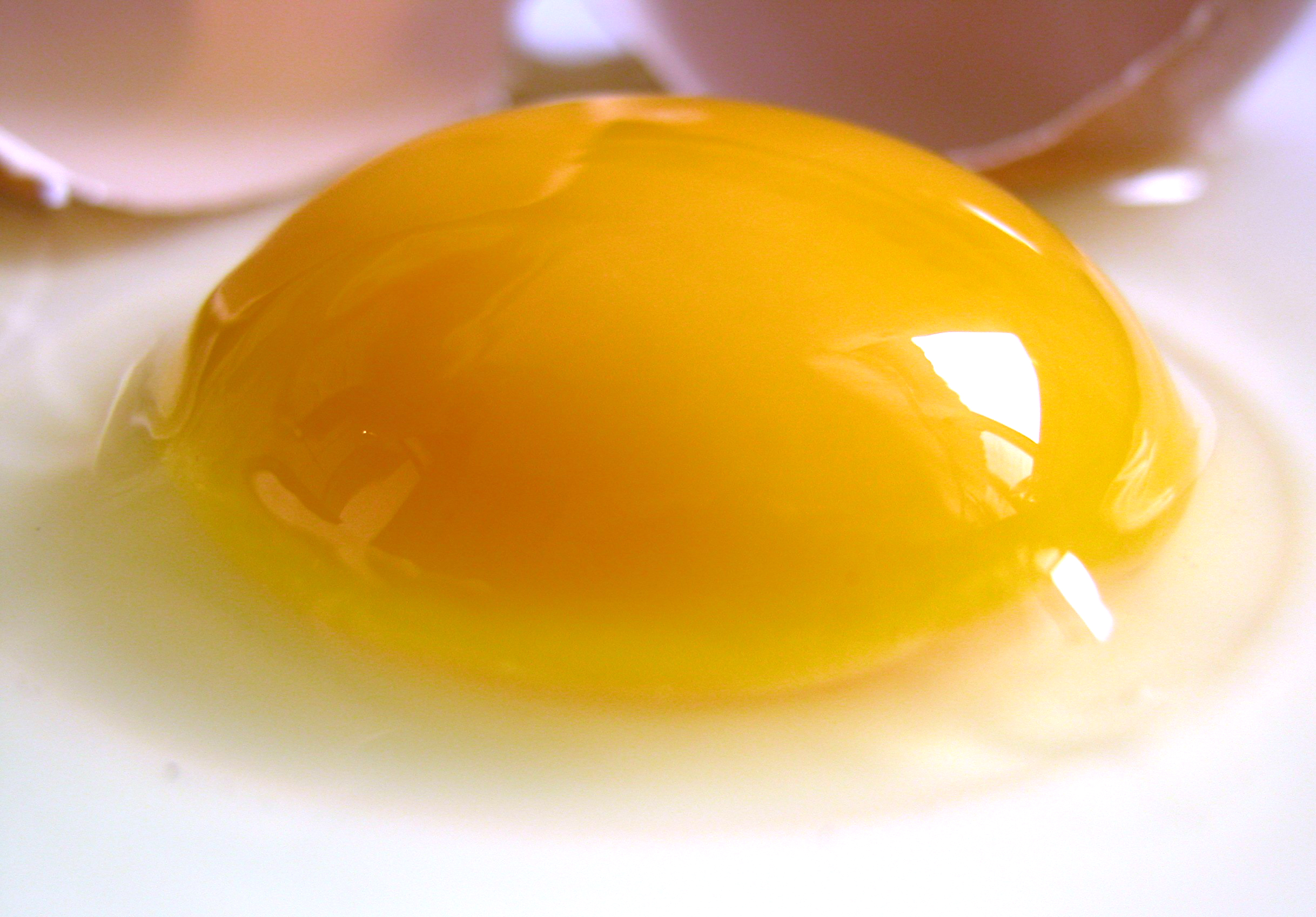
Among animals which produce eggs, the yolk (; also known as the vitellus) is the nutrient-bearing portion of the egg whose primary function is to supply food for the development of the embryo. Some types of egg contain no yolk, for example because they are laid in situations where the food supply is sufficient (such as in the body of the host of a parasitoid) or because the embryo develops in the parent's body, which supplies the food, usually through a placenta. Reproductive systems in which the mother's body supplies the embryo directly are said to be matrotrophic; those in which the embryo is supplied by yolk are said to be lecithotrophic. In many species, such as all birds, and most reptiles and insects, the yolk takes the form of a special storage organ constructed in the reproductive tract of the mother. In many other animals, especially very small species such as some fish and invertebrates, the yolk material is not in a special organ, but inside the egg cell. As sto ... [...More Info...] [...Related Items...] OR: [Wikipedia] [Google] [Baidu] |
Amnion
The amnion is a membrane that closely covers the human and various other embryos when first formed. It fills with amniotic fluid, which causes the amnion to expand and become the amniotic sac that provides a protective environment for the developing embryo. The amnion, along with the chorion, the yolk sac and the allantois protect the embryo. In birds, reptiles and monotremes, the protective sac is enclosed in a shell. In marsupials and placental mammals, it is enclosed in a uterus. The term is from Ancient Greek ἀμνίον 'little lamb', diminutive of ἀμνός 'lamb'. it is cognate with the English verb 'yean', bring forth young (usually lambs). The amnion is a feature of the vertebrate clade ''Amniota'', which includes reptiles, birds, and mammals. Amphibians and fish are not amniotes and thus lack the amnion. The amnion stems from the extra-embryonic somatic mesoderm on the outer side and the extra-embryonic ectoderm or trophoblast on the inner side. In humans In the ... [...More Info...] [...Related Items...] OR: [Wikipedia] [Google] [Baidu] |
Allantois
The allantois (plural ''allantoides'' or ''allantoises'') is a hollow sac-like structure filled with clear fluid that forms part of a developing amniote's conceptus (which consists of all embryonic and extraembryonic tissues). It helps the embryo exchange gases and handle liquid waste. The allantois, along with the amnion, chorion, and yolk sac (other extraembryonic membranes), identify humans and other mammals, birds, and other reptiles as amniotes. Fish and amphibians are anamniotes, and lack the allantois. In mammals the extraembryonic membranes are known as the fetal membranes. Function This sac-like structure, whose name is the New Latin equivalent of "sausage" (in reference to its shape when first formed) is primarily involved in nutrition and excretion, and is webbed with blood vessels. The function of the allantois is to collect liquid waste from the embryo, as well as to exchange gases used by the embryo. In mammals In mammals excluding egg-laying monotremes, the all ... [...More Info...] [...Related Items...] OR: [Wikipedia] [Google] [Baidu] |
Chorion
The chorion is the outermost fetal membrane around the embryo in mammals, birds and reptiles (amniotes). It develops from an outer fold on the surface of the yolk sac, which lies outside the zona pellucida (in mammals), known as the vitelline membrane in other animals. In insects it is developed by the follicle cells while the egg is in the ovary.Chapman, R.F. (1998) "The insects: structure and function", Section ''The egg and embryology''. Previewed in Google Bookon 26 Sep 2009. Structure In humans and other mammals (excluding monotremes), the chorion is one of the fetal membranes that exist during pregnancy between the developing fetus and mother. The chorion and the amnion together form the amniotic sac. In humans it is formed by extraembryonic mesoderm and the two layers of trophoblast that surround the embryo and other membranes; the chorionic villi emerge from the chorion, invade the endometrium, and allow the transfer of nutrients from maternal blood to fetal blood. ... [...More Info...] [...Related Items...] OR: [Wikipedia] [Google] [Baidu] |
Serosa
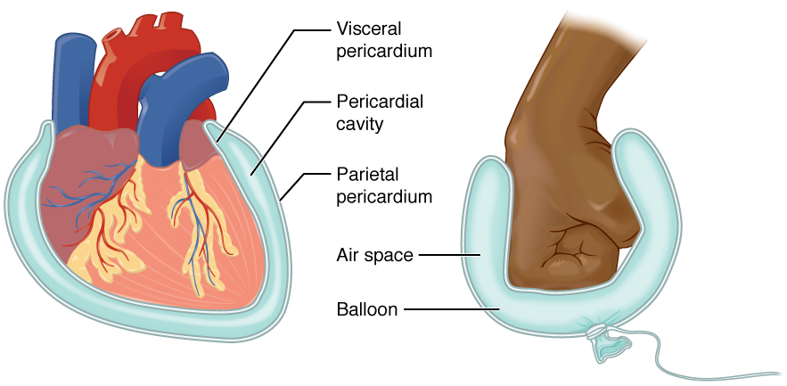
The serous membrane (or serosa) is a smooth tissue membrane of mesothelium lining the contents and inner walls of body cavities, which secrete serous fluid to allow lubricated sliding movements between opposing surfaces. The serous membrane that covers internal organs is called a ''visceral'' membrane; while the one that covers the cavity wall is called the ''parietal'' membrane. Between the two opposing serosal surfaces is often a potential space, mostly empty except for the small amount of serous fluid. The Latin anatomical name is '' tunica serosa''. Serous membranes line and enclose several body cavities, also known as serous cavities, where they secrete a lubricating fluid which reduces friction from movements. Serosa is entirely different from the adventitia, a connective tissue layer which binds together structures rather than reducing friction between them. The serous membrane covering the heart and lining the mediastinum is referred to as the pericardium, the serous ... [...More Info...] [...Related Items...] OR: [Wikipedia] [Google] [Baidu] |
Blastoderm
A blastoderm (germinal disc, blastodisc) is a single layer of embryonic epithelial tissue that makes up the blastula. It encloses the fluid filled blastocoel. Gastrulation follows blastoderm formation, where the tips of the blastoderm begins the formation of the ectoderm, mesoderm, and endoderm. Formation The blastoderm is formed when the oocyte plasma membrane begins cleaving by invagination, creating multiple cells that arrange themselves into an outer sleeve to the blastocoel. In oviparous In chicken eggs, the blastoderm represents a flat disc after embryonic fertilization. At the edge of the blastoderm is the site of active migration by most cells.{{cite book, last1=Bellairs, first1=Ruth, last2=Osmond, first2=Mark, title=Atlas of Chick Development, publisher=Atlas Press, page=15–28, edition=3 See also * Blastodisc *Embryology * Cleavage *Gastrulation Gastrulation is the stage in the early embryonic development of most animals, during which the blastula (a single-layered ho ... [...More Info...] [...Related Items...] OR: [Wikipedia] [Google] [Baidu] |
Zerknüllt
''Zerknüllt'' (zen, German for "crumpled") is a gene in the Antennapedia complex of ''Drosophila'' (fruit flies) and other insects, where it operates very differently from the canonical Hox genes in the same gene cluster. Comparison of Hox genes between species showed that the ''Zerknüllt'' gene evolved from one of the standard Hox genes (the 'paralogy group 3' Hox gene) in insects through accumulating many amino acid changes, changing expression pattern, losing ancestral function and gaining a new function. ''Zerknüllt'' codes for a homeoprotein regulates aspects of early embryogenesis in insects. Unlike the canonical Hox genes which are expressed in precise zones along the anteroposterior (head to tail) body axis, ''zerknüllt'' expression is restricted along the dorsoventral (back to belly) body axis. Expression of ''Zerknüllt'' is repressed in the ventral part of the embryo by a protein called Dorsal, and activated in the dorsal part of the embryo by the TGF beta signa ... [...More Info...] [...Related Items...] OR: [Wikipedia] [Google] [Baidu] |


_en_rotate_05.jpg)