|
Enamel Hypocalcification
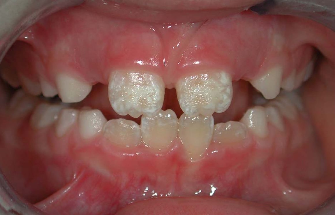
Enamel hypocalcification is a defect of tooth enamel in which normal amounts of enamel are produced but are hypomineralized. In this defect the enamel is softer than normal. Some areas in enamel are hypocalcified: enamel spindles, enamel tufts, and enamel lamellae. Causal factors may occur locally, affecting only a single tooth, or they may act systemically, affecting all teeth in which enamel is being formed. Local trauma Trauma most often refers to: * Major trauma, in physical medicine, severe physical injury caused by an external source * Psychological trauma, a type of damage to the psyche that occurs as a result of a severely distressing event *Traumatic i ... or abscess formation can adversely affect the ameloblasts overlying a developing Crown (tooth), crown, resulting in enamel hypocalcification or hypoplasia. Affected teeth may have areas of coronal discoloration, or they may have actual pits and irregularities. This is most commonly seen in permanent teeth in which ... [...More Info...] [...Related Items...] OR: [Wikipedia] [Google] [Baidu] |
Tooth
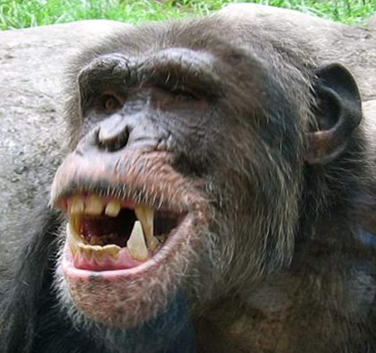
A tooth ( : teeth) is a hard, calcified structure found in the jaws (or mouths) of many vertebrates and used to break down food. Some animals, particularly carnivores and omnivores, also use teeth to help with capturing or wounding prey, tearing food, for defensive purposes, to intimidate other animals often including their own, or to carry prey or their young. The roots of teeth are covered by gums. Teeth are not made of bone, but rather of multiple tissues of varying density and hardness that originate from the embryonic germ layer, the ectoderm. The general structure of teeth is similar across the vertebrates, although there is considerable variation in their form and position. The teeth of mammals have deep roots, and this pattern is also found in some fish, and in crocodilians. In most teleost fish, however, the teeth are attached to the outer surface of the bone, while in lizards they are attached to the inner surface of the jaw by one side. In cartilaginous fish, s ... [...More Info...] [...Related Items...] OR: [Wikipedia] [Google] [Baidu] |
Tooth Enamel
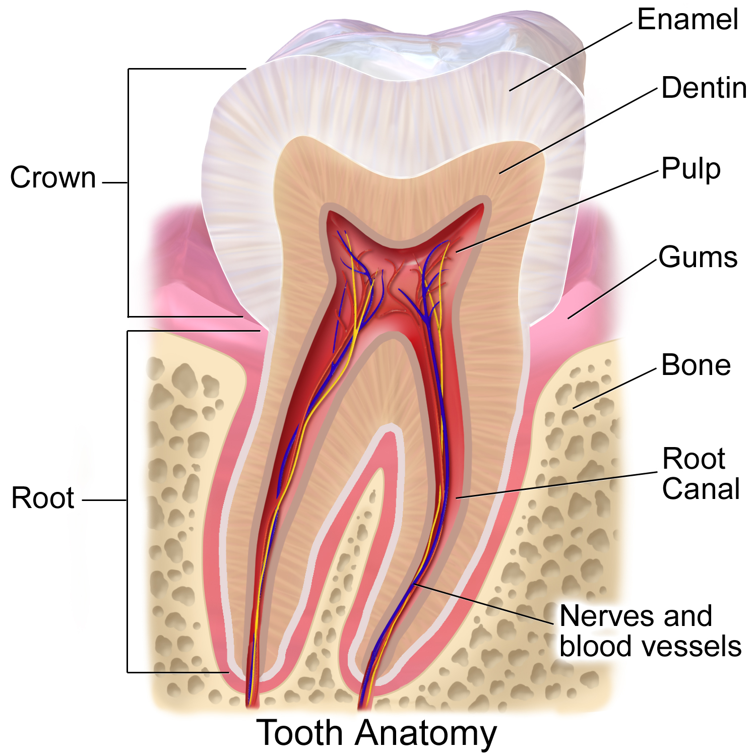
Tooth enamel is one of the four major Tissue (biology), tissues that make up the tooth in humans and many other animals, including some species of fish. It makes up the normally visible part of the tooth, covering the Crown (tooth), crown. The other major tissues are dentin, cementum, and Pulp (tooth), dental pulp. It is a very hard, white to off-white, highly mineralised substance that acts as a barrier to protect the tooth but can become susceptible to degradation, especially by acids from food and drink. Calcium hardens the tooth enamel. In rare circumstances enamel fails to form, leaving the underlying dentin exposed on the surface. Features Enamel is the hardest substance in the human body and contains the highest percentage of minerals (at 96%),Ross ''et al.'', p. 485 with water and organic material composing the rest.Ten Cate's Oral Histology, Nancy, Elsevier, pp. 70–94 The primary mineral is hydroxyapatite, which is a crystalline calcium phosphate. Enamel is formed o ... [...More Info...] [...Related Items...] OR: [Wikipedia] [Google] [Baidu] |
Trauma (medicine)
An injury is any physiological damage to living tissue caused by immediate physical stress. An injury can occur intentionally or unintentionally and may be caused by blunt trauma, penetrating trauma, burning, toxic exposure, asphyxiation, or overexertion. Injuries can occur in any part of the body, and different symptoms are associated with different injuries. Treatment of a major injury is typically carried out by a health professional and varies greatly depending on the nature of the injury. Traffic collisions are the most common cause of accidental injury and injury-related death among humans. Injuries are distinct from chronic conditions, psychological trauma, infections, or medical procedures, though injury can be a contributing factor to any of these. Several major health organizations have established systems for the classification and description of human injuries. Occurrence Injuries may be intentional or unintentional. Intentional injuries may be acts of vio ... [...More Info...] [...Related Items...] OR: [Wikipedia] [Google] [Baidu] |
Abscess
An abscess is a collection of pus that has built up within the tissue of the body. Signs and symptoms of abscesses include redness, pain, warmth, and swelling. The swelling may feel fluid-filled when pressed. The area of redness often extends beyond the swelling. Carbuncles and boils are types of abscess that often involve hair follicles, with carbuncles being larger. They are usually caused by a bacterial infection. Often many different types of bacteria are involved in a single infection. In many areas of the world, the most common bacteria present is ''methicillin-resistant Staphylococcus aureus''. Rarely, parasites can cause abscesses; this is more common in the developing world. Diagnosis of a skin abscess is usually made based on what it looks like and is confirmed by cutting it open. Ultrasound imaging may be useful in cases in which the diagnosis is not clear. In abscesses around the anus, computer tomography (CT) may be important to look for deeper infection. Standa ... [...More Info...] [...Related Items...] OR: [Wikipedia] [Google] [Baidu] |
Ameloblast
Ameloblasts are cells present only during tooth development that deposit tooth enamel, which is the hard outermost layer of the tooth forming the surface of the crown. Structure Each ameloblast is a columnar cell approximately 4 micrometers in diameter, 40 micrometers in length and is hexagonal in cross section. The secretory end of the ameloblast ends in a six-sided pyramid-like projection known as the Tomes' process. The angulation of the Tomes' process is significant in the orientation of enamel rods, the basic unit of tooth enamel. Distal terminal bars are junctional complexes that separate the Tomes' processes from ameloblast proper. Development Ameloblasts are derived from oral epithelium tissue of ectodermal origin. Their differentiation from preameloblasts (whose origin is from inner enamel epithelium) is a result of signaling from the ectomesenchymal cells of the dental papilla. Initially the preameloblasts will differentiate into presecretory ameloblasts and then into s ... [...More Info...] [...Related Items...] OR: [Wikipedia] [Google] [Baidu] |
Crown (tooth)
In dentistry, crown refers to the anatomical area of teeth, usually covered by enamel. The crown is usually visible in the mouth after developing below the gingiva The gums or gingiva (plural: ''gingivae'') consist of the mucosal tissue that lies over the mandible and maxilla inside the mouth. Gum health and disease can have an effect on general health. Structure The gums are part of the soft tissue lin ... and then erupting into place. If part of the tooth gets chipped or broken, a dentist can apply an artificial crown. Crowns are used most commonly to entirely cover a damaged tooth or cover an implant. Bridges are also used to cover a space if one or more teeth is missing. They are cemented to natural teeth or implants surrounding the space where the tooth once stood. There are various materials that can be used including a type of cement or stainless steel. The cement crowns look like regular teeth while the stainless steel crowns are silver or gold. References ... [...More Info...] [...Related Items...] OR: [Wikipedia] [Google] [Baidu] |
Hypocalcification
Enamel hypocalcification is a defect of tooth enamel in which normal amounts of enamel are produced but are hypomineralized. In this defect the enamel is softer than normal. Some areas in enamel are hypocalcified: enamel spindles, enamel tufts, and enamel lamellae. Causal factors may occur locally, affecting only a single tooth, or they may act systemically, affecting all teeth in which enamel is being formed. Local trauma or abscess formation can adversely affect the ameloblasts overlying a developing crown, resulting in enamel hypocalcification or hypoplasia. Affected teeth may have areas of coronal discoloration, or they may have actual pits and irregularities. This is most commonly seen in permanent teeth in which the overlying deciduous tooth becomes abscessed or is physically forced into the enamel organ of the permanent tooth Permanent teeth or adult teeth are the second set of teeth formed in diphyodont mammals. In humans and old world simians, there are thirty-two perma ... [...More Info...] [...Related Items...] OR: [Wikipedia] [Google] [Baidu] |
Hypoplasia
Hypoplasia (from Ancient Greek ὑπo- ''hypo-'' 'under' + πλάσις ''plasis'' 'formation'; adjective form ''hypoplastic'') is underdevelopment or incomplete development of a tissue or organ. Dictionary of Cell and Molecular Biology (11 March 2008) Although the term is not always used precisely, it properly refers to an inadequate or below-normal number of cells.Hypoplasia Stedman's Medical Dictionary. lww.com Hypoplasia is similar to |
Deciduous Teeth
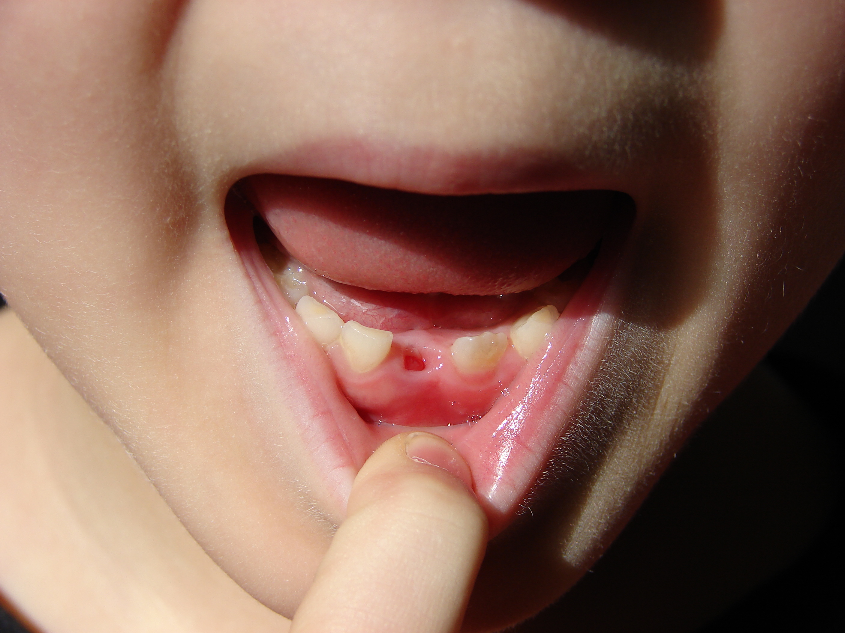
Deciduous teeth or primary teeth, also informally known as baby teeth, milk teeth, or temporary teeth,Illustrated Dental Embryology, Histology, and Anatomy, Bath-Balogh and Fehrenbach, Elsevier, 2011, page 255 are the first set of teeth in the growth and development of humans and other diphyodonts, which include most mammals but not elephants, kangaroos, or manatees which are polyphyodonts. Deciduous teeth develop during the embryonic stage of development and erupt (break through the gums and become visible in the mouth) during infancy. They are usually lost and replaced by permanent teeth, but in the absence of their permanent replacements, they can remain functional for many years into adulthood. Development Formation Primary teeth start to form during the embryonic phase of human life. The development of primary teeth starts at the sixth week of tooth development as the dental lamina. This process starts at the midline and then spreads back into the posterior region. B ... [...More Info...] [...Related Items...] OR: [Wikipedia] [Google] [Baidu] |
Enamel Organ
The enamel organ, also known as the dental organ, is a cellular aggregation seen in a developing tooth and it lies above the dental papilla. The enamel organ which is differentiated from the primitive oral epithelium lining the stomodeum.The enamel organ is responsible for the formation of enamel, initiation of dentine formation, establishment of the shape of a tooth's crown, and establishment of the dentoenamel junction. The enamel organ has four layers; the inner enamel epithelium, outer enamel epithelium, stratum intermedium, and the stellate reticulum. The dental papilla, the differentiated ectomesenchyme deep to the enamel organ, will produce dentin and the dental pulp. The surrounding ectomesenchyme tissue, the dental follicle, is the primitive cementum, periodontal ligament and alveolar bone beneath the tooth root. The site where the internal enamel epithelium and external enamel epithelium coalesce is the cervical root, important in proliferation of the dental root. Toot ... [...More Info...] [...Related Items...] OR: [Wikipedia] [Google] [Baidu] |
Permanent Teeth
Permanent teeth or adult teeth are the second set of teeth formed in diphyodont mammals. In humans and old world simians, there are thirty-two permanent teeth, consisting of six maxillary and six mandibular molars, four maxillary and four mandibular premolars, two maxillary and two mandibular canines, four maxillary and four mandibular incisors. Timeline The first permanent tooth usually appears in the mouth at around six years of age, and the mouth will then be in a transition time with both primary (or deciduous dentition) teeth and permanent teeth during the mixed dentition period until the last primary tooth is lost or shed. The first of the permanent teeth to erupt are the permanent first molars, right behind the last 'milk' molars of the primary dentition. These first permanent molars are important for the correct development of a permanent dentition. Up to thirteen years of age, 28 of the 32 permanent teeth will appear. The full permanent dentition is completed much late ... [...More Info...] [...Related Items...] OR: [Wikipedia] [Google] [Baidu] |
Turner's Tooth
Enamel hypoplasia is a defect of the teeth in which the enamel is deficient in quantity, caused by defective enamel matrix formation during enamel development, as a result of inherited and acquired systemic condition(s). It can be identified as missing tooth structure and may manifest as pits or grooves in the crown of the affected teeth, and in extreme cases, some portions of the crown of the tooth may have no enamel, exposing the dentin. It may be generalized across the dentition or localized to a few teeth. Defects are categorized by shape or location. Common categories are pit-form, plane-form, linear-form, and localised enamel hypoplasia. Hypoplastic lesions are found in areas of the teeth where the enamel was being actively formed during a systemic or local disturbance. Since the formation of enamel extends over a long period of time, defects may be confined to one well-defined area of the affected teeth. Knowledge of chronological development of deciduous and permanent ... [...More Info...] [...Related Items...] OR: [Wikipedia] [Google] [Baidu] |


